
Recommandé
Contenu connexe
Tendances
Tendances (20)
Similaire à INFERIOR ALVEOLAR NERVE BLOCK
Similaire à INFERIOR ALVEOLAR NERVE BLOCK (20)
Dernier
Dernier (20)
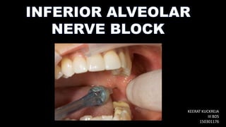
INFERIOR ALVEOLAR NERVE BLOCK
- 2. Inferior alveolar nerve block (IANB) is also known as the mandibular nerve block. Second most frequently used injection (after infiltration) in dentisty. It has the highest percentage of clinical failures. Useful technique in quadrant dentistry. Administration of bilateral IANBs is rarely indicated in dental treatments as : It produces discomfort, primarily from the lingual soft tissue anesthesia. Patient feels unable to swallow. Due to absence of all the sensations the patient is more like to self injure the soft tissues. A supplemental buccal nerve block is needed only when soft tissue anesthesia in the buccal posterior region is necessary.
- 3. 1. Inferior alveolar nerve 2. Incisive nerve 3. Mental nerve 4. Lingual nerve (commonly)
- 4. 1. Mandibular teeth to the midline. 2. Body of the mandible, inferior portion of the ramus 3. Buccal mucoperiosteum, mucus membrane anterior to mental foramen (mental nerve). 4. Anterior two thirds of the tongue and the floor of the oral cavity (lingual nerve). 5.Lingual soft tissues and periosteum (lingual nerve). 1. Procedures on multiple mandibular teeth in one quadrant 2. When buccal soft tissue anesthesia ( anterior to mental foramen) is necessary. 3. When lingual soft tissue anesthesia is necessary. 1. Infection or acute inflammation in the area of injection. 2. Patients who are more likely to bite their lip or tongue,like children or physically and mentally handicapped adult or child.
- 5. 1. A 25 gauge long needle is preferred. 2. Area of insertion : Mucous membrane on the medial side of the ramus of the mandible, at the intersection of two lines- • One horizontal representing the height of needle insertion • The other vertical ,representing the antero-posterior plane of injection. 3. Target area : inferior alveolar nerve as it passes downwards to the mandibular foramen but before it enters into the foramen. 4. Landmarks: • Coronoid notch (greatest concavity on the anterior border of the ramus) • Pterygomandibular raphe (vertical portion ) • Occlusal plane of the mandibular posterior teeth
- 6. 1. Assume the correct position • For a right IANB , 8 o’clock position facing the patient • For a left IANB, 10 o’clock position facing in the same direction as the patient 2. Position the patient in a supine or a semisupine position. Open the mouth wide to allow greater visibility and access 3. Three parameters must be considered during the administrations of IANB: • Height of injection • Anteroposterior placement of the needle • Depth of penetration
- 7. 1. Place the index finger or the thumb of your left hand in the coronoid notch 2. An imaginary line extends posteriorly from the fingertip in the coronoid notch to the deepest portion on the Pterygomandibular raphe. 3. This imaginary line should be parallel to the occlusal plane of the mandibular molar teeth and lies 6-10 mm above the occlusal plane in most patients 1. It is three fourths of the anteroposterior distance from the coronoid notch back to the deepest part of the Pterygomandibular raphe
- 8. Prepare tissue at the injection site and place the barrel of syringe in the corner of the mouth on the contralateral side , usually corresponding the premolars. 1. Slowly advance the needle until you can feel a bony resistance 2. The average depth of penetration to bony contact will be 20-25 mm, approx. 2/3 or ¾ the length of the long dental needle 3. Needle tip should be located slightly superior to the mandibular foramen
- 9. 4. When the bone is contacted , withdraw approx. 1mm to prevent subperiosteal injection. 5. Aspirate in two planes. If negative , slowly deposit 1.5ml of anesthetic over a minimum of 60 seconds 6. Slowly withdraw the syringe , and when half of its length remains within the tissues, reaspirate. If negative , deposit 0.2ml of the remaining solution to anesthetize the lingual nerve • In most patients this is not necessary as the LA from the IANB anesthetizes the lingual nerve 7. Withdraw the needle and make the needle safe.
- 11. • Tingling or numbness of lower lip (anesthesia of mental nerve) • Tingling or numbness of tongue (anesthesia of lingual nerve) • Using an electrical pulp tester • No pain is felt during dental therapy The needle contacts the bone, preventing over insertion and complications • Donot deposit local anesthesia if bone is not contacted • Avoid pain by not contacting the bone too forcefully
- 12. One injection provides wide area of anesthesia (useful for quadrant dentistry • Wide area of anesthesia not indicated for localised procedures • Rate of inadequate anesthesia is 31%- 81% • Intraoral landmarks not consistently reliable • Chances of positive aspiration are 10% - 15% • Lingual and lower lip anesthesia may sometimes lead to self inflicted soft tisse trauma • Partial anesthesia where bifid inferior alveolar nerve and mandibular canals present
- 13. The most common causes of absent or incomplete IANB are : 1. Deposition of anesthetic too low , below the mandibular foramen 2. Deposition of anesthetic too far anteriorly (laterally) on the ramus 3. Accessory innervation to the mandibular teeth • Several nerves provide the mandibular teeth with accessory innervation but mylohyoid nerve acts as the prime candidate • The Gow-Gates mandibular nerve block which blocks the mylohyoid nerve is not associated with problems of accessory iinervation 4. Incomplete anesthesia of central and lateral incisor • This is due to overlapping fibres of the contralateral inferior alveolar nerve
- 14. 1. HEMATOMA • Swelling of tissues on the medial side of the mandibular ramus after deposition of anesthetic 2. TRISMUS • Muscle soreness or limited movement • A slight degree of soreness when openening the mandible is common • More severe soreness is rare 3. TRANSIENT FACIAL NERVE PALSY • Produced by deposition of local anesthesia into the body of the parotid gland, blocking the VII cranial nerve. • Signs and symptoms include inability to close the lower eyelid and drooping of upper lip on the effected side
- 15. •Handbook of local anesthesia – Stanely F. Malamed •Google images