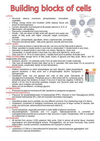
1. LIPIDS
o Structural: plasma membrane (phospholipids). Intracellular
organelles
o Energy: energy stores and insulation (white adipose tissue and
brown for thermogenesis)
o Carriers: absorption and transport of fat soluble vitamins A, D, E, K
o Messengers: cell signalling
o Precursors: cholesterol for many hormones.
o Simple – fats are esters of fatty acids with glycerol and waxes are
esters of fatty acids with higher molecular weight monohydric
alcohols.
o Complex – phospholipids, glycolipids, others: sulphonamides, aminolipids
o Precursor and derived lipids – fatty acids, glycerol, steroids and hormones.
FATTY ACIDS
o Occur mainly as esters in natural fats and oils, can occur as free fatty acids in plasma
o Either: saturated no double bonds in acyl chain or unsaturated >1 double bonds in acyl chain.
o Saturated: no double bonds in acyl chain, e.g. butyric. Named by –anoic acid.
o Unsaturated: >1 double bonds in acyl chain, e.g. oleic acid. Named by –enoic acid.
o Oleic acid 18:1:9. 18 no of carbon atoms, 1 double bond and 9 is position of double bond.
TRIGLYCERIDESMain storage forms of fatty acids. Esters of glycerol and fatty acids. Mono- and Di-
acyglycerides also exists.
o Synthesis: alcohol + 3x carboxylic acids. Form an ester bond and 3 water molecules.
o Fats have all available binding sites taken up by H, saturated. Oils have fewer H’s on account of
double bonds, unsaturated, liquids at room temperature.
PHOSPHOLIPIDS
o Platform (backbone) on which phospholipids are built: Glycerol, called phospholipids,
glycerol backbone, 2 fatty acids and a phosphorylated alcohol. Spingosine –
sphingomyelin.
o Substituted lipids, one unit glycerol, two units of fatty acids. Derivatives of
diacylglycerol-3-phosphate. Phosphate group is often linked with N2 containing group,
phosphate group has tendency to loose H ion-one O becomes negative. N tends to
attract a H ion one N becomes positively charged, end becomes polar and water
soluble, tail is nonpolar and insoluble.
o Base unit can be different, not always glycerol.
SPHINGOLIPIDS
o Prominent on plasma membrane of cells, backbone is spingosine not glycerol
PHOSPHATIDYLINOSITOLS
o Precursor for phosphatidylinositol 4,5-bisphosphate (PIP2). Cleaved to form Diacylglycerol (DAG)
and Inositol triphosphate (IP3), both are important secondary messengers.
STEROIDS
o Classified as lipids due to solubility, but very different structure. Four interlocking rings of C atoms.
o Cholesterol: constituent of biological membranes and precursor of large number of steroids, bile
acids, sex hormones, adrenocortical hormones and vitamin D.
GLYCOLIPIDS
o Found everywhere, especially prominent in outer leaflet of plasma membrane.
o Glycosphingolipids: Ceramine and 1-n sugars: Galactosylceramide is converted to
suphogalactoslyceramide and is present in high amounts in myelin. Gangliosides, GM1 is a receptor
for cholera toxin in intestine.
EICOSANOIDS
o All derived from eicosa (-C20) polyenoic fatty acids. Exist in almost all animal tissue, important
physiological and pharmacological actions: Prostaglandins, act as local hormones, they control
inflammation, muscle contraction. Leukotrienes are important in asthma.
LEUKOTRIENES
o Leukotriene (LT) B4 is a chemoattractant for leukocytes, induces leukocyte aggreagation and
adherence to vascular endothelium.
2. o LTC4, LTD4 and LTE4 are bronchoconstrictors, induce airway mucous secretion, increase vascular
permeability and are realeased from mast cells during an asthma attack.
PROPERTIES OF LIPIDS
o Melting points of even numbered carbon fatty acids increase with chain length and decrease with
increasing unsaturation.
o Membrane lipids are more unsaturated than storage lipids and are more fluidic. Cholesterol reduces
fluidity.
AMINO ACIDS
INTRODUCTION
o Polymers of 20 distinct amino acid monomers. Amino acids fall into 7 categories
based on their sidechain (R): aliphatic, aromatic, alcoholic, sulphur containing,
acidic, basic or amide. The amino side chain provides specific chemical property.
o Form a peptide bond between adjacent carboxylic acid and amino groups.
FUNCTIONS
o Catalysis: Proteins that catalyse chemical reactions in the body
o Storage and transport: Ferritin stores and transports iron in the body. Haemoglobin transports O.
o Mechanical support and shape: Collagen is a component of supportive tissue.
o Decoding information, gene expression: RNA polymerase synthesises RNA.
o Specialist functions: immunoglobulins (antibodies) and some hormones.
ALIPHATIC AMINO ACIDS
o Include simplest amino acid glycine where R=H. The hydrocarbon akyl side chains contain no
functional groups but are very hydrophobic. Include Gly-G, Ala-A, Val-V, Leu-L, Ile-I and Pro-P.
AROMATIC AMINO ACIDS
o Contain aromatic ring side chains. E.g. Phe-F, Trp-W and Tyr-Y all are quite
hydrophobic.
ALCOHOL CONTAINING AMINO ACIDS
o Side chains contain an alcohol functional group are chemically reactive and hydrophilic since they
can interact with water and other polar groups. E.g. Ser-S and Thr-T.
SULPHUR CONTAINING AMINO ACIDS
o Have side chains that contain sulphur, so are hydrophobic. –SH groups are very chemically
reactive. E.g. Cys-C and Met-M.
ACIDIC AMINO ACIDS
o Side chains contain –COOH groups, they are acidic and very hydrophilic. Chemically reactive and
found in active sites of enzymes. E.g. Asp-d and Glu-E.
BASIC (NITRGOGEN CONTAINING) AMINO ACIDS
o Side chains often contain nitrogen, so they are very hydrophilic, also often found in active sites of
enzymes. E.g. Lys-K, Arg-R and His-H.
AMIDE CONTAINING AMINO ACIDS
o Neutral and chemically less reactive than carboxylic acids. They are still very polar and therefore
hydrophilic. E.g. Asn-N and Gln-Q.
AMINO ACIDS
o Essential: Arg, His, Ile, Leu, Lys, Met, Phe, Thr, Trp and Val.
o Non-Essential: Ala, Asn, Asp, Cys, Glu, Gln, Gly, Pro, Ser, Tyr.
o Essential amino acids are biosynthesised by plants or microorganisms and must be obtained from
the diet. Non-essential can be biosynthesised within the body but mostly obtained from the diet.
o Used as starting materials for the biosynthesis of important molecules within the body. The
neurotransmitters Dopamine and Histamine are derived from amino acids.
ACIDITY/BASICITY OF AMINO ACIDS
o Human body is mostly aqueous and buffered at pH 7.4. In these
conditions, free amino acids exist as zwitterions, ionisation state varies
with pH. Side chain functional groups can also be effected by pH.
STEREOCHEMISTRY OF AMINO ACIDS
o Contain chiral centres. They can rotate plane polarized light in a clockwise (+)
or anti-clockwise (-) direction: they are optically active. They can exist as
enantiomers: compounds with the same molecular formula but differing in
their configuration in space.
o Pairs of amino acids are designated D or L. Reference compound is glyceraldehyde (a sugar). L-
amino acids have same configuration at their chiral carbon as L-glyceraldehyde. Glycine does not
have L-configuration but all other naturally occurring amino acids have it.
3. LABORATORY PROTEIN SYNTHESIS
o Condensation reaction between amino group and carboxyl group forming a peptide bond. A peptide
is a small protein of less than 50 amino acids. Peptides should be drawn from the amino (N)
terminal on the left to the carboxyl (C) terminal on the right.
o Steps – Protection: blocking of carboxyl and amino groups not involved in required peptide bond.
Activation: of carboxyl group involved in peptide bond
formation. The carboxyl group can be activated by
conversion to an acid chloride. Coupling: Reaction of free
amino group of the second amino acid with the activated
carboxyl group to form a new peptide bond. Deprotection:
Removal of the protecting groups.
USES OF PEPTIDES
o Aspartame is a synthetic dipeptide 200x sweeter than sugar. Β-endorphin is an endogenous 31
amino acid neuropeptide with analgesic effects. Eptifibatide is a cardiovascular antiplatelet drug.
o Ciclosporin is a immunosuppressant drug, isolated from natural sources that prevents rejection
following organ and tissue transplantation. It is a cyclic peptide that contains naturally occurring and
modified amino acids. Peptides are readily metabolised in the stomach.
BONDING IN PROTEINS
o Strong-Covalent, most common in backbone.
o Moderate-Ionic, attraction between charged groups, also called a salt bridge.
o Weak-Hydrogen bond, very important for protein structure.
o Very weak-hydrophobic, exclusion of water a driving force of this interaction.
PROTEINS
STRUCTURE
o Linear sequence of amino acids primary structure of a protein. Secondary structure is the 3D
arrangement of the primary amino acid sequence. Tertiary structure refers to 3D arrangement of
secondary structure. Quaternary structure composed of more than one polypeptide strand.
o Peptide bonds have an impact on the shape and function of
proteins. Peptide bond is planar, electron resonance gives 40%
double bond character. Peptide bond is the average of two
extreme resonance forms. The peptide bond is described as rigid and planar, rotation not possible.
Peptide bonds have a trans conformation.
o Only two free movements exist in a polypeptide chain: Rotation about the αC-N bond is called the
phi torsion angle, rotation about the αC-C bond is called psi torsion angle. Protein conformation
depends on phi and psi rotation. The flexibility of these bonds allows primary sequence to fold into
its native conformation. Rotation is limited by: Steric hindrance, bulky groups e.g. side chains
cannot approach each other. Rigidity of the peptide bond, ultimately restricts movement. Favourable
interactions, with other polypeptide chains.
α-HELIX
o The α-Helix results when consecutive amino acids have similar phi and psi angle values. Phi -57°
and Psi -47°. It is a single helix and 3.6 amino acids are required for each complete turn. Each
backbone carbonyl oxygen is hydrogen boded to the peptide nitrogen of the fourth residue along,
this stabilises the helix. Side chains are arranged on the outside. Receptors are proteins rich in α-
helices, usually contain a trans-membrane domain composed of entirely α-helices.
β-SHEET
o β-sheet is an elongated, flat structure. Inter-strand hydrogen bonds between backbone carbonyl
oxygen and amide nitrogens stabilize β-sheet, side chain interactions provide additional
stabilisation. Come in two varities, Antiparallel and Parallel. Antiparallel: linear hydrogen bonds, 2-
15 strands. Parallel: hydrogen bonds distorted, sheet less stable, no more than 5 strands
encountered. Avidin β-sheet, commonly found in egg whites and entirely composed of a β-sheet.
The β-turn occurs between β-strands. Hydrogen bonding stabilises a 180° change in direction.
FIBROUS PROTEINS
o Contain only α-Helix, simple, elongated structure resembling threads. Provide mechanical support in
skin, tendons and bones. Physically durable, chemically inert and water insoluble. Structure is
maintained by hydrogen bonding within the α-Helix. E.g. collagen.
GLOBULAR PROTEINS
o All enzymes are globular proteins. Water soluble, compact, roughly spherical, tightly folded peptide
chains, hydrophobic interior, hydrophilic surface, structure maintained by covalent and hydrogen
boding, non-covalent crosslinks and hydrophobic interactions, possess indents/clefts.
4. STABILISING TERTIARY STRUCTURE
o Hydrophobic effect - Proteins are more stable in water, non-polar side chains aggregate, causing
the protein to fold with non-polar sidechains inside and polar sidecahins outside the protein in
contact with water. Efficient packing maximises Van Der Waals interactions between non polar
residues and excludes water from interior of the protein. Structure controls function: Val, Leu, Ile,
Met, Phe and Ala rarely encountered on protein exterior.
o Non-covalent Interactions – Arranged so vitually all possible hydrogen bonds form. Polar sied
chains forced into interior so they can neutralize their polarity by forming hydrogen bonds or
electrostatic interactions.
o Covalent Interactions – Disulfide bonds are covalent cross links that form between adjacent
cysteine residues and help stabilise the conformations of some proteins. Especially common in
proteins that are secreted from cells.
o Effect of Temperature and pH – tertiary structure is responsible for biological activity. Tertiary
structure is maintained by weak interactions. Variations of pH/temperature disrupt the stabilising
interactions, causing changes to the tertiary structure. The protein is said to be denatured. Enzymes
have evolved to function at physiological conditions pH 7.4 and 37°C.
QUARTERNARY STRUCTURE
o Haemoglobin is composed of 4 subunits, tow identical α-units and two identical β-units, each unit
contains an iron atom vital for oxygen transport.
o Insulin a hormone that controls glucose metabolism consists of two peptide chains, linked and
maintained in biological active conformation by three disulphide bridges.
ENZYMES
o Catalysts: do not change the outcome of a reaction but effects the rate of a reaction by lowering the
activation energy for that reaction. Enzymes achieve catalysis by reducing the activation energy.
o Reactions are fast, specific for substrates-enzymes can distinguish between functional groups,
isomers and enantiomers, they are also efficient-minimise waste products.
o Most enzymes function at pH 7.4 and 37°C.
o Enzymes are globular proteins. Tertiary structure creates active site pockets, they can bind one or
more substances in an active site: substrate and cofactors may be required for the reactions.
NOMENCLATURE
o Classified according to reactions catalysed. Typically ‘-ase’ is added to substrate term for the type of
reaction. Categories enzymes according to 6 major groups according to class of chemical reactions
they catalyse. Oxidoreductases, Transferases, Hydrolases, Lyases, Isomerases and Ligases.
o Enzyme Commision assigns a unique code number to each enzyme. E.g. 3.2.1.4. First number is
the main class, second number is the sub-class, third number is the sub-sub class, the last number
is the serial number of the enzyme in the sub-sub class.
COFACTORS
o Cofactors supply chemical groups not otherwise found in active sites. Many enzymes are inactive
as a protein alone (apoenzymes) and cofactors are required for activity.
o Cosubstrates are weakly bound to the enzyme and are altered during the course of the reaction and
dissociate from the active site, (regenerated in another enzymic reaction and recycled.
o Prosthetic groups are tightly bound to the enzyme but must be regenerated each catalytic cycle.
ESSENTIAL IONS
o Nearly a third of proteins need a metal ion for activity, known as metalloproteins or metalloenzymes.
o Fe2+
found in haem (haemoglobin), Mg2+
found in many kinases (reactions involving
phosphorylations, Zn2+
found in many enzymes (oxidations/reductions, DNA recognitions)
CLASS 1 – OXIDOREDUCTASES
o Catalyse REDOX reactions. Enzymes in this class include
dehydrogenase, oxidases, peroxidases and reductases. E.g. alcohol
dehydrogenase oxidises thanol to ethanol, using a cofactor, NAD+
and a
zinc ion.
CLASS 2 – TRANSFERASES
o Catalyse functional group transfer reactions. Include kinases which catalyse
phosphorylation reactions.
o Transcarboxylase catalyses simultansous conversion of propionyl coA to methyl
malonyl CoA and oxaloacetate to pyruvate, which are important metabolic
reactions.
CLASS 3 – HYDROLASES
o Catalyse hydrolysis reactions.
5. o Trypsin mediates hydrolysis of Lys-aa or Arg-aa peptide bones. It is a
serine protease enzyme. It needs no cofactor, its active site contains
sufficient functionality to be able to perform the reaction unassisted.
o Trypsin mechanism of action: Serine residue attacks the substrate
peptide carbonyl bond, aided by Asp and His which promote the nuleophillic attack and stabilise the
resulting positive charge. The tetrahedral intermediate collapses, liberating R-NH2, but the carboxyl
residue remains bonded to the enzyme, via the serine. A water molecule, correctly orientated by
interaction with the His, attacks the ester carbonyl group. Reaction complete, the active amino acids
are unchanged and ready for further catalysis. New amino and carboxylic groups exposed to
hydrolysis.
CLASS 4 – LYASES
o Catalyse bond breaking reactions but not hydrolytic or oxidative
bond breaking. E.g. L-Dopa decarboxylase.
CLASS 5 – ISOMERASES
o Catalyse isomerisation reactions, i.e. the arrangement of groups within a substrate molecule. E.g.
alanine racemase, found in bacteria and catalyses the
interconversion of L and D alanine. D-alanine is
essential for bacterial cell wall.
o Enzyme removes H from one face of amino acid (A)
and replaces on opposite ace (B).
CLASS 6 – LIGASES
o Catalyse the ligation (joining) of two substrates. Pyruvate
carboxylase joins pyruvate with carbon dioxide to generate
oxaloacetate. This is an important step in metabolism/ the Krebs
Cycle.
NUCLEIC ACIDS
STRUCTURE
o A sugar molecule attached to a base and a phosphate.
o Bases: based on purine. E.g. adenine and Guanine or based on pyrmidine. E.g.
Cytosine and Thymine/Uracil.
o Sugars 2-deoxy-D-ribose in DNA and D-ribose in RNA.
o Monomers: Nucleoside-sugar and base, Nucleotide-sugar,
base and phosphate.
NUCLEOSIDES
o Numbering: Start at N attached to sugar, then try and give N’s the lowest possible number.
Anomeric position, is where base joins prime carbon.
o Synthesis: link base to sugar. Issues: base insolubility, sugar
polyfunctionality, reactivity and stereochemistry. TMS-Cl is used to
solubilise base. Stereochemistry is an issue with DEOXY systems
in DNA. Use a directing group so the base attaches at the top.
o Making deoxy nucleosides is much harder than ribonuclleosides
due to lack of NGP from the 2’-O-acyl group leading to an
alpha/beta mixture in the deoxy case.. N7 position is bad, N9 is good.
o Easiest are ribopyrmidines. Deoxypyrimidines and ribopurines are hard. Deoxypurines are hardest.
o Nucleoside analogues are used as drugs, e.g. AZT is used as an anti-HIV drug.
DNA
STRUCTURE
o Chargaff’s rule G=C and A=T due to complementary base pairing, not actually true as there are
regions of triple helices and single strands.
o Watson and Crick: identifies the structure as a double helix and published a paper on it in 1953.
o Double helix: two DNA chains running in opposite directions, coiled around a
common helical axis. At each nucleotide there is a rise of 3.4Å and a turn of
36Å.
o Base pairing: two strands are held together by hydrogen bonds between base
pairs on opposite strands.
REPLICATION
o Self-complementary nature also explains possible copying mechanism, the
6. structure is a pair of templates.
DNA POLYMERASE
o Discovered by Arthur Kornberg in 1955 in E.coli, it requires an old strand upon which the new strand
is built up. It requires all 4 deoxynucleoside triphosphates.
o 1st
step: ensures the correct match for the next base at the 3’
end of the primer (new) strand. When the base is correct the
second stage occurs. Enzyme deprotonates to make oxygen
more nucleophillic, diphosphate group is lost.
o Important: no synthesis without template strand, requires all 4
dNTPs, composition of primer is determined entirely by template composition. All synthesis takes
place towards 3’-end of primer (towards 5’ of template).
RNA
RNA POLYMERASE
o U replaces T in the RNA primer strand.
o Several types of RNA separated on their size and/or function. Simplest is mRNA the transcript of
DNA. Process of forming RNA form DNA is called transcription and is catalysed by RNA
polymerase.
o mRNA is vital, which entirely determines the sequence of amino acids in all of the cell peptides.
RNA TYPES
o Ribosomal rRNA, large 3D structures, site of protein biosynthesis.
o Transfer tRNA, small pieces of RNA involved in bringing amino acids to the rRNA.
o No base equivalences in RNA as there is a general lack of double helices.
tRNA
o Relatively small RNA molecules, normally 75-80 nucleotides long, responsible
for transfer of amino acid monomers to the rRNA machinery of protein
biosynthesis.
o Some regions of double stranded nature (Watson-crick paired), some single
stranded regions in the loops. A lot of odd modifies bases.
o Often drawn in clover leaf format, AA and RNA sites are far removed as the
loops cause a ‘bend’ in the structure.
o >20 different tRNAs in the cell, each carrying a different amino acid at its 3’-end, each one has a
different base sequence including in the crucial anticodon loop.
PROTEIN BIOSYNTHESIS
o First, mRNA binds to the ribosome, then an amino acyl tRNA will bind to the ribosome and try to
base pair its anti-codon loop to the codon on the mRNA. If the match is correct the tRNA remains.
o Then the second codon on the mRNA is read by another amino acyl tRNA until the match is found.
The free amino group on the new amino acid attacks the 3’-ester link on the ‘old’ amino
acid to make a peptide bond. The ribosome slides along the mRNA and the old (free)
tRNA departs.
o The next codon on the mRNA is read until the correct match is found. The free amino
group on the new amino acid attacks the 3’ ester link on the old dipeptide to make a
new peptide bond. This continues, the peptide growing on the new tRNA at each step.
o Eventually a stop codon is read on the mRNA and peptide synthesis stops and the
protein is released.
o Codon/Anticodon: recognition of mRNA by tRNA is on Watson-Crick basis. Which
amino acid the tRNA carries with this anticodon is encoded in the genetic code.
3'