GrosS LARGE BOWEL DR N P TIWARI
•Télécharger en tant que PPTX, PDF•
12 j'aime•4,740 vues

This document provides information about gross presentation and examination of bowel loop specimens. It discusses the anatomy of the large bowel and types of bowel resections that can be performed. It describes the procedure for examining a specimen, which includes weighing, measuring, sampling lymph nodes, and options for fixation. Key details to describe include any lesions, wall thickness, involvement of serosa or blood vessels. For tumors, characteristics like size, shape, extent and invasion should be documented. Sections for histology include samples from the tumor, surgical margins, and lymph nodes. Various pathologies that may be seen are also listed.

Recommandé
Contenu connexe
Tendances
Tendances (20)
Similaire à GrosS LARGE BOWEL DR N P TIWARI
Similaire à GrosS LARGE BOWEL DR N P TIWARI (20)
Plus de Narmada Tiwari
Plus de Narmada Tiwari (16)
Dernier
Dernier (20)
GrosS LARGE BOWEL DR N P TIWARI
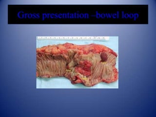
- 1. Gross presentation –bowel loop
- 2. Anatomy Large bowel • Forms three sided frame around SI leaving inferior area open to the pelvis. • Approx. 1.5 m in length & extend from lleum to anus. • Diameter dec from caecum (7cm) to sigmoid colon (2.5cm). • Divided into 4 segmentscecum,colon,rectum & anus.
- 3. Types of bowel resection • • • • • • Total colectomy. Right hemicolectomy. Transverse colectomy. Left hemicolectomy. Low anterior resection. Abdominoperineal resection.
- 4. Procedure • Weigh & measure the specimen • Sample lymph nodes, & remove the mesentry while the specimen is fresh. • Two options are there:A) Open the bowel longitudinally, pin on a corkboard & fix it overnight. B)Injecting formalin through one end when the other end is tied, then tying off the injected end.
- 5. • Take photographs . • In cases with deep penetration by tumor, dissect the veins carefully for possible tumor invasion.
- 6. Description • Part of bowel removed & length of specimen . • Mucosa- type of lesion,extent,ulceration(linear or transverse),depth, pseudoplyps,hemorrhage,fissures. • Wall thickening (focal or diffuse),atrophy ,fibrosis, necrosis. • Serosa- fibrin, pus,fibrosis,adherence of mesentry. • Diverticulum- number, size, location in relation to teniae,content, evidence of inflammation, hemorrhage or perforation.
- 7. Description for tumor Tumor • size (including thickness). • Shape (fungating,flat,ulcerating) • Extent through bowel wall • Serosal involvement,satellite nodules. • Areas of necrosis & hemorrhage. • Evidence of blood vessel invasion & invasion of adjacent organs.
- 8. • Distance of tumor to each line of resection. • Estimate the no. of lymph nodes found, whether or not nodes appear to be involved by tumor, size of largest node.
- 9. Sections for histology For non tumoral conditions:• As many as necessary to sample abnormal areas. • Proximal & distal lines of resection in cases of colitis. • Appendix, if included in specimen.
- 10. For tumoral conditions:• 3 sections from tumor. • Representative section of subserosal connective tissue, fat & blood vessel around tumor. • Both surgical margins. • Bowel b/w tm & distal line of resection. • Appendix if included in the specimen.
- 11. • Lymph nodes:A) around tumor. B)distal to tumor. C)proximal to tumor. D)at high point of resection(areas surrounding ligated vessels) • In abdominoperineal resections:- anorectal junction.
- 13. Crohns disease
- 19. Amebic dysentery
- 22. MELANOSIS COLI
- 23. Endometriosis
- 28. THANK YOU SPEAKER DR N. P. TIWARI
Notes de l'éditeur
- This opened segment of iteumshows a wide diverticutum. about 2 cm in diameter. lined by rathersmooth mucosa. Meckel's diverticutum is a congenital malformationrepresenting a remnant of the vitello-intestinat duct. Usually foundabout 60 cm from the ileocaecat valve, it affects about 2% of thepoputation. White it may become inflamed or obstructed, ectopicgastr ic mucosa is present in some cases, which may tead to pepticulceration. Other ectopic epithelia which are often found includepancreatic, duodenal and cotonic types.
- Crohn's disease. This opened segment of large bowelshows two quite separate 'skip' lesions, characterised by floridmucosat ulceration. The tesion on the teft has induced markedluminal stenosis with obvious proximat dilatation. Up to 15% ofpatients with Crohn's disease show large bowel involvement, with orwithout small intestinat disease. There is a definite increased risk ofcolonic adenocarcinoma, but th is is much less marked than in ulcerativecotitis. Crohn's disease. This opened length of small bowelshows the typical 'cobbtestone' appearance of the mucosa, eachnodule being separated by ulcerated fissures . Crahn's disease is anidiopathic granulomatous condition which may affecl any site in thealimentary tract but shows a predilection for the terminat iteum . Itpresents most often in the 2nd to 4th decades. Postulated aetiologicalagents include various micro-organisms and fine particutatematter, which have induced an abnormal immunotogical respon se.Multifocal invotvement, giving rise to 'skip' lesions, is characteristicand inflammation of the full thickness of the bowel wall causes deepfissuring , fistula formation and fibros is.
- Typhoid ulceration.These segments of small intestinehave been opened to showseveral ovoid ulcers lyingparallel to the bowel wall (cf . Fig.329) The ulceration hasoccurred al the site of necrosisof Peyer's patches. Typhoidfever remains endemic in someparts of the world, especiallyAsia and the Far East. It is due toingestion of food or drink contaminatedwith Salmonella typhi,usually from an asymptomaticcarner. Important local complicationsinclude perforationand haemorrhage. Followinginvasion of the bloodstream,excretion of Salmonellae in bilemay tead to chronic gallbladderinfection (whence the carrier Intestinal tuberculosis. In contrast to Fig . 3.28. thisulceration. while still originating in Peyer's patches. extends transverselyaround the bowel wall following the lines of lymphatic drainage.Intestinal tuberculosis may be primary. re sulting from ingestionof unpasteurised milk. or secondary. as a consequence of swallowinginfected sputum from pulmonary disease. Adjacent lymphnodes are usually involved and may later undergo dystrophic· ~ Icification . Peritoneal involvement may lead to ascites.
- Mesenteric embolism. The superior mesenteric artery istotally occluded by thrombus which has emboli sed from the lettatrium in this patient with atrial fibrillation. Proximal occlusion. such asthi s, results in infarction of almost the entire small bowel and isinvariably fatal. Small Intestinal ischaemia. This loop of bowel is dilated1I 11 111 11 111\C 'llly congested. This is the appearance of infarction of theI " ,w,,1 w.t ll, ill II lesser degrees of ischaemia may result only inII! II ''' lI ilIIIi .lllAlion. II most commonly results from an embolus.I,' I, Illy •. "Ii h.ll; III o ri ~ lin , occluding a branch of the superior mesent-I I, 11 1. 111/ I HI II !I Ciluses include severe hypotension. thrombosis inIII 11111 ' I'"l"h ll l(. v,,~~ se l , retrograde infarction due to mesenteric" . • 11 ' II I II 'II Ii J(/~ i l: i c)r diqltalis therapy . Carcinoid tumour. The terminal ileum and caecum areshown here. Originating in the ileocaecal valve is a wellcircumscribed, yellow tumour in the submucosa. In the adjacentmesenteric fat, a lymph node containing metastatic tumour can beseen (arrowed) . Carcinoid tumours arise from neuroendocrine APUDcells and are usually found in the appendix or small intestine. Tumoursin the appendix tend to be solitary and affect young adults, whilethose in the small bowel may be multiple and usually present in oldpeople . The appendiceat neoplasms almost never metastasise, butsmall bowel tumours frequently spread to lymph nodes and the liver