Ventilator And Nursing
•Télécharger en tant que PPTX, PDF•
29 j'aime•10,604 vues

Lecture held in king saud hospital ICU

Recommandé
Contenu connexe
Tendances
Tendances (20)
En vedette
En vedette (20)
Similaire à Ventilator And Nursing
Similaire à Ventilator And Nursing (20)
Dernier
Dernier (20)
Ventilator And Nursing
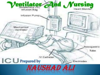
- 1. Ventilator And Nursing Prepared by Naushadali
- 2. Defination:Mechanical ventilation is the use of a mechanical device (machine) to inflate and deflate the lungs. Purpose: Mechanical ventilation provides the force needed to deliver air to the lungs in a patient whose own ventilatory abilities are diminished or lost.
- 3. The nurse must be able to do the following 1. Identify the indications for mechanical ventilation. 2. List the steps in preparing a patient for intubation. 3. Determine the FIO2, tidal volume, rate and mode of ventilation on a given ventilator. 4. Describe the various modes of ventilation and their implications. 5. Describe at least two complications associated with patient’s response to mechanical ventilation and their signs and symptoms.
- 4. 6. Describe the causes and nursing measures taken when trouble-shooting ventilator alarms. 7. Describe preventative measures aimed at preventing selected other complications related to endotracheal intubation. 8. Give rationale for selected nursing interventions in the plan of care for the ventilated patient. 9. Complete the care of the ventilated patient checklist. 10. Complete the suctioning checklist. (A) To review indications for and basic modes of mechanical ventilation, possible complications that can occur, and nursing observations and procedures to detect and/or prevent such complications. (B) To provide a systematic nursing assessment procedure to ensure early detection of complications associated with mechanical ventilation.
- 5. Indication for Intubation Acute respiratory failure evidenced by the lungs inability to maintain arterial oxygenation or eliminate carbon dioxide leading to tissue hypoxia in spite of low-flow or high-flow oxygen delivery devices. (Impaired gas exchange, airway obstruction or ventilation-perfusion abnormalities). 2. In a patient with previously normal ABGs, the ABG results will be as follows: PaO2 > 50 mm Hg pH < 7.25 PaO2 < 50 mm Hg on 60% FIO2 : restlessness, dyspnea, confusion, anxiety, tachypnea, tachycardia, and diaphoresis PaCO2 > 50 mm Hg : hypertension, irritability, somnolence (late), cyanosis (late), and LOC (late)
- 6. 3. Neuromuscular or neurogenic loss of respiratory regulation. (Impaired ventilation) 4. Usual reasons for intubation: Airway maintenance, Secretion control, Oxygenation and Ventilation. Types of intubation: Orotracheal, Nasotracheal, Tracheostomy
- 7. Preparing for Intubation 1. Recognize the need for intubation. 2. Notify physician and respiratory therapist. Ensure consent obtained if not emergency. 3. Gather all necessary equipment: a. Suction canister with regulator and connecting tubing b. Sterile 14 Fr.(Adult) suction catheter or closed in-line suction catheter c. Sterile gloves d. Normal saline e. Yankuer suction-tip catheter and nasogastric tube
- 8. f. Intubation equipment: Manual resuscitator bag (MRB), Laryngoscope and blade, Wire guide, (Stylet), Water soluble lubricant, Xylocaine spray g. Endotracheal attachment device (E-tad) or tape h. Get order for initial ventilator settings i. Sedation as required j. Soft wrist restraints as required k. Call for chest x-ray to confirm position of endotracheal tube l. Provide emotional support as needed/ ensure family notified of change in condition
- 9. Intubation Types of Ventilators Ventilator Settings Modes of Mechanical Ventilation
- 10. 1. Associated with patient’s response to mechanical ventilation: A. Decreased Cardiac Output Cause - venous return to the right atrium impeded by the dramatically increased intrathoracic pressures during inspiration from positive pressure ventilation. Also reduced sympatho-adrenal stimulation leading to a decrease in peripheral vascular resistance and reduced blood pressure. 2. Symptoms – increased heart rate, decreased blood pressure and perfusion to vital organs, decreased CVP, and cool clammy skin. 3. Treatment – aimed at increasing preload (e.g. fluid administration) and decreasing the airway pressures exerted during mechanicalventilation by decreasing inspiratory flow rates and TV, or using other methods to decrease airway pressures (e.g. different modes of ventilation).
- 11. B. Barotraumas 1. Cause – damage to pulmonary system due to alveolar rupture from excessive airway pressures and/or over distention of alveoli. 2. Symptoms – may result in pneumothorax, pneumomediastinum, pneumoperitoneum, or subcutaneous emphysema. 3. Treatment - aimed at reducing TV, cautious use of PEEP, and avoidance of high airway pressures resulting in development of auto-PEEP in high risk patients (patients with obstructive lung diseases (asthma, bronchospasm), unevenly distributed lung diseases (lobar pneumonia), or hyper inflated lungs (emphysema).
- 12. C. Nosocomial Pneumonia Cause – invasive device in critically ill patients becomes colonized with pathological bacteria within 24 hours in almost all patients. 20-60% of these, develop nosocomial pneumonia. 2. Treatment – aimed at prevention by the following: a.Avoid cross-contamination by frequent handwashing
- 13. b.Decrease risk of aspiration (cuff occlusion of trachea, positioning, use of small-bore NG tubes) c.Suction only when clinically indicated, using sterile technique d.Maintain closed system setup on ventilator circuitry and avoid pooling of condensation in the tubing e.Ensure adequate nutrition d.Avoid neutralization of gastric contents with antacids and H2 blockers
- 14. D. Positive Water Balance 1. Syndrome of Inappropriate Antidiuretic Hormone (SIADH) – due to vagal stretch receptors in right atrium sensing a decrease in venous return and see it as hypovolemia, leading to a release of ADH from the posterior pituitary gland and retention of sodium and water. Treatment is aimed at decreasing fluid intake. 2. Decrease of normal insensible water loss due to closed ventilator circuit preventing water loss from lungs. This fluid overload evidenced by decreased urine specific gravity, dilutionalhyponatremia, increased heart rate and BP.
- 15. E. Decreased Renal Perfusion – can be treated with low dose dopamine therapy. F. Increased Intracranial Pressure (ICP) – reduce PEEP G. Hepatic congestion – reduce PEEP H. Worsening of intracardiac shunts –reduce PEEP
- 16. 2. Associated with ventilator malfunction: A. Alarms turned off or nonfunctional – may lead to apnea and respiratory arrest Troubleshooting Ventilator Alarms Low exhaled volume: Cuff leak, Tubing disconnect, Patient disconnected Evaluate cuff; reinflateprn; if ruptured, tube will need to be replaced. Evaluate connections; tighten or replace as needed; check ETT placement, Reconnect to ventilator High pressure: Secretions in airway, Patient biting tubing, Tube kinked, Cuff herniation, Increased airway resistance/decreased lung compliance (caused by bronchospasm, right mainstem bronchus intubation, pneumothorax, pneumonia), Patient coughing and/or fighting the ventilator; anxiety; fear; pain.
- 17. Suction patient, Insert bite block, Reposition patient’s head/neck; check all tubing lengths, Deflate and reinflate cuff, Auscultate breath sounds, Evaluate compliance and tube position; stabilize tube, Explain all procedures to patient in calm, reassuring manner, Sedate/medicate as necessary Low oxygen pressure: Oxygen malfunction Disconnect patient from ventilator; manually bag with ambu; call R.T
- 18. 3. Other complications related to endotracheal intubation. A. Sinusitis and nasal injury – obstruction of paranasal sinus drainage; pressure necrosis of nares 1.Prevention: avoid nasal intubations; cushion nares from tube and tape/ties. 2. Treatment: remove all tubes from nasal passages; administer antibiotics.
- 19. B. Tracheoesophageal fistula – pressure necrosis of posterior tracheal wall resulting from overinflated cuff and rigid nasogastric tube 1. Prevention: inflate cuff with minimal amount of air necessary; monitor cuff pressures q. 8 h. 2. Treatment: position cuff of tube distal to fistula; place gastrostomy tube for enteral feedings; place esophageal tube for secretion clearance proximal to fistula. C. Mucosal lesions – pressure at tube and mucosal interface 1. Prevention: Inflate cuff with minimal amount of air necessary; monitor cuff pressure q. 8 h.; use appropriate size tube. 2. Treatment: may resolve spontaneously; perform surgical interventions.
- 20. D. Laryngeal or tracheal stenosis– injury to area from end of tube or cuff, resulting in scar tissue formation and narrowing of airway 1. Prevention: inflate cuff with minimal amount of air necessary; monitor cuff pressure q. 8.h.; suction area above cuff frequently. 2. Treatment: perform tracheostomy; place laryngeal stent; perform surgical repair.
- 21. E. Cricoidabcess– mucosal injury with bacterial invasion 1. Prevention: inflate cuff with minimal amount of air necessary; monitor cuff pressure q. 8 h.; suction area above cuff frequently. 2. Treatment: perform incision and drainage of area; administer antibiotics. 4. Other common potential problems related to mechanical ventilation: Aspiration, GI bleeding, Inappropriate ventilation (respiratory acidosis or alkalosis, Thick secretions, Patient discomfort due to pulling or jarring of ETT or tracheostomy, High PaO2, Low PaO2, Anxiety and fear, Dysrhythmias or vagal reactions during or after suctioning, Incorrect PEEP setting, Inability to tolerate ventilator mode.
- 22. PLAN OF CARE FOR THE VENTILATED PATIENT Patient Goals: Patient will have effective breathing pattern. Patient will have adequate gas exchange. Patient’s nutritional status will be maintained to meet body needs. Patient will not develop a pulmonary infection. Patient will not develop problems related to immobility. Patient and/or family will indicate understanding of the purpose for mechanical ventilation.
- 23. ThankYou nau