

Recommandé
Contenu connexe
Tendances
Tendances (20)
En vedette
En vedette (15)
Similaire à Anatomy of renal system
Similaire à Anatomy of renal system (20)
Dernier
Dernier (20)
Anatomy of renal system
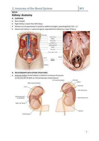
- 1. 2. Anatomy of the Renal System W1 RENAL 1 Kidney: Anatomy A. OVERVIEW Bean-shaped Right kidney is lower than left kidney Kidneys are retroperitoneal in posterior abdominal region, extending levels T12 – L3 Above each kidney i a suprarenal gland, separated from kidney by a layer of fascia B. RELATIONSHIPS WITH OTHER STRUCTURES Anterior surface of each kidney is related to numerous structures (1) Directly OR (2) With an intervening layer of peritoneum
- 2. 2. Anatomy of the Renal System W1 RENAL 2 Posterior surface of each kidney is related to similar structures C. RENAL FAT and FASCIA From innermost to outermost: 1. Renal capsule 2. Perinephric fat: - Extraperitoneal fat - Completely surrounds kidneys 3. Renal fascia: - Membranous condensation of Extraperitoneal fascia 4. Paranephric fat: - Accumulates posterior and posterolateral to kidneys
- 3. 2. Anatomy of the Renal System W1 RENAL 3 D. KIDNEY STRUCTURE Hilum: - On medial margin of each kidney - Entrance and exit to renal vessels, lymphatics and nerves - Internally continuous with Renal sinus - Perinephric fat continues into hilum and sinus and surrounds all structures Outer renal cortex: - Continuous band of pale tissue Inner renal medulla: - Divided into Renal pyramids (Discontinuous aggregations of triangular-shaped tissue): o Renal pyramids are separated by Cortical columns of Bertin o Bases (Corticomedullary Junction) are directed outwards, towards renal cortex o Apex (Renal papilla) are directed inwards, towards renal sinus - Contains Ducts of Bellini - Empty urine into surrounding Minor calyces via area cribosa - Several minor calyces unite to form a Major calyces - Several major calyces unite to form Renal pelvis (continuous with Ureter) E. RENAL VASCULATURE Renal artery (lateral branch of abdominal aorta, inferior to origin of superior mesenteric artery L1-L2) supply about 10% of the total blood volume to each kidney Right renal artery is longer, passes posterior to IVC
- 4. 2. Anatomy of the Renal System W1 RENAL 4 Renal artery divides into 5 separate Segmental arteries: - Supply different segments of kidneys - Do not anastomose with each other - I.e. Result in distinct vascular segmentation of kidney, with each being surgically resectable Renal vasculature follows the following pathway
- 5. 2. Anatomy of the Renal System W1 RENAL 5 F. RENAL INNERVATION 1. OUTPUT: Visceral efferent fibres Sympathetic nervous system: via Renal plexus - Formed by filaments from: (1) Coeliac plexus (2) Aorticorenal ganglion (3) Aortic plexus - Joined by the least splanchnic nerve - Enter kidney alongside renal artery - Triggers renal vasoconstriction, reducing renal blood flow Parasympathetic nervous system 2. INPUT: Visceral afferent fibres Parasympathetic nervous system Sensory Input: - Follow sympathetic fibres, entering spinal cord at T11-L2 G. RENAL LYMPHATICS Most renal lymphatic drainage is into Lumbar lymph nodes (which surround abdominal aorta)
- 6. 2. Anatomy of the Renal System W1 RENAL 6 URETERS: ANATOMY A. OVERVIEW Ureters: - Muscular tubes - Transport urine from kidneys to bladder Superiorly continuous with renal pelvis at Ureteropelvic junction. Inferior to this junction, ureters descend retroperitoneally on medial aspect of psoas major. On pelvic prim, ureters cross end of Common iliac arteries, entering pelvic cavity > Bladder B. CONSTRICTION OF THE LUMEN There are 3 points along course of ureters where their lumen is constricted: (1) Ureteropelvic junction (2) Where ureters cross common iliac vessels at pelvic brim (3) Where ureters enter bladder wall Kidneys stones can become lodged at these constriction
- 7. 2. Anatomy of the Renal System W1 RENAL 7 C. URETERIC VASCULATURE Ureters receive arterial branches from adjacent vessels as they pass towards the bladder: (1) Upper end: Supplied by renal arteries (2) Middle part: Supplied by - abdominal aorta - testicular/ovarian arteries - common iliac arteries (3) In pelvic cavity: Supplied by internal iliac arteries D. URETERIC INNERVATION Involves (1) Renal (2) Aortic (3) Superior Hypogastric (4) Inferior Hypogastric plexuses Visceral efferent fibres: From sympathetic and parasympathetic sources Visceral afferent fibres: Return to T11-L2 spinal cord I.e. Ureteric pain is referred to cutaneous area supplied by T11-L2, including: - Posterior & Lateral abdominal wall (below ribs and above iliac crest) - Pubic region - Scrotum in males/Labia majora in females - Proximal anterior aspect of the thigh
- 8. 2. Anatomy of the Renal System W1 RENAL 8 SUPRARENAL GLANDS: ANATOMY A. OVERVIEW Associated with superior pole of each kidney Structure: (1) Outer cortex (2) Inner medulla Right gland is pyramidal, left gland is semilunar and larger Suprarenal gland is surrounded by Perinephric fat, enclosed in Renal fascia B. SUPRARENAL VASCULATURE Arterial Supply: Arises from 3 primary sources: (1) Superior suprarenal arteries: From Inferior phrenic arteries (2) Middle suprarenal artery: From Abdominal aorta (3) Inferior branches: From Renal arteries Venous Drainage: (1) Right suprarenal vein: - Short - Almost immediately enters IVC (2) Left suprarenal vein: - Passes inferiorly to enter left renal vein
- 9. 2. Anatomy of the Renal System W1 RENAL 9 GLOSSARY Resection: Surgical removal of structures, organs Retroperitoneal: Situated behind the peritoneum