
Recommandé
Contenu connexe
Tendances
Tendances (20)
Similaire à Diseases of oral cavity and ludwig’s angina ug,18.07.16 dr.davis thomas
Similaire à Diseases of oral cavity and ludwig’s angina ug,18.07.16 dr.davis thomas (20)
Plus de ophthalmgmcri
Plus de ophthalmgmcri (20)
Dernier
Dernier (20)
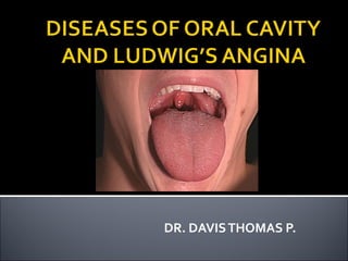
Diseases of oral cavity and ludwig’s angina ug,18.07.16 dr.davis thomas
- 2. Oral cavity Lips Tongue Floor of Mouth Buccal mucosa Palate Retromolar trigone
- 3. Reactive lesions Inflammatory lesions Oral cancer Precancerous lesions (Leukoplakia & erythroplakia) Benign Tumors of Oral Cavity
- 4. 1-Irritation fibroma : Most common 61 % of all the reactive lesions Can occur throughout the oral cavity Most common along the "bite line." Microscopically: fibrous tissue covered by squamous mucosa.
- 6. 2-Pyogenic granulomas 12 % Highly vascular lesions similar to granulation tissue. 3-Peripheral giant cell granuloma (giant cell epulis) 5% Aggregation of multinucleated foreign body-like giant cells Separated by fibroangiomatous stroma. Giant cell epulis
- 7. Epulis is a clinical term applied to swellings at the gum margin. Most of them are granulomas associated with chronic gingivitis A few are true neoplasms
- 8. Reactive lesions Inflammatory lesions Oral cancer Precancerous lesions (Leukoplakia & erythroplakia) Benign Tumors of Oral Cavity
- 9. 1. Viral infection 2. Fungal infection 3. Bacterial infection A - Vincent’s angina B - Syphilis C - Oral tuberculosis 4. Aphthous ulcers (aphthous stomatitis) 5. Dermatoses
- 10. Inflammation of the mouth (Stomatitis)(Stomatitis) Inflammation of the Lips (Cheilitis)(Cheilitis) Inflammation of the soft tissues around teeth typically resulting from inadequate oral hygiene (Gingivitis)(Gingivitis) Inflammmation of the tongue (Glossitis).(Glossitis). GlossitisGlossitis more commonly applied to the "beefy-red" tongues"beefy-red" tongues of certain deficiency states (e.g.; vitamin B12, and iron, deficiencies).(e.g.; vitamin B12, and iron, deficiencies). Other causes of glossitis:Other causes of glossitis: hot and spicy foods, chronic irritation by excessive smoking, ragged tooth or syphilitic inflammation
- 11. Herpes simplex virus (usually type 1) infection causes "cold sores" The virus infects the mouth in children. Most adults have had HSV1 infection, but it remains latent and produces this small sore: During periods of stress From local trauma Environmental changes Cold sores consist of numerous vesicles and shallow ulcerations. Cold sore of lower lip (herpes labialis) Sore = abraded or painful area of the body
- 13. Treatment Antipyretics, analgesics, hydration Valacyclovir and famciclovir inhibit viral DNA polymerase – help to suppress and controlhelp to suppress and control symptoms,symptoms, but does not curebut does not cure (given for 1 week) If catch in the prodrome - 5% acyclovir cream for 1 week has shown to shorten course or completely abort reactivation altogether KEY TO DIAGNOSIS – Clinical + Fluid analysis (PCR)Clinical + Fluid analysis (PCR) and/or serology (Elisa, Western Blot)and/or serology (Elisa, Western Blot)
- 14. Coxackie A virus causes herpangia Acute vesiculo-ulcertaive mucosal lesion Occurs in epidemics Affects children Begins in tonsils, soft palate &tonsils, soft palate & uvulauvula Painful Heal spontaneously within few days Koplik’s spots are a feature of measles Koplik’s spots Herpangia
- 16. Herpangina
- 17. Candida albicans is an oral commensal in 20-40%oral commensal in 20-40% of population. Infection occurs in: Infants Patients on broad spectrum antibiotics, steriod or cytotoxic therapy Diabetes Neutropenia Immunodeficiency (AIDS) Presents as superficial gray-white inflammatory membranes comprising fungus in a fibrinosuppurative exudate. White exudate can be removed by scraping Exudate bleeds on removal
- 19. Treatment Mild, acute formsMild, acute forms – topical Nystatin Mild, chronicMild, chronic – topical Nystatin + Clotrimazole troches (troche=lozenge) Refractory or immunocomprimised WITHOUTRefractory or immunocomprimised WITHOUT systemic involvementsystemic involvement – add oral Fluconazole Severe forms (systemicsystemic) – IV Amphotericin B with or without Fluconazole KEY TO DIAGNOSIS: Clinical + KOH Prep; culture and serum (1,3)β-D-glucan detection assay if unclear
- 20. Caused by Borellia vincenti and fusiform bacilli Both are normal inhabitants of oral cavity Decreased resistance (inadequate nutrition, immunofeciency) is a predisposing factor to infection Punched out erosions ulceration→ spreads invovles all gingival→ → margin, which become covered by a necrotic pseudomembrane
- 22. A-Ulcerated chancre B-Ulcerated mucous patches (snail track ulcers) C-Gummatous ulcer C - Tuberculosis of The Tongue
- 25. Apthous ulcers are extremely common lesions (up to 20% of population) They are painful, multiple, small, shallow, recurrent ulcerations Presented clinically as white lesions (1<,1> CM) Etiology is unknown Aphtha = Whitish spot
- 26. ○ Most common cause of non-traumatic ulcerations of the oral cavity ○ Etiology unclear ○ 10-20% of general population ○ Diagnosis of exclusion ○ Classifications Minor aphthous ulcerMinor aphthous ulcer - < 1cm< 1cm in diameter - Located on freely mobile oral mucosa - Appears as a well-delineated white lesion with an erythematous halo - Prodrome of burning or tingling in area prior to ulcer’s appearance - Resolve in 7-10 days - Never scars Major aphthous ulcerMajor aphthous ulcer - > 1cm> 1cm in diameter - Involves freely mobile mucosa, tongue, and palate - Last much longer – 6 weeks or more - Typically scar upon healing
- 27. Herpetiform ulcers - Small, 1-3mm in diameter ulcerations appearing in crops of 20-200 ulcers - Typically located on mobile oral mucosa, tongue, and palate - Last 1-2 weeks - Called herpetiform because ulcerations resemble those of HSV,Called herpetiform because ulcerations resemble those of HSV, but there isbut there is no vesicular phaseno vesicular phase Treatment Topical tetracycline solution for 5-7 days has shown good results Topical steroids shown to shorten disease duration Sucralfate suspension shown to improve pain as well as shorten disease duration Major aphthous ulcers or more severe forms of disease require 2Major aphthous ulcers or more severe forms of disease require 2 week course of systemic steroidsweek course of systemic steroids KEY TO DIAGNOSIS: Diagnosis of exclusion; clinical appearance/course
- 29. Lichen planus White plaques
- 30. whitish linear lesions in lacy patternwhitish linear lesions in lacy pattern
- 31. Reactive lesions Inflammatory lesions Oral cancer Precancerous lesions (Leukoplakia & erythroplakia) Benign Tumors of Oral Cavity
- 32. Incidence: Geographic variation: Accounts for 2% of cancers in UK Commoner in S. East Asia Ages & sex : Old Men (50-60 years) • Site : 1.Lip (lower lip) 2.Tongue (anterior )⅔ 3.Mouth floor 4.Tonsil and Fauces Squamous Cell Carcinoma constitutes 95% of oral cancers
- 33. Aetiology: 1-Tobacco and alcohol are the most common associations: Smokers can have 15-fold greater risk ( than nonsmokers ) of malignancy. Chewing tobacco and betel nuts are important causes in India and parts of Asia. 2- Leukoplakia and Erythroplakia 3- Human papilloma virus (HPV) (type16) 4- Genetic factors may also play a role (deletions in chromosomes 18q, lap, 8p, and 3p are implicated). 5- Exposure to ultra-violet light (cancer of the lip).
- 34. Gross: Ulcerated nodule with raised everted edges Often on lower lip Histologically: Well differentiated squamous carcinomas Spread: Growth is relatively slow Submandibular nodes Deeper cervical lymph nodes
- 35. More aggressive than tumors of the lips Grossly starts as a nodule malignant ulcer→ Spread: 1-Local Local infiltration to floor of the mouth, facuces and pharynx leads to fixation the tongue, interfering with speech and swallowing. Local spreads into the medullary cavity of the mandible. 2-lymphatic spread (occurs early) → deep cervical lymph nodes.
- 36. Perform incisional Bx in any oral lesion persist for more than 2wks
- 37. Prognosis is best with lip lesions Poorest with mouth floor and tongue base lesions (20%-30% 5-year survival rate ).
- 38. Malignant melanoma Lymphomas Leukemic infiltration Adenocarcinoma of minor salivary glands Sarcomas Acute Leukemia: gum involvement
- 39. Reactive lesions Inflammatory lesions Oral cancer Precancerous lesions Benign Tumors of Oral Cavity
- 40. Premalignant lesions ○ Leukoplakia Whitish plaque that cannot be scrapped off 5-20% malignant potential5-20% malignant potential Microscopic examination reveals hyperkeratosis and atypia Lesions on lateral tongue, lower lip, and floor of mouth more likely to progress to malignancy ○ Erythroplakia Red patch or macule with soft, velvety texture Much higher chance of harboring malignancy – 60-90%– 60-90% of untreated cases Treatment is surgical excision or laser ablation
- 41. Causes include: 1- Chronic tobacco use (pipe - smoking). 2- Chronic irritation (e.g.; dentures). 2- Alcohol abuse.
- 42. An oral lesion seen in HIV infected, AIDS patients Caused by Epstein-Barr virus (EBV) infection, often with superimposed candida Lesions are white patches of fluffy ("hairy") hyperkeratosis on tongue lateral borders.
- 44. It is an insidious chronic disease affecting any part of the oral cavity and sometimes the pharynx. Although occasionally preceded by and /or associated with vesicle formation ,it is always associated with juxta-epithelial inflammatory reaction followed by a fibro-elastic changes of the lamina propria with epithelial atrophy leading to stiffness of the oral mucosa and causing trismus and inability to eat. DEFINITION
- 45. Etiology of OSMF: Exact etiology is unknown. The suggested factors are, 1. Chronic Irritation Chilies Lime Betel nut Tobacco Chewing 2. Deficiency disease. 3. Defective iron metabolism 4. Bacterial Infection 5. Collagen disorder 6. Immunological disorders 7. Genetic disorder.
- 46. MULTIFACTORIAL PATHOGENESIS ARECANUT TOBACCO LIME VOLATILE OILSVOLATILE LIQUIDS TANNIN& AFLOTOXIN ARECOLINE DEGRADATION OF COLLAGEN INCREASED SYNTHESIS OF COLLAGEN MECHANICAL TRAUMA CHEMICAL BURN HYPERSENSITIVITY ALTERED IMMUNITY GENETIC REDISPOSITION FIBROBLAST FORMATION IRREVERSIBLE FIBROSIS CACINOMAEXPOSURE CONTINOUS
- 47. Prodromal symptoms : Onset is insidious. The most common initial symptoms are: Burning sensation on eating spicy food Blisters on the palate Ulceration or recurrent stomatitis Excessive salivation Defective gustatory sensation Dryness of mouth.
- 48. Later, Difficulty in opening mouth Inability to whistle, blow Difficulty in swallowing When fibrosis involves pharynx- referred pain to the ear. Changes in tone of the voice due to vocal cord involvement Some times deafness due to occlusion of eustachian tubes
- 49. COMMON SITES INVOLVED Buccal mucosa, faucial pillars ,soft palate, lips and hard palate. The fibrous bands in the buccal mucosa run in a vertical direction ,sometimes so marked that the cheeks are almost immovable. In the soft palate the fibrous bands radiate from the pterygomandibular raphe or the faucial pillars and have a sear like appearance
- 50. The uvula is markedly involved , shrinks and appears as a small fibrous bud. The faucial pillars become thick , short, and extremely hard. The tonsils may be pressed between the fibrosed pillars The lips are often affected and upon palpation , a circular band can be felt around the entire rima oris When gingiva is affected , it is fibrotic, blanched and devoid of its normal stippled appearance.
- 58. MANAGEMENT Various modalities of treatment have been tried. 1.Restriction of habits/ Behavioral therapy 2.Medicinal therapy 3.Surgical therapy. 4.Oral Physiotherapy
- 59. Reactive lesions Inflammatory lesions Oral cancer Precancerous lesions (Leukoplakia & erythroplakia) Benign Tumors of Oral Cavity
- 60. 1-Squamous cell papilloma.**** 2-Capillary hemangioma 3-Cavernous hemangioma & lymphangioma → macrochelia & macroglossia 4-leiomyoma 5-Schwannoma Cavernous hemangioma
- 62. Extension of localized periapical infection Anterior mandibular → Sublingual Posterior mandibular (molar) → Submandibular Fascial planes
- 63. Recent dental extraction or work Dental caries Fever Swelling of mouth, face, neck Compromised host Co-morbidities (diabetes)
- 64. Toxicity Brawny bilateral boardlike edema Submandibular, submental, sublingual Trismus Tongue elevation No fluctuance
- 65. A, Ludwig angina may initially appear benign. B, In Ludwig angina, rapid progression may compromise the airway in a few hours.
- 66. Streptococcus Staphylococcus Mixed aerobic/anaerobic infection B. Fragilis ß-lactamase resistance (<= 40%)
- 67. Clinical CT scan
- 68. Airway control - EARLY Fiberoptic Deterioration may be rapid Cricothyrotomy or tracheostomy may be necessary Surgical consultation mandatory Oral maxillofacial surgeon or ENT Definitive surgical drainage and debridement ICU
- 69. Extended spectrum penicillins Ampicillin/Sulbactam (Unasyn) Ticarcillin/Clauvulate (Timentin) Piperacillin/Tazobactam (Zosyn) Clindamycin + Cipro (PCN allergy) Flagyl (B. Fragilis)
- 70. Reduce edema “Used routinely when airway compromise suspected” (Larawin et al.) Dexamethasone 10-20 mg IV Then 4-6 mg Q6 for 8 doses (Busch)
- 71. Serious deep space infection Potentially fatal Aggressive manage airway as indicated Surgical consultation Antibiotics and steroids ICU
Notes de l'éditeur
- Figure 66-32 A, Ludwig angina may initially appear benign. B, In Ludwig angina, rapid progression may compromise the airway in a few hours.
- Airway - deterioration may be rapid, control aggressively The most important therapeutic treatment is surgical debridement
- Adjunctive therapy to surgical management
- Marple BF. Ludwig angina: a review of current airway management. Arch Otolaryngol Head Neck Surg. 1999;125:596-599. Busch RF. Ludwig angina: early aggressive therapy. Arch Otolaryngol Head Neck Surg. 1999 Nov;125(11):1283-4. Upon admission to the emergency department, patients are given an immediate dose of 10 to 20 mg of dexamethasone, followed by 4 to 6 mg every 6 hours for a maximum of 8 doses. Antibiotic therapy consists of ampicillin-sulbactam, 3.0 g administered intravenously every 6 hours, in addition to clindamycin, 600 mg administered intravenously every 6 hours. For patients who are allergic to penicillin, ciprofloxacin, 400 mg administered intravenously every 12 hours, is used instead of ampicillin-sulbactam in addition to the clindamycin therapy. Within 24 to 48 hours, patients are taken to the operating room, where decompression of the sublingual and submandibular spaces is accomplished through the submental space. A through-and-through Penrose drain is placed in the midline adjacent to the lingual aspect of the mandible between and anterior to the submandibular duct orifices. This drain is removed in the clinic 1 week later. At the time of surgical decompression, any offending teeth are removed and any additional involved spaces are drained.