Cystocele physical examination
•Télécharger en tant que PPT, PDF•
4 j'aime•6,564 vues

Grade 2 or 3 cystocele can be diagnosed through symptoms and vaginal exam where the bladder reaches or protrudes through the vaginal opening. Further tests like cystogram, pelvic fluoroscopy, MRI, or urodynamics can provide more detail on cystocele and urinary problems by visualizing soft tissues and measuring bladder function and residual urine. Cystocele occurs when the pubocervical fascia is attenuated, causing the bladder to bulge into the anterior vaginal wall.

Recommandé
Contenu connexe
Tendances
Tendances (20)
En vedette
En vedette (17)
Similaire à Cystocele physical examination
Similaire à Cystocele physical examination (20)
Plus de Regi Septian
Plus de Regi Septian (9)
Cystocele physical examination
- 3. We may be able to diagnose a grade 2 or grade 3 cystocele from a description of the symptoms and physical examination of the vagina. 1. grade 1 (mild) : The bladder protrudes only to the upper (internal) vagina 2. grade 2(moderated) : The bladder reaches the vaginal opening 3. grade 3 (most advanced) : The bladder bulges directly through the vaginal opening
- 4. We may press a speculum against the vaginal side walls and ask the individual to straincystocele will seen bulging into the anterior vaginal wall. The central cystocele results from attenuation or separation of the pubocervical fasciaprotrusion will results in the classic appearance of a loss of the rugae or vaginal The fallen part of the bladder may be visible upon examination of the vagina; a smooth, bulging mass will be seen below the level of the cervix
- 7. If a woman has difficulty emptying her bladder, we may measure the amount of postvoid residualwith a bladder ultrasound or use a catheter to measure the amount of remaining urine.
- 8. 1. Voiding Cystourethrogram 2. Pelvic floor fluoroscopy 3. Magnetic resonance imaging (MRI) 4. Urodynamic testing 5. Fluoroscopic examination (cinefluorography) 6. Cystometry measures bladder capacity and control 7. Uroflowmeter
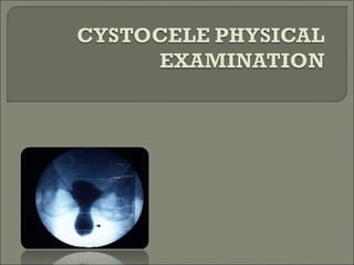
- 9. 1.Voiding Cystourethrogram A woman gets a voiding cystourethrogram while urinating. The x-ray images show the shape of the woman’s bladder to see any problems that might block normal urine flow.
- 10. In such circumstances, an upright resting and straining cystogram can be helpful in determining the presence and degree of cystocele.
- 11. 2. Pelvic floor fluoroscopy To measures perineal descent, helping to determine which organ has herniated into the vagina: it is especially useful if problems with defecation have been reported. 3. Magnetic resonance imaging (MRI) can reveal similar information, and has the added advantage of allowing Cystocele visualization soft tissue of the pelvic floor in greater detail.
- 12. 4. Urodynamic testing is performed if incontinence accompanies pelvic organ prolapse.This testing involves taking x-rays of the bladder during urination (voiding cystourethrogram) to reveal the shape of the bladder and problems that might block the normal flow of urine. 5. Fluoroscopic examination (cinefluorography) while voiding, may be needed to rule out bladder abnormalities and other problems in the urinary system 6. Cystometry measures bladder capacity and control 7.Uroflowmetery