
Recommandé
Contenu connexe
Similaire à chapter 4 PsychopharmacologyOutline· ■ Principles of Psy.docx
Similaire à chapter 4 PsychopharmacologyOutline· ■ Principles of Psy.docx (20)
Plus de robertad6
Plus de robertad6 (20)
Dernier
Dernier (20)
chapter 4 PsychopharmacologyOutline· ■ Principles of Psy.docx
- 1. chapter 4 Psychopharmacology Outline · ■ Principles of Psychopharmacology Pharmacokinetics Drug Effectiveness Effects of Repeated Administration Placebo Effects Section Summary · ■ Sites of Drug Action Effects on Production of Neurotransmitters Effects on Storage and Release of Neurotransmitters Effects on Receptors Effects on Reuptake or Destruction of Neurotransmitters Section Summary · ■ Neurotransmitters and Neuromodulators Acetylcholine The Monoamines Dopamine Amino Acids Peptides Lipids Nucleosides Soluble Gases Section Summary Several years ago I spent the academic year in a neurological research center affiliated with the teaching hospital at a medical center. One morning as I was having breakfast, I read a brief item in the newspaper about a man who had been hospitalized for botulism. Later that morning, I attended a weekly meeting during which the chief of neurology discussed interesting cases presented by the neurological residents. I was surprised to see
- 2. that we would visit the man with botulism. We entered the intensive care unit and saw that the man was clearly on his way to recovery. His face was pale and his voice was weak, but he was no longer on a respirator. There wasn’t much to see, so we went back to the lounge and discussed his case. Just before dinner a few days earlier, Mr. F. had opened a jar of asparagus that his family had canned. He noted right away that it smelled funny. Because his family had grown the asparagus in their own garden, he was reluctant to throw it away. However, he decided that he wouldn’t take any chances. He dipped a spoon into the liquid in the jar and touched it to his tongue. It didn’t taste right, so he didn’t swallow it. Instead, he stuck his tongue out and rinsed it under a stream of water from the faucet at the kitchen sink. He dumped the asparagus into the garbage disposal. About an hour later, as the family was finishing dinner, Mr. F. discovered that he was seeing double. Alarmed, he asked his wife to drive him to the hospital. When he arrived at the emergency room, he was seen by one of the neurological residents, who asked him, “Mr. F., you haven’t eaten some home-canned foods recently, have you?” Learning that he had indeed let some liquid from a suspect jar of asparagus touch his tongue, the resident ordered a vial of botulinum antitoxin from the pharmacy. Meanwhile, he took a blood sample from Mr. F.’s vein and sent it to the lab for some in vivo testing in mice. He then administered the antitoxin to Mr. F., but already he could see that it was too late: The patient was showing obvious signs of muscular weakness and was having some difficulty breathing. He was immediately sent to the intensive care unit, where he was put on a respirator. Although he became completely paralyzed, the life support
- 3. system did what its name indicates, and he regained control of his muscles. What fascinated me the most was the in vivo testing procedure for the presence of botulinum toxin in Mr. F.’s blood. Plasma extracted from the blood was injected into several mice, half of which had been pretreated with botulinum antitoxin. The pretreated mice survived; the others died. Just think: Mr. F. had touched only a few drops of the contaminated liquid on his tongue and then rinsed it off immediately, but enough of the toxin entered his bloodstream that a small amount of his blood plasma could kill a mouse. By the way, we will examine the pharmacological effect of botulinum toxin later in this chapter. Chapter 2 introduced you to the cells of the nervous system, and Chapter 3 described its basic structure. Now it is time to build on this information by introducing the field of psychopharmacology. Psychopharmacology is the study of the effects of drugs on the nervous system and (of course) on behavior. (Pharmakon is the Greek word for “drug.”) psychopharmacology The study of the effects of drugs on the nervous system and on behavior. But what is a drug? Like many words, this one has several different meanings. In one context it refers to a medication that we would obtain from a pharmacist—a chemical that has a therapeutic effect on a disease or its symptoms. In another context the word refers to a chemical that people are likely to abuse, such as heroin or cocaine. The meaning that will be used in this book (and the one generally accepted by pharmacologists) is “an exogenous chemical not necessary for normal cellular functioning that significantly alters the functions of certain cells of the body when taken in relatively low doses.” Because the topic of this chapter is psycho pharmacology, we will concern ourselves here only
- 4. with chemicals that alter the functions of cells within the nervous system. The word exogenous rules out chemical messengers produced by the body, such as neurotransmitters, neuromodulators, or hormones. (Exogenous means “produced from without”—that is, from outside the body.) Chemical messengers produced by the body are not drugs, although synthetic chemicals that mimic their effects are classified as drugs. The definition of a drug also rules out essential nutrients, such as proteins, fats, carbohydrates, minerals, and vitamins that are a necessary constituent of a healthy diet. Finally, it states that drugs are effective in low doses. This qualification is important, because large quantities of almost any substance— even common ones such as table salt—will alter the functions of cells. As we will see in this chapter, drugs have effects and sites of action.Drug effects are the changes we can observe in an animal’s physiological processes and behavior. For example, the effects of morphine, heroin, and other opiates include decreased sensitivity to pain, slowing of the digestive system, sedation, muscular relaxation, constriction of the pupils, and euphoria. The sites of action of drugs are the points at which molecules of drugs interact with molecules located on or in cells of the body, thus affecting some biochemical processes of these cells. For example, the sites of action of the opiates are specialized receptors situated in the membrane of some neurons. When molecules of opiates attach to and activate these receptors, the drugs alter the activity of these neurons and produce their effects. This chapter considers both the effects of drugs and their sites of action. drug effect The changes a drug produces in an animal’s physiological processes and behavior. sites of action The locations at which molecules of drugs interact with molecules located on or in cells of the body, thus
- 5. affecting some biochemical processes of these cells. Psychopharmacology is an important field of neuroscience. It has been responsible for the development of psychotherapeutic drugs, which are used to treat psychological and behavioral disorders. It has also provided tools that have enabled other investigators to study the functions of cells of the nervous system and the behaviors controlled by particular neural circuits. This chapter does not contain all this book has to say about the subject of psychopharmacology. Throughout the book you will learn about the use of drugs to investigate the nature of neural circuits involved in the control of perception, memory, and behavior. In addition, Chapters 16 and 17 discuss the use of drugs to study and treat mental disorders such as schizophrenia, depression, and the anxiety disorders, and Chapter 18 discusses the physiology of drug abuse. Principles of Psychopharmacology This chapter begins with a description of the basic principles of psychopharmacology: the routes of administration of drugs and their fate in the body. The second section discusses the sites of drug actions. The final section discusses specific neurotransmitters and neuromodulators and the physiological and behavioral effects of specific drugs that interact with them. Pharmacokinetics To be effective, a drug must reach its sites of action. To do so, molecules of the drug must enter the body and then enter the bloodstream so that they can be carried to the organ (or organs) on which they act. Once there, they must leave the bloodstream and come into contact with the molecules with which they interact. For almost all of the drugs we are interested in, this
- 6. means that the molecules of the drug must enter the central nervous system (CNS). Some behaviorally active drugs exert their effects on the peripheral nervous system, but these drugs are less important to us than the drugs that affect cells of the CNS. Molecules of drugs must cross several barriers to enter the body and find their way to their sites of action. Some molecules pass through these barriers easily and quickly; others do so very slowly. And once molecules of drugs enter the body, they begin to be metabolized—broken down by enzymes—or excreted in the urine (or both). In time, the molecules either disappear or are transformed into inactive fragments. The process by which drugs are absorbed, distributed within the body, metabolized, and excreted is referred to as pharmacokinetics (“movements of drugs”). pharmacokinetics The process by which drugs are absorbed, distributed within the body, metabolized, and excreted. ROUTES OF ADMINISTRATION First, let’s consider the routes by which drugs can be administered. For laboratory animals the most common route is injection. The drug is dissolved in a liquid (or, in some cases, suspended in a liquid in the form of fine particles) and injected through a hypodermic needle. The fastest route is intravenous (IV) injection—injection into a vein. The drug enters the bloodstream immediately and reaches the brain within a few seconds. The disadvantages of IV injections are the increased care and skill they require in comparison to most other forms of injection and the fact that the entire dose reaches the bloodstream at once. If an animal is especially sensitive to the drug, there may be little time to administer another drug to counteract its effects.
- 7. intravenous (IV) injection Injection of a substance directly into a vein. An intraperitoneal (IP) injection is rapid but not as rapid as an IV injection. The drug is injected through the abdominal wall into the peritoneal cavity—the space that surrounds the stomach, intestines, liver, and other abdominal organs. IP injection is the most common route for administering drugs to small laboratory animals. An intramuscular (IM) injection is made directly into a large muscle, such as those found in the upper arm, thigh, or buttocks. The drug is absorbed into the bloodstream through the capillaries that supply the muscle. If very slow absorption is desirable, the drug can be mixed with another drug (such as ephedrine) that constricts blood vessels and retards the flow of blood through the muscle. A drug can also be injected into the space beneath the skin by means of a subcutaneous (SC) injection. A subcutaneous injection is useful only if small amounts of drug need to be administered, because injecting large amounts would be painful. Some fat- soluble drugs can be dissolved in vegetable oil and administered subcutaneously. In this case, molecules of the drug will slowly leave the deposit of oil over a period of several days. If very slow and prolonged absorption of a drug is desirable, the drug can be formed into a dry pellet or placed in a sealed silicone rubber capsule and implanted beneath the skin. intraperitoneal (IP) injection (in tra pair i toeneeul) Injection of a substance into the peritoneal cavity—the space that surrounds the stomach, intestines, liver, and other abdominal organs. intramuscular (IM) injection Injection of a substance into a muscle. subcutaneous (SC) injection Injection of a substance into the space beneath the skin.
- 8. Oral administration is the most common form of administering medicinal drugs to humans. Because of the difficulty of getting laboratory animals to eat something that does not taste good to them, researchers seldom use this route. Some chemicals cannot be administered orally because they will be destroyed by stomach acid or digestive enzymes or because they are not absorbed from the digestive system into the bloodstream. For example, insulin, a peptide hormone, must be injected. Sublingual administration of certain drugs can be accomplished by placing them beneath the tongue. The drug is absorbed into the bloodstream by the capillaries that supply the mucous membrane that lines the mouth. (Obviously, this method works only with humans, who will cooperate and leave the capsule beneath their tongue.) Nitroglycerine, a drug that causes blood vessels to dilate, is taken sublingually by people who suffer the pains of angina pectoris, caused by obstructions in the coronary arteries. oral administration Administration of a substance into the mouth so that it is swallowed. sublingual administration (sublingwul) Administration of a substance by placing it beneath the tongue. Drugs can also be administered at the opposite end of the digestive tract, in the form of suppositories. Intrarectal administration is rarely used to give drugs to experimental animals. For obvious reasons this process would be difficult with a small animal. In addition, when agitated, small animals such as rats tend to defecate, which would mean that the drug would not remain in place long enough to be absorbed. And I’m not sure I would want to try to administer a rectal suppository to a large animal. Rectal suppositories are most commonly used to administer drugs that might upset a person’s stomach. intrarectal administration Administration of a substance into the
- 9. rectum. The lungs provide another route for drug administration: inhalation. Nicotine, freebase cocaine, and marijuana are usually smoked. In addition, drugs used to treat lung disorders are often inhaled in the form of a vapor or fine mist, and many general anesthetics are gasses that are administered through inhalation. The route from the lungs to the brain is very short, and drugs administered this way have very rapid effects. inhalation Administration of a vaporous substance into the lungs. Some drugs can be absorbed directly through the skin, so they can be given by means of topical administration. Natural or artificial steroid hormones can be administered in this way, as can nicotine (as a treatment to make it easier for a person to stop smoking). The mucous membrane lining the nasal passages also provides a route for topical administration. Commonly abused drugs such as cocaine hydrochloride are often sniffed so that they come into contact with the nasal mucosa. This route delivers the drug to the brain very rapidly. (The technical, rarely used name for this route is insufflation. And note that sniffing is not the same as inhalation; when powdered cocaine is sniffed, it ends up in the mucous membrane of the nasal passages, not in the lungs.) topical administration Administration of a substance directly onto the skin or mucous membrane. Finally, drugs can be administered directly into the brain. As we saw in Chapter 2, the blood–brain barrier prevents certain chemicals from leaving capillaries and entering the brain. Some drugs cannot cross the blood–brain barrier. If these drugs are to reach the brain, they must be injected directly into the brain or
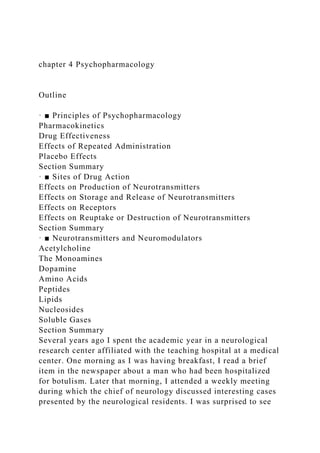
- 10. into the cerebrospinal fluid in the brain’s ventricular system. To study the effects of a drug in a specific region of the brain (for example, in a particular nucleus of the hypothalamus), a researcher will inject a very small amount of the drug directly into the brain. This procedure, known as intracerebral administration, is described in more detail in Chapter 5. To achieve a widespread distribution of a drug in the brain, a researcher will get past the blood–brain barrier by injecting the drug into a cerebral ventricle. The drug is then absorbed into the brain tissue, where it can exert its effects. This route, intracerebroventricular (ICV) administration, is used very rarely in humans—primarily to deliver antibiotics directly to the brain to treat certain types of infections. intracerebral administration Administration of a substance directly into the brain. intracerebroventricular (ICV) administration Administration of a substance into one of the cerebral ventricles. Figure 4.1 shows the time course of blood levels of a commonly abused drug, cocaine, after intravenous injection, inhalation, oral administration, and sniffing. The amounts received were not identical, but the graph illustrates the relative rapidity with which the drug reaches the blood. (See Figure 4.1.) FIGURE 4.1 Cocaine in Blood Plasma The graph shows the concentration of cocaine in blood plasma after intravenous injection, inhalation, oral administration, and sniffing. (Adapted from Feldman, R. S., Meyer, J. S., and Quenzer, L. F. Principles of Neuropsychopharmacology. Sunderland, MA: Sinauer Associates, 1997; after Jones, R. T. NIDA Research
- 11. Monographs, 1990, 99, 30–41.) ENTRY OF DRUGS INTO THE BRAIN As we saw, drugs exert their effects only when they reach their sites of action. In the case of drugs that affect behavior, most of these sites are located on or in particular cells in the central nervous system. The previous section described the routes by which drugs can be introduced into the body. With the exception of intracerebral or intracerebroventricular administration, the routes of drug administration vary only in the rate at which a drug reaches the blood plasma (that is, the liquid part of the blood). But what happens next? All the sites of action of drugs of interest to psychopharmacologists lie outside the blood vessels. The most important factor that determines the rate at which a drug in the bloodstream reaches sites of action within the brain is lipid solubility. The blood–brain barrier is a barrier only for water-soluble molecules. Molecules that are soluble in lipids pass through the cells that line the capillaries in the central nervous system, and they rapidly distribute themselves throughout the brain. For example, diacetylmorphine (more commonly known as heroin) is more lipid soluble than morphine is. Thus, an intravenous injection of heroin produces more rapid effects than does one of morphine. Even though the molecules of the two drugs are equally effective when they reach their sites of action in the brain, the fact that heroin molecules get there faster means that they produce a more intense “rush,” and this explains why drug addicts prefer heroin to morphine. INACTIVATION AND EXCRETION Drugs do not remain in the body indefinitely. Many are deactivated by enzymes, and all are eventually excreted, primarily by the kidneys. The liver plays an especially active
- 12. role in enzymatic deactivation of drugs, but some deactivating enzymes are also found in the blood. The brain also contains enzymes that destroy some drugs. In some cases, enzymes transform molecules of a drug into other forms that themselves are biologically active. Occasionally, the transformed molecule is even more active than the one that is administered. In such cases the effects of a drug can have a very long duration. Drug Effectiveness Drugs vary widely in their effectiveness. The effects of a small dose of a relatively effective drug can equal or exceed the effects of larger amounts of a relatively ineffective drug. The best way to measure the effectiveness of a drug is to plot a dose-response curve. To do this, subjects are given various doses of a drug, usually defined as milligrams of drug per kilogram of a subject’s body weight, and the effects of the drug are plotted. Because the molecules of most drugs distribute themselves throughout the blood and then throughout the rest of the body, a heavier subject (human or laboratory animal) will require a larger quantity of a drug to achieve the same concentration as a smaller quantity will produce in a smaller subject. As Figure 4.2 shows, increasingly stronger doses of a drug cause increasingly larger effects until the point of maximum effect is reached. At this point, increasing the dose of the drug does not produce any more effect. (See Figure 4.2.) dose-response curve A graph of the magnitude of an effect of a drug as a function of the amount of drug administered. FIGURE 4.2 A Dose-Response Curve Increasingly stronger doses of the drug produce increasingly larger effects until the maximum effect is reached. After that point, increments in the dose do not produce any increments in
- 13. the drug’s effect. However, the risk of adverse side effects increases. Most drugs have more than one effect. Opiates such as morphine and codeine produce analgesia (reduced sensitivity to pain), but they also depress the activity of neurons in the medulla that control heart rate and respiration. A physician who prescribes an opiate to relieve a patient’s pain wants to administer a dose that is large enough to produce analgesia but not large enough to depress heart rate and respiration—effects that could be fatal. Figure 4.3 shows two dose-response curves, one for the analgesic effects of a painkiller and one for the drug’s depressant effects on respiration. The difference between these curves indicates the drug’s margin of safety. Obviously, the most desirable drugs have a large margin of safety. (See Figure 4.3.) FIGURE 4.3 Dose-Response Curves for Morphine The dose-response curve on the left shows the analgesic effect of morphine, and the curve on the right shows one of the drug’s adverse side effects: its depressant effect on respiration. A drug’s margin of safety is reflected by the difference between the dose-response curve for its therapeutic effects and that for its adverse side effects. One measure of a drug’s margin of safety is its therapeutic index. This measure is obtained by administering varying doses of the drug to a group of laboratory animals such as mice. Two numbers are obtained: the dose that produces the desired effects in 50 percent of the animals and the dose that produces toxic effects in 50 percent of the animals. The therapeutic index is the ratio of these two numbers. For example, if the toxic dose is five times higher than the effective dose, then the therapeutic index is 5.0. The lower the therapeutic index, the more care
- 14. must be taken in prescribing the drug. For example, barbiturates have relatively low therapeutic indexes—as low as 2 or 3. In contrast, tranquilizers such as Valium have therapeutic indexes of well over 100. As a consequence, an accidental overdose of a barbiturate is much more likely to have tragic effects than a similar overdose of Valium. therapeutic index The ratio between the dose that produces the desired effect in 50 percent of the animals and the dose that produces toxic effects in 50 percent of the animals. Why do drugs vary in their effectiveness? There are two reasons. First, different drugs—even those with the same behavioral effects—may have different sites of action. For example, both morphine and aspirin have analgesic effects, but morphine suppresses the activity of neurons in the spinal cord and brain that are involved in pain perception, whereas aspirin reduces the production of a chemical involved in transmitting information from damaged tissue to pain-sensitive neurons. Because the drugs act very differently, a given dose of morphine (expressed in terms of milligrams of drug per kilogram of body weight) produces much more pain reduction than the same dose of aspirin does. The second reason that drugs vary in their effectiveness has to do with the affinity of the drug with its site of action. As we will see in the next major section of this chapter, most drugs of interest to psychopharmacologists exert their effects by binding with other molecules located in the central nervous system— with presynaptic or postsynaptic receptors, with transporter molecules, or with enzymes involved in the production or deactivation of neurotransmitters. Drugs vary widely in their affinity for the molecules to which they attach—the readiness with which the two molecules join together. A drug with a high affinity will produce effects at a relatively low concentration, whereas a drug with a low affinity must be
- 15. administered in higher doses. Thus, even two drugs with identical sites of action can vary widely in their effectiveness if they have different affinities for their binding sites. In addition, because most drugs have multiple effects, a drug can have high affinities for some of its sites of action and low affinities for others. The most desirable drug has a high affinity for sites of action that produce therapeutic effects and a low affinity for sites of action that produce toxic side effects. One of the goals of research by drug companies is to find chemicals with just this pattern of effects. affinity The readiness with which two molecules join together. Effects of Repeated Administration Often, when a drug is administered repeatedly, its effects will not remain constant. In most cases its effects will diminish—a phenomenon known as tolerance. In other cases a drug becomes more and more effective—a phenomenon known as sensitization. tolerance A decrease in the effectiveness of a drug that is administered repeatedly. sensitization An increase in the effectiveness of a drug that is administered repeatedly. Let’s consider tolerance first. Tolerance is seen in many drugs that are commonly abused. For example, a regular user of heroin must take larger and larger amounts of the drug for it to be effective. And once a person has taken heroin regularly enough to develop tolerance, that individual will suffer withdrawal symptoms if he or she suddenly stops taking the drug. Withdrawal symptoms are primarily the opposite of the effects of the drug itself. For example, heroin produces euphoria; withdrawal from it produces dysphoria—a feeling of anxious
- 16. misery. (Euphoria and dysphoria mean “easy to bear” and “hard to bear,” respectively.) Heroin produces constipation; withdrawal from it produces nausea and cramping. Heroin produces relaxation; withdrawal from it produces agitation. withdrawal symptom The appearance of symptoms opposite to those produced by a drug when the drug is administered repeatedly and then suddenly no longer taken. Withdrawal symptoms are caused by the same mechanisms that are responsible for tolerance. Tolerance is the result of the body’s attempt to compensate for the effects of the drug. That is, most systems of the body, including those controlled by the brain, are regulated so that they stay at an optimal value. When the effects of a drug alter these systems for a prolonged time, compensatory mechanisms begin to produce the opposite reaction, at least partially compensating for the disturbance from the optimal value. These mechanisms account for the fact that more and more of the drug must be taken to achieve a given level of effects. Then, when the person stops taking the drug, the compensatory mechanisms make themselves felt, unopposed by the action of the drug. Research suggests that there are several types of compensatory mechanisms. As we will see, many drugs that affect the brain do so by binding with receptors and activating them. The first compensatory mechanism involves a decrease in the effectiveness of such binding. Either the receptors become less sensitive to the drug (that is, their affinity for the drug decreases), or the receptors decrease in number. The second compensatory mechanism involves the process that couples the receptors to ion channels in the membrane or to the production of second messengers. After prolonged stimulation of the receptors, one or more steps in the coupling process become less effective. (Of course, both effects can occur.) The details of these compensatory mechanisms are described in Chapter 18,
- 17. which discusses the causes and effects of drug abuse. As we saw, many drugs have several different sites of action and thus produce several different effects. This means that some of the effects of a drug may show tolerance but others may not. For example, barbiturates cause sedation and also depress neurons that control respiration. The sedative effects show tolerance, but the respiratory depression does not. This means that if larger and larger doses of a barbiturate are taken to achieve the same level of sedation, the person begins to run the risk of taking a dangerously large dose of the drug. Sensitization is, of course, the exact opposite of tolerance: Repeated doses of a drug produce larger and larger effects. Because compensatory mechanisms tend to correct for deviations away from the optimal values of physiological processes, sensitization is less common than tolerance. And some of the effects of a drug may show sensitization while others show tolerance. For example, repeated injections of cocaine become more and more likely to produce movement disorders and convulsions, whereas the euphoric effects of the drug do not show sensitization—and may even show tolerance. Placebo Effects A placebo is an innocuous substance that has no specific physiological effect. The word comes from the Latin placere, “to please.” A physician may sometimes give a placebo to anxious patients to placate them. (You can see that the word placate also has the same root.) But although placebos have no specificphysiological effect, it is incorrect to say that they have no effect. If a person thinks that a placebo has a physiological effect, then administration of the placebo may actually produce that effect. placebo (plaseeboh) An inert substance that is given to an
- 18. organism in lieu of a physiologically active drug; used experimentally to control for the effects of mere administration of a drug. When experimenters want to investigate the behavioral effects of drugs in humans, they must use control groups whose members receive placebos, or they cannot be sure that the behavioral effects they observe were caused by specific effects of the drug. Studies with laboratory animals must also use placebos, even though we need not worry about the animals’ “beliefs” about the effects of the drugs we give them. Consider what you must do to give a rat an intraperitoneal injection of a drug. You reach into the animal’s cage, pick the animal up, hold it in such a way that its abdomen is exposed and its head is positioned to prevent it from biting you, insert a hypodermic needle through its abdominal wall, press the plunger of the syringe, and replace the animal in its cage, being sure to let go of it quickly so that it cannot turn and bite you. Even if the substance you inject is innocuous, the experience of receiving the injection would activate the animal’s autonomic nervous system, cause the secretion of stress hormones, and have other physiological effects. If we want to know what the behavioral effects of a drug are, we must compare the drug-treated animals with other animals who receive a placebo, administered in exactly the same way as the drug. (By the way, a skilled and experienced researcher can handle a rat so gently that it shows very little reaction to a hypodermic injection.) SECTION SUMMARY: Principles of Psychopharmacology Psychopharmacology is the study of the effects of drugs on the nervous system and behavior. Drugs are exogenous chemicals that are not necessary for normal cellular functioning that significantly alter the functions of certain cells of the body when taken in relatively low doses. Drugs have effects, physiological and behavioral, and they have sites of action—
- 19. molecules located somewhere in that body with which they interact to produce these effects. Pharmacokinetics is the fate of a drug as it is absorbed into the body, circulates throughout the body, and reaches its sites of action. Drugs may be administered by intravenous, intraperitoneal, intramuscular, and subcutaneous injection; they may be administered orally, sublingually, intrarectally, by inhalation, and topically (on skin or mucous membrane); and they may be injected intracerebrally or intracerebroventricularly. Lipid-soluble drugs easily pass through the blood–brain barrier, whereas others pass this barrier slowly or not at all. The time courses of various routes of drug administration are different. Eventually, drugs disappear from the body. Some are deactivated by enzymes, especially in the liver, and others are simply excreted. The dose-response curve represents a drug’s effectiveness; it relates the amount administered (usually in milligrams per kilogram of the subject’s body weight) to the resulting effect. Most drugs have more than one site of action and thus more than one effect. The safety of a drug is measured by the difference between doses that produce desirable effects and those that produce toxic side effects. Drugs vary in their effectiveness because of the nature of their sites of actions and the affinity between molecules of the drug and these sites of action. Repeated administration of a drug can cause either tolerance, often resulting in withdrawal symptoms, or sensitization. Tolerance can be caused by decreased affinity of a drug with its receptors, by decreased numbers of receptors, or by decreased coupling of receptors with the biochemical steps it controls. Some of the effects of a drug may show tolerance, while others
- 20. may not—or may even show sensitization. ■ THOUGHT QUESTIONS 1. Choose a drug whose effects you are familiar with and suggest where in the body the sites of action of that drug might be. 2. Some drugs can cause liver damage if large doses are taken for an extended period of time. What aspect of the pharmacokinetics of these drugs might cause the liver damage? Sites of Drug Action Throughout the history of our species, people have discovered that plants—and some animals—produce chemicals that act on the nervous system. (Of course, the people who discovered these chemicals knew nothing about neurons and synapses.) Some of these chemicals have been used for their pleasurable effects; others have been used to treat illness, reduce pain, or poison other animals (or enemies). More recently, scientists have learned to produce completely artificial drugs, some with potencies far greater than those of the naturally occurring drugs. The traditional uses of drugs remain, but in addition they can be used in research laboratories to investigate the operations of the nervous system. Most drugs that affect behavior do so by affecting synaptic transmission. Drugs that affect synaptic transmission are classified into two general categories. Those that block or inhibit the postsynaptic effects are called antagonists. Those that facilitate them are called agonists. (The Greek word agon means “contest.” Thus, an agonist is one who takes part in the contest.) antagonist A drug that opposes or inhibits the effects of a
- 21. particular neurotransmitter on the postsynaptic cell. agonist A drug that facilitates the effects of a particular neurotransmitter on the postsynaptic cell. This section will describe the basic effects of drugs on synaptic activity. Recall from Chapter 2 that the sequence of synaptic activity goes like this: Neurotransmitters are synthesized and stored in synaptic vesicles. The synaptic vesicles travel to the presynaptic membrane, where they become docked. When an axon fires, voltage-dependent calcium channels in the presynaptic membrane open, permitting the entry of calcium ions. The calcium ions interact with the docking proteins and initiate the release of the neurotransmitters into the synaptic cleft. Molecules of the neurotransmitter bind with postsynaptic receptors, causing particular ion channels to open, which produces excitatory or inhibitory postsynaptic potentials. The effects of the neurotransmitter are kept relatively brief by their reuptake by transporter molecules in the presynaptic membrane or by their destruction by enzymes. In addition, the stimulation of presynaptic autoreceptors on the terminal buttons regulates the synthesis and release of the neurotransmitter. The discussion of the effects of drugs in this section follows the same basic sequence. All of the effects I will describe are summarized in Figure 4.4, with some details shown in additional figures. I should warn you that some of the effects are complex, so the discussion that follows bears careful reading. I recommend that you Simulate actions of drugs on MyPsychLab, which reviews this material. Effects on Production of Neurotransmitters The first step is the synthesis of the neurotransmitter from its precursors. In some cases the rate of synthesis and release of a neurotransmitter is increased when a precursor is administered;
- 22. in these cases the precursor itself serves as an agonist. (See step 1 in Figure 4.4.) The steps in the synthesis of neurotransmitters are controlled by enzymes. Therefore, if a drug inactivates one of these enzymes, it will prevent the neurotransmitter from being produced. Such a drug serves as an antagonist. (See step 2 in Figure 4.4.) FIGURE 4.4 Drug Effects on Synaptic Transmission The figure summarizes the ways in which drugs can affect the synaptic transmission (AGO = agonist; ANT = antagonist; NT = neurotransmitter). Drugs that act as agonists are marked in blue; drugs that act as antagonists are marked in red. Effects on Storage and Release of Neurotransmitters Neurotransmitters are stored in synaptic vesicles, which are transported to the presynaptic membrane, where the chemicals are released. The storage of neurotransmitters in vesicles is accomplished by the same kind of transporter molecules that are responsible for reuptake of a neurotransmitter into a terminal button. The transporter molecules are located in the membrane of synaptic vesicles, and their action is to pump molecules of the neurotransmitter across the membrane, filling the vesicles. Some of the transporter molecules that fill synaptic vesicles are capable of being blocked by a drug. Molecules of the drug bind with a particular site on the transporter and inactivate it. Because the synaptic vesicles remain empty, nothing is released when the vesicles eventually rupture against the presynaptic membrane. The drug serves as an antagonist. (See step 3 in Figure 4.4.) Some drugs act as antagonists by preventing the release of neurotransmitters from the terminal button. They do so by
- 23. deactivating the proteins that cause docked synaptic vesicles to fuse with the presynaptic membrane and expel their contents into the synaptic cleft. Other drugs have just the opposite effect: They act as agonists by binding with these proteins and directly triggering release of the neurotransmitter. (See steps 4 and 5 in Figure 4.4.) Effects on Receptors The most important—and most complex—site of action of drugs in the nervous system is on receptors, both presynaptic and postsynaptic. Let’s consider postsynaptic receptors first. (Here is where the careful reading should begin.) Once a neurotransmitter is released, it must stimulate the postsynaptic receptors. Some drugs bind with these receptors, just as the neurotransmitter does. Once a drug has bound with the receptor, it can serve as either an agonist or an antagonist. A drug that mimics the effects of a neurotransmitter acts as a direct agonist. Molecules of the drug attach to the binding site to which the neurotransmitter normally attaches. This binding causes ion channels controlled by the receptor to open, just as they do when the neurotransmitter is present. Ions then pass through these channels and produce postsynaptic potentials. (See step 6 in Figure 4.4.) direct agonist A drug that binds with and activates a receptor. Drugs that bind with postsynaptic receptors can also serve as antagonists. Molecules of such drugs bind with the receptors but do not open the ion channel. Because they occupy the receptor’s binding site, they prevent the neurotransmitter from opening the ion channel. These drugs are called receptor blockers or direct antagonists. (See step 7 in Figure 4.4.) receptor blocker A drug that binds with a receptor but does not
- 24. activate it; prevents the natural ligand from binding with the receptor. direct antagonist A synonym for receptor blocker. FIGURE 4.5 Drug Actions at Binding Sites (a) Competitive binding: Direct agonists and antagonists act directly on the neurotransmitter binding site. (b) Noncompetitive binding: Indirect agonists and antagonists act on an alternative binding site and modify the effects of the neurotransmitter on opening of the ion channel. Some receptors have multiple binding sites, to which different ligands can attach. Molecules of the neurotransmitter bind with one site, and other substances (such as neuromodulators and various drugs) bind with the others. Binding of a molecule with one of these alternative sites is referred to as noncompetitive binding, because the molecule does not compete with molecules of the neurotransmitter for the same binding site. If a drug attaches to one of these alternative sites and prevents the ion channel from opening, the drug is said to be an indirect antagonist. The ultimate effect of an indirect antagonist is similar to that of a direct antagonist, but its site of action is different. If a drug attaches to one of the alternative sites and facilitates the opening of the ion channel, it is said to be an indirect agonist. (See Figure 4.5.) noncompetitive binding Binding of a drug to a site on a receptor; does not interfere with the binding site for the principal ligand. indirect antagonist A drug that attaches to a binding site on a receptor and interferes with the action of the receptor; does not interfere with the binding site for the principal ligand.
- 25. indirect agonist A drug that attaches to a binding site on a receptor and facilitates the action of the receptor; does not interfere with the binding site for the principal ligand. FIGURE 4.6 Presynaptic Heteroreceptors Presynaptic facilitation is caused by activation of receptors that facilitate the opening of calcium channels near the active zone of the postsynaptic terminal button, which promotes release of the neurotransmitter. Presynaptic inhibition is caused by activation of receptors that inhibit the opening of these calcium channels. As we saw in Chapter 2, the presynaptic membranes of some neurons contain autoreceptors that regulate the amount of neurotransmitter that is released. Because stimulation of these receptors causes less neurotransmitter to be released, drugs that selectively activate presynaptic receptors act as antagonists. Drugs that block presynaptic autoreceptors have the opposite effect: They increase the release of the neurotransmitter, acting as agonists. (Refer to steps 8 and 9 in Figure 4.4.) We also saw in Chapter 2 that some terminal buttons form axoaxonic synapses—synapses of one terminal button with another. Activation of the first terminal button causes presynaptic inhibition or facilitation of the second one. The second terminal button contains presynaptic heteroreceptors, which are sensitive to the neurotransmitter released by the first one. (Auto means “self”; hetero means “other.”) Presynaptic heteroreceptors that produce presynaptic inhibition do so by inhibiting the release of the neurotransmitter. Conversely, presynaptic heteroreceptors responsible for presynaptic facilitation facilitate the release of the neurotransmitter. So drugs can block or facilitate presynaptic inhibition or
- 26. facilitation, depending on whether they block or activate presynaptic heteroreceptors. (See Figure 4.6.) presynaptic heteroreceptor A receptor located in the membrane of a terminal button that receives input from another terminal button by means of an axoaxonic synapse; binds with the neurotransmitter released by the presynaptic terminal button. Finally (yes, this is the last site of action I will describe in this subsection), you will recall from Chapter 2that autoreceptors are located in the membrane of dendrites of some neurons. When these neurons become active, their dendrites, as well as their terminal buttons, release neurotransmitter. The neurotransmitter released by the dendrites stimulates autoreceptors located on these same dendrites, which decrease neural firing by producing hyperpolarizations. This mechanism has a regulatory effect, serving to prevent these neurons from becoming too active. Thus, drugs that bind with and activatedendritic autoreceptors will serve as antagonists. Those that bind with and block dendritic autoreceptors will serve as agonists, because they will prevent the inhibitory hyperpolarizations. (See Figure 4.7.) As you will surely realize, the effects of a particular drug that binds with a particular type of receptor can be very complex. The effects depend on where the receptor is located, what its normal effects are, and whether the drug activates the receptor or blocks its actions. FIGURE 4.7 Dendritic Autoreceptors The dendrites of certain neurons release some neurotransmitter when the cell is active. Activation of dendritic autoreceptors by the neurotransmitter (or by a drug that binds with these receptors) hyperpolarizes the membrane, reducing the neuron’s
- 27. rate of firing. Blocking of dendritic autoreceptors by a drug prevents this effect. Effects on Reuptake or Destruction of Neurotransmitters The next step after stimulation of the postsynaptic receptor is termination of the postsynaptic potential. Two processes accomplish that task: Molecules of the neurotransmitter are taken back into the terminal button through the process of reuptake, or they are destroyed by an enzyme. Drugs can interfere with either of these processes. In the first case, molecules of the drug attach to the transporter molecules responsible for reuptake and inactivate them, thus blocking reuptake. In the second case, molecules of the drug bind with the enzyme that normally destroys the neurotransmitter and prevents the enzymes from working. The most important example of such an enzyme is acetylcholinesterase, which destroys acetylcholine. Because both types of drugs prolong the presence of molecules of the neurotransmitter in the synaptic cleft (and hence in a location where these molecules can stimulate postsynaptic receptors), they serve as agonists. (Refer to steps 10 and 11 in Figure 4.4.) SECTION SUMMARY: Sites of Drug Action The process of synaptic transmission entails the synthesis of the neurotransmitter, its storage in synaptic vesicles, its release into the synaptic cleft, its interaction with postsynaptic receptors, and the consequent opening of ion channels in the postsynaptic membrane. The effects of the neurotransmitter are then terminated by reuptake into the terminal button or by enzymatic deactivation. Each of the steps necessary for synaptic transmission can be interfered with by drugs that serve as antagonists, and a few can be stimulated by drugs that serve as agonists. Thus, drugs can
- 28. increase the pool of available precursor, block a biosynthetic enzyme, prevent the storage of neurotransmitter in synaptic vesicles, stimulate or block the release of the neurotransmitter, stimulate or block presynaptic or postsynaptic receptors, retard reuptake, or deactivate enzymes that destroy the neurotransmitter. A drug that activates postsynaptic receptors serves as an agonist, whereas one that activates presynaptic or dendritic autoreceptors serves as an antagonist. A drug that blocks postsynaptic receptors serves as an antagonist, whereas one that blocks autoreceptors serves as an agonist. A drug that activates or blocks presynaptic heteroreceptors serves as an agonist or antagonist, depending on whether the heteroreceptors are responsible for presynaptic facilitation or inhibition. ■ THOUGHT QUESTION Explain how a drug that blocks receptors can serve as an agonist. Neurotransmitters and Neuromodulators Because neurotransmitters have two general effects on postsynaptic membranes—depolarization (EPSP) or hyperpolarization (IPSP)—one might expect that there would be two kinds of neurotransmitters, excitatory and inhibitory. Instead, there are many different kinds—several dozen at least. In the brain most synaptic communication is accomplished by two neurotransmitters: one with excitatory effects (glutamate) and one with inhibitory effects (GABA). (Another inhibitory neurotransmitter, glycine, is found in the spinal cord and lower brain stem.) Most of the activity of local circuits of neurons involves balances between the excitatory and inhibitory effects of these chemicals, which are responsible for most of the information transmitted from place to place within the brain. In fact, there are probably no neurons in the brain that do not receive excitatory input from glutamate-secreting terminal buttons and inhibitory input from neurons that secrete either
- 29. GABA or glycine. And with the exception of neurons that detect painful stimuli, all sensory organs transmit information to the brain through axons whose terminals release glutamate. (Pain- detecting neurons secrete a peptide.) What do all the other neurotransmitters do? In general, they have modulating effects rather than information-transmitting effects. That is, the release of neurotransmitters other than glutamate and GABA tends to activate or inhibit entire circuits of neurons that are involved in particular brain functions. For example, secretion of acetylcholine activates the cerebral cortex and facilitates learning, but the information that is learned and remembered is transmitted by neurons that secrete glutamate and GABA. Secretion of norepinephrine increases vigilance and enhances readiness to act when a signal is detected. Secretion of histamine enhances wakefulness. Secretion of serotonin suppresses certain categories of species-typical behaviors and reduces the likelihood that the animal acts impulsively. Secretion of dopamine in some regions of the brain generally activates voluntary movements but does not specify which movements will occur. In other regions, secretion of dopamine reinforces ongoing behaviors and makes them more likely to occur at a later time. Because particular drugs can selectively affect neurons that secrete particular neurotransmitters, they can have specific effects on behavior. This section introduces the most important neurotransmitters, discusses some of their behavioral functions, and describes the drugs that interact with them. As we saw in the previous section of this chapter, drugs have many different sites of action. Fortunately for your information-processing capacity (and perhaps your sanity), not all types of neurons are affected by all types of drugs. As you will see, that still leaves a good number of drugs to be mentioned by name. Obviously, some are more important than others. Those whose effects I describe in some detail are more important than those I mention in passing.
- 30. Acetylcholine Acetylcholine is the primary neurotransmitter secreted by efferent axons of the peripheral nervous system. All muscular movement is accomplished by the release of acetylcholine, and ACh is also found in the ganglia of the autonomic nervous system and at the target organs of the parasympathetic branch of the ANS. Because ACh is found outside the central nervous system in locations that are easy to study, this neurotransmitter was the first to be discovered, and it has received much attention from neuroscientists. Some terminology: These synapses are said to be acetylcholinergic. Ergon is the Greek word for “work.” Thus, dopaminergic synapses release dopamine, serotonergic synapses release serotonin, and so on. (The suffix -ergic is pronounced “urjik”.) The axons and terminal buttons of acetylcholinergic neurons are distributed widely throughout the brain. Three systems have received the most attention from neuroscientists: those originating in the dorsolateral pons, the basal forebrain, and the medial septum. The effects of ACh release in the brain are generally facilitatory. The acetylcholinergic neurons located in the dorsolateral pons play a role in REM sleep (the phase of sleep during which dreaming occurs). Those located in the basal forebrain are involved in activating the cerebral cortex and facilitating learning, especially perceptual learning. Those located in the medial septum control the electrical rhythms of the hippocampus and modulate its functions, which include the formation of particular kinds of memories. Figure 4.8 shows a schematic midsagittal view of a rat brain. On it are indicated the most important sites of acetylcholinergic cell bodies and the regions served by the branches of their axons. The figure illustrates a rat brain because most of the neuroanatomical tracing studies have been performed with rats.
- 31. Presumably, the location and projections of acetylcholinergic neurons in the human brain resemble those found in the rat brain, but we cannot yet be certain. The methods used for tracing particular systems of neurons in the brain and the difficulty of doing such studies with the human brain are described in Chapter 5. (See Figure 4.8.) Acetylcholine is composed of two components: choline, a substance derived from the breakdown of lipids, and acetate, the anion found in vinegar, also called acetic acid. Acetate cannot be attached directly to choline; instead, it is transferred from a molecule of acetyl-CoA. CoA (coenzyme A) is a complex molecule, consisting in part of the vitamin pantothenic acid (one of the B vitamins). CoA is produced by the mitochondria, and it takes part in many reactions in the body. Acetyl-CoA is simply CoA with an acetate ion attached to it. ACh is produced by the following reaction: In the presence of the enzyme choline acetyltransferase (ChAT), the acetate ion is transferred from the acetyl-CoA molecule to the choline molecule, yielding a molecule of ACh and one of ordinary CoA. (See Figure 4.9.) acetyl-CoA (aseetul) A cofactor that supplies acetate for the synthesis of acetylcholine. choline acetyltransferase (ChAT) (kohleen a see tultransfer ace) The enzyme that transfers the acetate ion from acetyl coenzyme A to choline, producing the neurotransmitter acetylcholine. FIGURE 4.8 Acetylcholinergic Pathways in a Rat Brain This schematic figure shows the locations of the most important groups of acetylcholinergic neurons and the distribution of their axons and terminal buttons.
- 32. (Adapted from Woolf, N. J. Progress in Neurobiology, 1991, 37, 475–524.) FIGURE 4.9 Biosynthesis of Acetylcholine A simple analogy will illustrate the role of coenzymes in chemical reactions. Think of acetate as a hot dog and choline as a bun. The task of the person (enzyme) who operates the hot dog vending stand is to put a hot dog into the bun (make acetylcholine). To do so, the vendor needs a fork (coenzyme) to remove the hot dog from the boiling water. The vendor inserts the fork into the hot dog (attaches acetate to CoA) and transfers the hot dog from fork to bun. Two drugs, botulinum toxin and the venom of the black widow spider, affect the release of acetylcholine. Botulinum toxin is produced by clostridium botulinum, a bacterium that can grow in improperly canned food. This drug prevents the release of ACh (step 5 of Figure 4.4). As we saw in this chapter’s opening case, botulinum toxin drug is an extremely potent poison because the paralysis it can cause leads to suffocation. In contrast, black widow spider venom has the opposite effect: It stimulates the release of ACh (step 4 of Figure 4.4). Although the effects of black widow spider venom can also be fatal, the venom is much less toxic than botulinum toxin. In fact, most healthy adults would have to receive several bites, but infants or frail elderly people would be more susceptible. botulinum toxin (bot youlinum) An acetylcholine antagonist; prevents release by terminal buttons. black widow spider venom A poison produced by the black widow spider that triggers the release of acetylcholine.
- 33. FIGURE 4.10 Destruction of Acetylcholine (ACh) by Acetylcholinesterase (AChE) You may have been wondering why double vision was the first symptom of botulism in the opening case. The answer is that the delicate balance among the muscles that move the eyes is upset by any interference with acetylcholinergic transmission. You undoubtedly know that botox treatment has become fashionable. A very dilute (obviously!) solution of botulinum toxin is injec ted into people’s facial muscles to stop muscular contractions that are causing wrinkles in the skin. I’m not planning on getting a botox treatment, but if I did, I would want to be sure that the solution was sufficiently dilute. You will recall from Chapter 2 that after being released by the terminal button, ACh is deactivated by the enzyme acetylcholinesterase (AChE), which is present in the postsynaptic membrane. (See Figure 4.10.) Drugs that deactivate AChE (step 11 of Figure 4.4) are used for several purposes. Some are used as insectici des. These drugs readily kill insects but not humans and other mammals, because our blood contains enzymes that destroy them. (Insects lack the enzyme.) Other AChE inhibitors are used medically. For example, a hereditary disorder called myasthenia gravis is caused by an attack of a person’s immune system against acetylch oline receptors located on skeletal muscles. (Kathryn D., whose symptoms were described in the opening case of Chapter 2, had this disorder.) The person becomes weaker and weaker as the muscles become less responsive to the neurotransmitter. If the person is given an AChE inhibitor such as neostigmine, the person will regain some strength because the acetylcholine that is released has a more prolonged effect on the remaining receptors. (Neostigmine cannot cross the blood– brain barrier, so it does not affect the AChE found in the central nervous system.)
- 34. neostigmine (nee ostigmeen) A drug that inhibits the activity of acetylcholinesterase. There are two types of ACh receptors: one ionotropic and one metabotropic. These receptors were identified when investigators discovered that different drugs activated them (step 6 of Figure 4.4). The ionotropic ACh receptor is stimulated by nicotine, a drug found in tobacco leaves. (The Latin name of the plant is Nicotiniana tabacum.) The metabotropic ACh receptor is stimulated by muscarine, a drug found in the poison mushroom Amanita muscaria. Consequently, these two ACh receptors are referred to as nicotinic receptors and muscarinic receptors, respectively. Because muscle fibers must be able to contract rapidly, they contain the rapid, ionotropic nicotinic receptors. nicotinic receptor An ionotropic acetylcholine receptor that is stimulated by nicotine and blocked by curare. muscarinic receptor (muss karinic) A metabotropic acetylcholine receptor that is stimulated by muscarine and blocked by atropine. Because muscarinic receptors are metabotropic in nature and thus control ion channels through the production of second messengers, their actions are slower and more prolonged than those of nicotinic receptors. The central nervous system contains both kinds of ACh receptors, but muscarinic receptors predominate. Some nicotinic receptors are found at axoaxonic synapses in the brain, where they produce presynaptic facilitation. Activation of these receptors is responsible for the addictive effect of the nicotine found in tobacco smoke. Just as two different drugs stimulate the two classes of acetylcholine receptors, two different drugs blockthem (step 7
- 35. of Figure 4.4). Both drugs were discovered in nature long ago, and both are still used by modern medicine. The first, atropine, blocks muscarinic receptors. The drug is named after Atropos, the Greek fate who cut the thread of life (which a sufficient dose of atropine will certainly do). Atropine is one of several belladonna alkaloids extracted from a plant called the deadly nightshade, and therein lies a tale. Many years ago, women who wanted to increase their attractiveness to men put drops containing belladonna alkaloids into their eyes. In fact, belladonna means “pretty lady.” Why was the drug used this way? One of the unconscious responses that occurs when we are interested in something is dilation of our pupils. By blocking the effects of acetylcholine on the pupil, belladonna alkaloids such as atropine make the pupils dilate. This change makes a woman appear more interested in a man when she looks at him, and, of course, this apparent sign of interest makes him regard her as more attractive. atropine (a tro peen) A drug that blocks muscarinic acetylcholine receptors. Another drug, curare, blocks nicotinic receptors. Because these receptors are the ones found on muscles, curare, like botulinum toxin, causes paralysis. However, the effects of curare are much faster. The drug is extracted from several different species of plants found in South America, where it was discovered long ago by people who used it to coat the tips of arrows and darts. Within minutes of being struck by one of these points, an animal collapses, ceases breathing, and dies. Nowadays, curare (and other drugs with the same site of action) are used to paralyze patients who are to undergo surgery so that their muscles will relax completely and not contract when they are cut with a scalpel. An anesthetic must also be used, because a person who receives only curare will remain perfectly conscious and sensitive to pain, even though paralyzed. And, of course, a respirator must be used to supply air to the lungs.
- 36. curare (kew rahr ee) A drug that blocks nicotinic acetylcholine receptors. The Monoamines Dopamine, norepinephrine, epinephrine, serotonin, and histamine are five chemicals that belong to a family of compounds called monoamines. Because the molecular structures of these substances are similar, some drugs affect the activity of all of them to some degree. The first three— dopamine, norepinephrine, and epinephrine—belong to a subclass of monoamines called catecholamines. It is worthwhile learning the terms in Table 4.1, because they will be used throughout the rest of this book. (See Table 4.1.) monoamine (mahno a meen) A class of amines that includes indolamines, such as serotonin; and catecholamines, such as dopamine, norepinephrine, and epinephrine. catecholamine (cat akohla meen) A class of amines that includes the neurotransmitters dopamine, norepinephrine, and epinephrine. The monoamines are produced by several systems of neurons in the brain. Most of these systems consist of a relatively small number of cell bodies located in the brain stem, whose axons branch repeatedly and give rise to an enormous number of terminal buttons distributed throughout many regions of the brain. Monoaminergic neurons thus serve to modulate the function of widespread regions of the brain, increasing or decreasing the activities of particular brain functions. DOPAMINE TABLE 4.1 Classification of the Monoamine Neurotransmitters
- 37. Catecholamines Indolamine Ethylamine Dopamine Serotonin Histamine Norepinephrine Epinephrine The first catecholamine in Table 4.1, dopamine (DA), produces both excitatory and inhibitory postsynaptic potentials, depending on the postsynaptic receptor. Dopamine is one of the more interesting neurotransmitters because it has been implicated in several important functions, including movement, attention, learning, and the reinforcing effects of drugs that people tend to abuse. It is discussed in Chapters 8, 9, 13, 16, and 18. The synthesis of the catecholamines is somewhat more complicated than that of ACh, but each step is a simple one. The precursor molecule is modified slightly, step by step, until it achieves its final shape. Each step is controlled by a different enzyme, which causes a small part to be added or taken off. The precursor for the two major catecholamine neurotransmitters (dopamine and norepinephrine) is tyrosine, an essential amino acid that we must obtain from our diet. Tyrosine receives a hydroxyl group (OH—an oxygen atom and a hydrogen atom) and becomes L-DOPA (L-3,4-dihydroxyphenylalanine). The enzyme that adds the hydroxyl group is called tyrosine hydroxylase. L-DOPA then loses a carboxyl group (COOH—one carbon atom, two oxygen atoms, and one hydrogen atom) through the activity of the enzyme DOPA decarboxylase and becomes dopamine. Finally, the enzyme dopamine β- hydroxylase attaches a hydroxyl group to dopamine, which
- 38. becomes norepinephrine. These reactions are shown in Figure 4.11. dopamine (DA) (dopea meen) A neurotransmitter; one of the catecholamines. L-DOPA (elldopea) The levorotatory form of DOPA; the precursor of the catecholamines; often used to treat Parkinson’s disease because of its effect as a dopamine agonist. The brain contains several systems of dopaminergic neurons. The three most important of these originate in the midbrain: in the substantia nigra and in the ventral tegmental area. (The substantia nigra was shown in Figure 3.21; the ventral tegmental area is located just below this region.) The cell bodies of neurons of the nigrostriatal system are located in the substantia nigra and project their axons to the neostriatum: the caudate nucleus and the putamen. The neostriatum is an important part of the basal ganglia, which is involved in the control of movement. The cell bodies of neurons of the mesolimbic system are located in the ventral tegmental area and project their axons to several parts of the limbic system, including the nucleus accumbens, amygdala, and hippocampus. The nucleus accumbens plays an important role in the reinforcing (rewarding) effects of certain categories of stimuli, including those of drugs that people abuse. The cell bodies of neurons of the mesocortical system are also located in the ventral tegmental area. Their axons project to the prefrontal cortex. These neurons have an excitatory effect on the frontal cortex and affect such functions as formation of short-term memories, planning, and strategy preparation for problem solving. These three systems of dopaminergic neurons are shown in Figure 4.12. nigrostriatal system (nigh grow stryaytul) A system of neurons originating in the substantia nigra and terminating in the neostriatum (caudate nucleus and putamen).
- 39. mesolimbic system (mee zolimbik) A system of dopaminergic neurons originating in the ventral tegmental area and terminating in the nucleus accumbens, amygdala, and hippocampus. mesocortical system (mee zokorti kul) A system of dopaminergic neurons originating in the ventral tegmental area and terminating in the prefrontal cortex. FIGURE 4.11 Biosynthesis of the Catecholamines Degeneration of dopaminergic neurons that connect the substantia nigra with the caudate nucleus causes Parkinson’s disease, a movement disorder characterized by tremors, rigidity of the limbs, poor balance, and difficulty in initiating movements. The cell bodies of these neurons are located in a region of the brain called the substantia nigra (“black substance”). This region is normally stained black with melanin, the substance that gives color to skin. This compound is produced by the breakdown of dopamine. (The brain damage that causes Parkinson’s disease was discovered by pathologists who observed that the substantia nigra of a deceased person who had had this disorder was pale rather than black.) People with Parkinson’s disease are given L-DOPA, the precursor to dopamine. Although dopamine cannot cross the blood–brain barrier, L-DOPA can. Once L-DOPA reaches the brain, it is taken up by dopaminergic neurons and is converted to dopamine (step 1 of Figure 4.4). The increased synthesis of dopamine causes more dopamine to be released by the surviving dopaminergic neurons in patients with Parkinson’s disease. As a consequence, the patients’ symptoms are alleviated. Parkinson’s disease A neurological disease characterized by tremors, rigidity of the limbs, poor balance, and difficulty in initiating movements; caused by degeneration of the
- 40. nigrostriatal system. Another drug, AMPT (or α-methyl-p-tyrosine), inactivates tyrosine hydroxylase, the enzyme that converts tyrosine to L- DOPA (step 2 of Figure 4.4). Because this drug interferes with the synthesis of dopamine (and of norepinephrine as well), it serves as a catecholamine antagonist. The drug is not normally used medically, but it has been used as a research tool in laboratory animals. AMPT A drug that blocks the activity of tyrosine hydroxylase and thus interferes with the synthesis of the catecholamines. The drug reserpine prevents the storage of monoamines in synaptic vesicles by blocking the transporters in the membrane of vesicles of monoaminergic neurons (step 3 of Figure 4.4). Because the synaptic vesicles remain empty, no neurotransmitter is released when an action potential reaches the terminal button. Reserpine, then, is a monoamine antagonist. The drug, which comes from the root of a shrub, was discovered over 3000 years ago in India, where it was found to be useful in treating snakebite and seemed to have a calming effect. Pieces of the root are still sold in markets in rural areas of India. In Western medicine, reserpine was previously used to treat high blood pressure, but it has been replaced by drugs with fewer side effects. reserpine (reesurpeen) A drug that interferes with the storage of monoamines in synaptic vesicles. Several different types of dopamine receptors have been identified, all metabotropic. Of these, two are the most common: D1 receptors and D2 receptors. It appears that D1 receptors are exclusively postsynaptic, whereas D2 receptors are found both presynaptically and postsynaptically in the brain. Stimulation of D1receptors increases the production of the
- 41. second messenger cyclic AMP, whereas stimulation of D2receptors decreases it, as does stimulation of D3 and D4 receptors. Several drugs stimulate or block specific types of dopamine receptors. FIGURE 4.12 Dopaminergic Pathways in a Rat Brain This schematic figure shows the locations of the most important groups of dopaminergic neurons and the distribution of their axons and terminal buttons. (Adapted from Fuxe, K., Agnati, L. F., Kalia, M., et al., in Basic and Clinical Aspects of Neuroscience: The Dopaminergic System, edited by E. Fluckinger, E. E. Muller, and M. O. Thomas. Berlin: Springer–Verlag, 1985.) Autoreceptors are found in the dendrites, soma, and terminal buttons of dopaminergic neurons. Activation of the autoreceptors in the dendritic and somatic membrane decreases neural firing by producing hyperpolarizations. The presynaptic autoreceptors located in the terminal buttons suppress the activity of the enzyme tyrosine hydroxylase and thus decrease the production of dopamine—and ultimately its release. Dopamine autoreceptors resemble D2 receptors, but there seem to be some differences. For example, the drug apomorphine is a D2 agonist, but it seems to have a greater affinity for presynaptic D2 receptors than for postsynaptic D2 receptors. A low dose of apomorphine acts as an antagonist, because it stimulates the presynaptic receptors and inhibits the production and release of dopamine. Higher doses begin to stimulate postsynaptic D2 receptors, and the drug begins to act as a direct agonist. (See Figure 4.13.) apomorphine (ap omorefeen) A drug that blocks dopamine autoreceptors at low doses; at higher doses, blocks postsynaptic
- 42. receptors as well. Several drugs inhibit the reuptake of dopamine, thus serving as potent dopamine agonists (step 10 of Figure 4.4). The best known of these drugs are amphetamine, cocaine, and methylphenidate. Amphetamine has an interesting effect: It causes the release of both dopamine and norepinephrine by causing the transporters for these neurotransmitters to run in reverse, propelling DA and NE into the synaptic cleft. Of course, this action also blocks reuptake of these neurotransmitters. Cocaine and methylphenidate simply block dopamine reuptake. Because cocaine also blocks voltage- dependent sodium channels, it is sometimes used as a topical anesthetic, especially in the form of eye drops for eye surgery. Methylphenidate (Ritalin) is used to treat children who have attention deficit disorder. methylphenidate (meth ulfeni date) A drug that inhibits the reuptake of dopamine. The production of the catecholamines is regulated by an enzyme called monoamine oxidase (MAO). This enzyme is found within monoaminergic terminal buttons, where it destroys excessive amounts of neurotransmitter. A drug called deprenyl destroys the particular form of monoamine oxidase (MAO-B) that is found in dopaminergic terminal buttons. Because deprenyl prevents the destruction of dopamine, more dopamine is released when an action potential reaches the terminal button. Thus, deprenyl serves as a dopamine agonist. (See Figure 4.14.) monoamine oxidase (MAO) (mahno a meen) A class of enzymes that destroy the monoamines: dopamine, norepinephrine, and serotonin. deprenyl (deppra nil) A drug that blocks the activity of MAO-B; acts as a dopamine agonist.
- 43. FIGURE 4.13 Effects of Low and High Doses of Apomorphine At low doses, apomorphine serves as a dopamine antagonist; at high doses, it serves as an agonist. MAO is also found in the blood, where it deactivates amines that are present in foods such as chocolate and cheese. Without such deactivation these amines could cause dangerous increases in blood pressure. Dopamine has been implicated as a neurotransmitter that might be involved in schizophrenia, a serious mental disorder whose symptoms include hallucinations, delusions, and disruption of normal, logical thought processes. Drugs such as chlorpromazine, which block D2 receptors, alleviate these symptoms (step 7 of Figure 4.4). Hence, investigators have speculated that schizophrenia is produced by overactivity of dopaminergic neurons. More recently discovered drugs—the so- called atypical antipsychotics—have more complicated actions, which are discussed in Chapter 16. chlorpromazine (klorprohma zeen) A drug that reduces the symptoms of schizophrenia by blocking dopamine D2 receptors. FIGURE 4.14 Role of Monoamine Oxidase This schematic shows the role of monoamine oxidase in dopaminergic terminal buttons and the action of deprenyl. FIGURE 4.15 Noradrenergic Pathways in a Rat Brain This schematic figure shows the locations of the most important
- 44. groups of noradrenergic neurons and the distribution of their axons and terminal buttons. (Adapted from Cotman, C. W. and McGaugh, J. L. Behavioral Neuroscience: An Introduction. New York: Academic Press, 1980.) NOREPINEPHRINE Because norepinephrine (NE), like ACh, is found in neurons in the autonomic nervous system, this neurotransmitter has received much experimental attention. I should note that the terms Adrenalin and epinephrine are synonymous, as are noradrenalin and norepinephrine. Let me explain why. Epinephrineis a hormone produced by the adrenal medulla, the central core of the adrenal glands, located just above the kidneys. Epinephrine also serves as a neurotransmitter in the brain, but it is of minor importance compared with norepinephrine. Ad renal is Latin for “toward the kidney.” In Greek, one would say epi nephron (“upon the kidney”), hence the term epinephrine. The latter term has been adopted by pharmacologists, probably because the word Adrenalin was appropriated by a drug company as a proprietary name; therefore, to be consistent with general usage, I will refer to the neurotransmitter as norepinephrine. The accepted adjectival form is noradrenergic; I suppose that norepinephrinergic never caught on because it takes so long to pronounce. norepinephrine (NE) (nor epp ineffrin) One of the catecholamines; a neurotransmitter found in the brain and in the sympathetic division of the autonomic nervous system. epinephrine (epp ineffrin) One of the catecholamines; a hormone secreted by the adrenal medulla; serves also as a neurotransmitter in the brain.
- 45. We have already seen the biosynthetic pathway for norepinephrine in Figure 4.11. The drug AMPT, which prevents the conversion of tyrosine to L-DOPA, blocks the production of norepinephrine as well as dopamine (step 2 of Figure 4.4). Most neurotransmitters are synthesized in the cytoplasm of the terminal button and then stored in newly formed synaptic vesicles. However, for norepinephrine the final step of synthesis occurs inside the vesicles themselves. The vesicles are first filled with dopamine. Then the dopamine is converted to norepinephrine through the action of the enzyme dopamine β- hydroxylase located within the vesicles. The drug fusaric acid inhibits the activity of the enzyme dopamine-β-hydroxylase and thus blocks the production of norepinephrine without affecting the production of dopamine. fusaric acid (fewsahrik) A drug that inhibits the activity of the enzyme dopamine-β-hydroxylase and thus blocks the production of norepinephrine. Excess norepinephrine in the terminal buttons is destroyed by monoamine oxidase, type A. The drug moclobemide specifically blocks MAO-A and hence serves as a noradrenergic agonist. moclobemide (mok lowbemide) A drug that blocks the activity of MAO-A; acts as a noradrenergic agonist. Almost every region of the brain receives input from noradrenergic neurons. The cell bodies of most of these neurons are located in seven regions of the pons and medulla and one region of the thalamus. The cell bodies of the most important noradrenergic system begin in the locus coeruleus, a nucleus located in the dorsal pons. The axons of these neurons project to the regions shown in Figure 4.15. As we will see later, the primary effect of activation of these neurons is an increase in vigilance—attentiveness to events in the environment.
- 46. (See Figure 4.15.) locus coeruleus (suroolee us) A dark-colored group of noradrenergic cell bodies located in the pons near the rostral end of the floor of the fourth ventricle. Most neurons that release norepinephrine do not do so through terminal buttons on the ends of axonal branches. Instead, they usually release them through axonal varicosities, beadlike swellings of the axonal branches. These varicosities give the axonal branches of catecholaminergic neurons the appearance of beaded chains. axonal varicosity An enlarged region along the length of an axon that contains synaptic vesicles and releases a neurotransmitter or neuromodulator. There are several types of noradrenergic receptors, identified by their differing sensitivities to various drugs. Actually, these receptors are usually called adrenergic receptors rather than noradrenergicreceptors, because they are sensitive to epinephrine (Adrenalin) as well as norepinephrine. Neurons in the central nervous system contain α1- and α2-adrenergic receptors and β1- and β2-adrenergic receptors.All four kinds of receptors are also found in various organs of the body besides the brain and are responsible for the effects of epinephrine and norepinephrine when they act as hormones outside the central nervous system. In the brain, all autoreceptors appear to be of the α2 type. (The drug idazoxanblocks α2 autoreceptors and hence acts as an agonist.) All adrenergic receptors are metabotropic, coupled to G proteins that control the production of second messengers. idazoxan A drug that blocks presynaptic noradrenergic α2 receptors and hence acts as an agonist, facilitating the synthesis and release of NE.
- 47. Adrenergic receptors produce both excitatory and inhibitory effects. In general, the behavioral effects of the release of NE are excitatory. In the brain, α1 receptors produce a slow depolarizing (excitatory) effect on the postsynaptic membrane, while α2 receptors produce a slow hyperpolarization. Both types of b receptors increase the responsiveness of the postsynaptic neuron to its excitatory inputs, which presumably related to the role this neurotransmitter plays in vigilance. Noradrenergic neurons—in particular, α2 receptors—are also involved in sexual behavior and in the control of appetite. SEROTONIN The third monoamine neurotransmitter, serotonin (also called 5- HT, or 5-hydroxytryptamine), has also received much experimental attention. Its behavioral effects are complex. Serotonin plays a role in the regulation of mood; in the control of eating, sleep, and arousal; and in the regulation of pain. Serotonergic neurons are involved somehow in the control of dreaming. serotonin (5-HT) (sair atoenin) An indolamine neurotransmitter; also called 5-hydroxytryptamine. The precursor for serotonin is the amino acid tryptophan. The enzyme tryptophan hydroxylase adds a hydroxyl group, producing 5-HTP (5-hydroxytryptophan). The enzyme 5-HTP decarboxylase removes a carboxyl group from 5-HTP, and the result is 5-HT (serotonin). (See Figure 4.16.) The drug PCPA (p-chlorophenylalanine) blocks the activity of tryptophan hydroxylase and thus serves as a serotonergic antagonist. PCPA A drug that inhibits the activity of tryptophan hydroxylase and thus interferes with the synthesis of 5-HT.
- 48. The cell bodies of serotonergic neurons are found in nine clusters, most of which are located in the raphe nuclei of the midbrain, pons, and medulla. Like norepinephrine, 5-HT is released from varicosities rather than terminal buttons. The two most important clusters of serotonergic cell bodies are found in the dorsal and medial raphe nuclei, and I will restrict my discussion to these clusters. The word raphe means “seam” or “crease” and refers to the fact that most of the raphe nuclei are found at or near the midline of the brain stem. Both the dorsal and median raphe nuclei project axons to the cerebral cortex. In addition, neurons in the dorsal raphe innervate the basal ganglia, and those in the median raphe innervate the dentate gyrus, a part of the hippocampal formation. These and other connections are shown in Figure 4.17. FIGURE 4.16 Biosynthesis of Serotonin (5-Hydroxytryptamine, or 5-HT) Investigators have identified at least nine different types of serotonin receptors: 5-HT1A-1B, 5-HT1D-1F, 5-HT2A-2C, and 5-HT3. Of these the 5-HT1B and 5-HT1D receptors serve as presynaptic autoreceptors. In the dorsal and median raphe nuclei, 5-HT1A receptors serve as autoreceptors in the membrane of dendrites and soma. All 5-HT receptors are metabotropic except for the 5-HT3 receptor, which is ionotropic. The 5-HT3 receptor controls a chloride channel, which means that it produces inhibitory postsynaptic potentials. These receptors appear to play a role in nausea and vomiting, because 5-HT3 antagonists have been found to be useful in treating the side effects of chemotherapy and radiotherapy for the treatment of cancer. Pharmacologists have discovered drugs that serve as agonists or antagonists for some, but not all, of the types of 5-HT receptors.
- 49. FIGURE 4.17 Serotonergic Pathways in a Rat Brain This schematic figure shows the locations of the most important groups of serotonergic neurons and the distribution of their axons and terminal buttons. (Adapted from Consolazione, A. and Cuello, A. C. CNS serotonin pathways. In Biology of Serotonergic Transmission, edited by N. N. Osborne. Chichester: England: Wiley & Sons, 1982.) Drugs that inhibit the reuptake of serotonin have found a very important place in the treatment of mental disorders. The best known of these, fluoxetine (Prozac), is used to treat depression, some forms of anxiety disorders, and obsessive-compulsive disorder. These disorders—and their treatment—are discussed in Chapters 16 and 17. Another drug, fenfluramine, which causes the release of serotonin as well as inhibits its reuptake, was formerly used as an appetite suppressant in the treatment of obesity. Chapter 12discusses the topic of obesity and its control by means of drugs. fluoxetine (floooxi teen) A drug that inhibits the reuptake of 5- HT. fenfluramine (fenfluori meen) A drug that stimulates the release of 5-HT. Several hallucinogenic drugs produce their effects by interacting with serotonergic transmission. LSD(lysergic acid diethylamide) produces distortions of visual perceptions that some people find awesome and fascinating but that simply frighten other people. This drug, which is effective in extremely small doses, is a direct agonist for postsynaptic 5- HT2A receptors in the forebrain. Another drug, MDMA(methylenedioxymethamphetamine), is both a
- 50. noradrenergic and a serotonergic agonist and has both excitatory and hallucinogenic effects. Like its relative amphetamine, MDMA (popularly called “ecstasy”) causes noradrenergic transporters to run backwards, thus causing the release of NE and inhibiting its reuptake. This site of action is apparently responsible for the drug’s excitatory effect. MDMA also causes serotonergic transporters to run backwards, and this site of action is apparently responsible for the drug’s hallucinogenic effects. Unfortunately, research indicates that MDMA can damage serotonergic neurons and cause cognitive deficits. LSD A drug that stimulates 5-HT2A receptors. MDMA A drug that serves as a noradrenergic and serotonergic agonist, also known as “ecstasy”; has excitatory and hallucinogenic effects. HISTAMINE Histamine is produced from histidine—an amino acid—by the action of the enzyme histidine decarboxylase. The cell bodies of histaminergic neurons are found in only one place in the brain: the tuberomammillary nucleus, located in the posterior hypothalamus. Histaminergic neurons send their axons to widespread regions of the cerebral cortex and brain stem. Histamine plays an important role in wakefulness. In fact, the activity of histaminergic neurons is strongly correlated with the states of sleep and wakefulness, and drugs that block histamine receptors cause drowsiness. Histamine also plays a role in control of the digestive system and immune system and is essential for the development of allergic symptoms. Histaminergic H1 receptors are responsible for the itching produced by histamine and for the constriction of the bronchi seen in asthma attacks, H2 receptors stimulate gastric secretions, and both H2and H4 receptors are involved in
- 51. immune reactions. Cimetidine, an H2 antagonist, blocks gastric acid secretion. H3 receptors serve as autoreceptors on the terminals of histaminergic neurons in the brain; thus, the drug ciproxifan, an H3 antagonist, increases the release of histamine. All types of histamine receptors are found in the central nervous system. histamine A neurotransmitter that plays an important role in stimulating wakefulness. The older antihistamines (H1 antagonists) such as diphenhydramine produced drowsiness. In fact, some over- the-counter sleep aids contain these drugs for that very reason. Modern antihistamines that are used to treat the symptoms of allergies do not cross the blood–brain barrier, so they have no direct effects on the brain. Amino Acids So far, all of the neurotransmitters I have described are synthesized within neurons: acetylcholine from choline, the catecholamines from the amino acid tyrosine, and serotonin from the amino acid tryptophan. Some neurons secrete simple amino acids as neurotransmitters. Because amino acids are used for protein synthesis by all cells of the brain, it is difficult to prove that a particular amino acid is a neurotransmitter. However, investigators suspect that at least eight amino acids may serve as neurotransmitters in the mammalian central nervous system. As we saw in the introduction to this section, three of them are especially important because they are the most common neurotransmitters in the CNS: glutamate, gamma- aminobutyric acid (GABA), and glycine. GLUTAMATE Because glutamate (also called glutamic acid) and GABA are
- 52. found in very simple organisms, many investigators believe that these neurotransmitters were the first to have evolved. Besides producing postsynaptic potentials by activating postsynaptic receptors, they also have direct excitatory effects (glutamic acid) and inhibitory effects (GABA) on axons; they raise or lower the threshold of excitation, thus affecting the rate at which action potentials occur. These direct effects suggest that these substances had a general modulating role even before the evolutionary development of specific receptor molecules. glutamate An amino acid; the most important excitatory neurotransmitter in the brain. Glutamate is the principal excitatory neurotransmitter in the brain and spinal cord. It is produced in abundance by the cells’ metabolic processes. There is no effective way to prevent its synthesis without disrupting other activities of the cell. Investigators have discovered four major types of glutamate receptors. Three of these receptors are ionotropic and are named after the artificial ligands that stimulate them: the NMDA receptor, the AMPA receptor, and the kainate receptor. The other glutamate receptor—the metabotropic glutamate receptor—is (obviously!) metabotropic. Actually, there appear to be at least eight subtypes of metabotropic glutamate receptors, but little is known about their functions except that some of them serve as presynaptic autoreceptors. The AMPA receptor is the most common glutamate receptor. It controls a sodium channel, so when glutamate attaches to the binding site, it produces EPSPs. The kainate receptor has similar effects. NMDA receptor A specialized ionotropic glutamate receptor that controls a calcium channel that is normally blocked by Mg2+ ions; has several other binding sites. AMPA receptor An ionotropic glutamate receptor that controls a
- 53. sodium channel; stimulated by AMPA. kainate receptor (kayin ate) An ionotropic glutamate receptor that controls a sodium channel; stimulated by kainic acid. metabotropic glutamate receptor (meh tab atrohpik) A category of metabotropic receptors that are sensitive to glutamate. The NMDA receptor has some special—and very important— characteristics. It contains at least six different binding sites: four located on the exterior of the receptor and two located deep within the ion channel. When it is open, the ion channel controlled by the NMDA receptor permits both sodium and calcium ions to enter the cell. The influx of both of these ions causes a depolarization, of course, but the entry of calcium (Ca2+) is especially important. Calcium serves as a second messenger, binding with—and activating—various enzymes within the cell. These enzymes have profound effects on the biochemical and structural properties of the cell. As we shall see, one important result is alteration in the characteristics of the synapse that provide one of the building blocks of a newly formed memory. These effects of NMDA receptors will be discussed in much more detail in Chapter 13. The drug AP5 (2- amino-5-phosphonopentanoate) blocks the glutamate binding site on the NMDA receptor and impairs synaptic plasticity and certain forms of learning. AP5 (2-amino-5-phosphonopentanoate) A drug that blocks the glutamate binding site on NMDA receptors. Figure 4.18 presents a schematic diagram of an NMDA receptor and its binding sites. Obviously, glutamate binds with one of these sites, or we would not call it a glutamate receptor. However, glutamate by itself cannot open the calcium channel. For that to happen, a molecule of glycine must be attached to the glycine binding site, located on the outside of the receptor.
- 54. (We do not yet understand why glycine—which also serves as an inhibitory neurotransmitter in some parts of the central nervous system—is required for this ion channel to open.) (See Figure 4.18.) An additional requirement for the opening of the calcium channel is that a magnesium ion not be attached to the magnesium binding site, located deep within the channel. Under normal conditions, when the postsynaptic membrane is at the resting potential, a magnesium ion (Mg2+) is attracted to the magnesium binding site and blocks the calcium channel. If a molecule of glutamate attaches to its binding site, the channel widens, but the magnesium ion still blocks it, so no calcium can enter the postsynaptic neuron. However, if the postsynaptic membrane is partially depolarized, the magnesium ion is repelled from its binding site. Thus, the NMDA receptor opens only if glutamate is present and the postsynaptic membrane is depolarized. The NMDA receptor, then, is a voltage- and neurotransmitter-dependent ion channel. (See Figure 4.18.) FIGURE 4.18 NMDA Receptor This schematic illustration of an NMDA receptor shows its binding sites. What about the other three binding sites? If a zinc ion (Zn2+) binds with the zinc binding site, the activity of the NMDA receptor is decreased. On the other hand, the polyamine site has a facilitatory effect. (Polyamines are chemicals that have been shown to be important for tissue growth and development. The significance of the polyamine binding site is not yet understood.) The PCP site, located deep within the ion channel near the magnesium binding site, binds with a hallucinogenic drug, PCP (phencyclidine, also known as “angel dust”). PCP serves as an indirect antagonist; when it attaches to its binding
- 55. site, calcium ions cannot pass through the ion channel. PCP is a synthetic drug and is not produced by the brain. Thus, it is not the natural ligand of the PCP binding site. What that ligand is and what useful functions it serves are not yet known. PCP Phencyclidine; a drug that binds with the PCP binding site of the NMDA receptor and serves as an indirect antagonist. Several drugs affect glutamatergic synapses. As you already know, NMDA, AMPA, and kainate (more precisely, kainic acid) serve as direct agonists at the receptors named after them. In addition, one of the most common drugs—alcohol—serves as an antagonist of NMDA receptors. As we will see in Chapter 18, this effect is responsible for the seizures that can be provoked by sudden withdrawal from heavy long-term alcohol intake. GABA GABA (gamma-aminobutyric acid) is produced from glutamic acid by the action of an enzyme (glutamic acid decarboxylase, or GAD) that removes a carboxyl group. The drug allylglycine inactivates GAD and thus prevents the synthesis of GABA (step 2 of Figure 4.4). GABA is an inhibitory neurotransmitter, and it appears to have a widespread distribution throughout the brain and spinal cord. Two GABA receptors have been identified: GABAA and GABAB. The GABAA receptor is ionotropic and controls a chloride channel; the GABAB receptor is metabotropic and controls a potassium channel. GABA An amino acid; the most important inhibitory neurotransmitter in the brain. allylglycine A drug that inhibits the activity of GAD and thus blocks the synthesis of GABA.
- 56. As you know, neurons in the brain are greatly interconnected. Without the activity of inhibitory synapses these interconnections would make the brain unstable. That is, through excitatory synapses neurons would excite their neighbors, which would then excite their neighbors, which would then excite the originally active neurons, and so on, until most of the neurons in the brain would be firing uncontrollably. In fact, this event does sometimes occur, and we refer to it as a seizure. (Epilepsy is a neurological disorder characterized by the presence of seizures.) Normally, an inhibitory influence is supplied by GABA-secreting neurons, which are present in large numbers in the brain. Some investigators believe that one of the causes of epilepsy is an abnormality in the biochemistry of GABA-secreting neurons or in GABA receptors. Like NMDA receptors, GABAA receptors are complex; they contain at least five different binding sites. The primary binding site is, of course, for GABA. The drug muscimol (derived from the ACh agonist muscarine) serves as a direct agonist for this site (step 6 of Figure 4.4). Another drug, bicuculline, blocks this GABA binding site, serving as a direct antagonist (step 7 of Figure 4.4). A second site on the GABAAreceptor binds with a class of tranquilizing drugs called the benzodiazepines. These drugs include diazepam (Valium) and chlordiazepoxide (Librium), which are used to reduce anxiety, promote sleep, reduce seizure activity, and produce muscle relaxation. The third site binds with barbiturates. The fourth site binds with various steroids, including some steroids used to produce general anesthesia. The fifth site binds with picrotoxin, a poison found in an East Indian shrub. In addition, alcohol binds with an as-yet unknown site on the GABAA receptor. (See Figure 4.19.) muscimol (muski mawl) A direct agonist for the GABA binding site on the GABAA receptor. bicuculline (bykewkew leen) A direct antagonist for the GABA
- 57. binding site on the GABAA receptor. benzodiazepine (ben zoe dyazza peen) A category of anxiolytic drugs; an indirect agonist for the GABAA receptor. FIGURE 4.19 GABAA Receptor This schematic illustration of a GABAA receptor shows its binding sites. Barbiturates, drugs that bind to the steroid site, and benzodiazepines all promote the activity of the GABAA receptor; thus, all these drugs serve as indirect agonists. The benzodiazepines are very effective anxiolytics, or “anxiety-dissolving” drugs. They are often used to treat people with anxiety disorders. In addition, some benzodiazepines serve as effective sleep medications, and others are used to treat some types of seizure disorder. anxiolytic (angz ee ohlitik) An anxiety-reducing effect. Picrotoxin has effects opposite to those of benzodiazepines and barbiturates: It inhibits the activity of the GABAA receptor, thus serving as an indirect antagonist. In high enough doses, this drug causes convulsions. Various steroid hormones are normally produced in the body, and some hormones related to progesterone (the principal pregnancy hormone) act on the steroid binding site of the GABAA receptor, producing a relaxing, anxiolytic sedative effect. However, the brain does not produce Valium, barbiturates, or picrotoxin. The natural ligands for these binding sites have not yet been discovered. What about the GABAB receptor? This metabotropic receptor,
- 58. coupled to a G protein, serves as both a postsynaptic receptor and a presynaptic autoreceptor. A GABAB agonist, baclofen, serves as a muscle relaxant. Another drug, CGP 335348, serves as an antagonist. The activation of GABAB receptors opens potassium channels, producing hyperpolarizing inhibitory postsynaptic potentials. GLYCINE The amino acid glycine appears to be the inhibitory neurotransmitter in the spinal cord and lower portions of the brain. Little is known about its biosynthetic pathway; there are several possible routes, but not enough is known to decide how neurons produce glycine. The bacteria that cause tetanus (lockjaw) release a chemical that prevents the release of glycine (and GABA as well); the removal of the inhibitory effect of these synapses causes muscles to contract continuously. glycine (glyseen) An amino acid; an important inhibitory neurotransmitter in the lower brain stem and spinal cord. The glycine receptor is ionotropic, and it controls a chloride channel. Thus, when it is active, it produces inhibitory postsynaptic potentials. The drug strychnine, an alkaloid found in the seeds of the Strychnos nux vomica, a tree found in India, serves as a glycine antagonist. Strychnine is very toxic, and even relatively small doses cause convulsions and death. No drugs have yet been found that serve as specific glycine agonists. strychnine (strik neen) A direct antagonist for the glycine receptor. Researchers have discovered that some terminal buttons in the brain release both glycine and GABA (Jonas, Bischofberger, and Sandkühler, 1998; Nicoll and Malenka, 1998). The apparent