2. 23226 Engineering of Monomeric Streptavidin
TABLE I
Mutagenic primers for the construction of streptavidin mutants
Mutated codons are bolded and underlined. Restriction enzymes in parentheses after the mutants refer to the set of enzymes used for cloning.
V125R,V125T (ScaI/SphI)
Forward primer SAVV125RTF
5Ј-GGAAAAGTACTCTTA(C/G)AGGACATGATACATTTAC-3Ј
Backward primer pUB18H3
5Ј-GATTTCATACACGGTGCCTG-3Ј
V55R,V55T (XbaI/SphI)
Forward primer SAVV55RTF
5Ј-GAATCTAGATACA(C/G)ACTTACAGGAAGATATG-3Ј
Backward primer pUB18H3
T76R (BamHI/SphI)
Forward primer SAVT76RF
5Ј-GTGGATCCGGAACAGCACTTGGATGGAGAGTT-3Ј
Backward primer pUB18H3
D61A,W120K (XbaI/ScaI)
Forward primer SAVD61AF
5Ј-CATCTAGATACGTGCTTACAGGAAGATATGCATCTGCACCT-3Ј
Backward primer SAVW120KB
5Ј-CAAGAGTACTTTTTTTTGCATTTGCTTC-3Ј
T76R,L109T (BamHI/ScaI)
Forward primer SAVT76RF
Backward primer SAVL109TB
5Ј-GAGAGTACTTTTCCATGCATTTGCTTCTGTTGTTCCAGATGTTAATGTCCATTGTGTG-3Ј
Downloaded from www.jbc.org by Juan Slebe on August 14, 2007
based oligonucleotide-directed mutagenesis. Five mutants (V125R, containing 5 mM CaCl2, pH 8.0. The reaction was stopped by precipita-
V125T, V55R, V55T, and T76R), each bearing a single mutation that tion with trichloroacetic acid (18). Boiled samples of precipitated pro-
results in the change of an amino acid residue as the name suggests, teins were resolved by reducing SDS-PAGE. The same analysis was
were constructed using pSSAV-Tcry as the template and the primers performed with streptavidin samples treated with biotin (1 mM final
listed in Table I. The amplified products were digested with the pair of concentration) prior to proteinase K digestion.
enzymes listed in Table I and cloned into pSSAV-Tcry. Five plasmids Cross-linking Reactions—Cross-linking of streptavidin and its mu-
(pV125R, pV125T, pV55R, pV55T, and pT76R) resulted. teins was carried out using ethylene glycol bis(sulfosuccinimidyl succi-
Two double mutants (M2 and AK) were also constructed. For M2 nate) (sulfo-EGS) (Pierce) as the cross-linker. A typical reaction mixture
(T76R,V125R), a ScaI/NheI-digested fragment of pV125R was used to (20 l) contained the purified mutein (0.25 mg/ml) and sulfo-EGS (10-
replace the corresponding fragment in pT76R. For AK (D61A,W120K), fold molar excess over the protein) in PBS. After 30 min at room
the fragment bearing the two mutations was amplified by PCR using temperature, the reaction was quenched with Tris-HCl (30 mM, pH 7.5).
the primers SAVD61AF and SAVW120KB (Table I) and the template Aliquots of the cross-linking reaction samples were boiled and exam-
pSSAV-Tcry. The amplified fragment was digested by XbaI/ScaI and ined by SDS-PAGE. Lysozyme (Sigma, 0.25 mg/ml) was included in the
used to replace the corresponding fragment in pSSAV-Tcry. study to help establish the optimal reaction conditions.
The construction of M4 (T76R,V125R,V55T,L109T) involved two Kinetic Analysis of Streptavidin Muteins—The kinetic parameters
steps (Supplemental Fig. S1). First, a 164-bp fragment bearing two (both on and off rates for interaction with biotin) of streptavidin mu-
mutations (T76R,L109T) was amplified using SAVT76RF and teins were determined in real time using the surface plasmon reso-
SAVL109TB (Table I) as primers and pSSAV-Tcry as template. The nance-based BIAcoreX biosensor. Biotin-conjugated bovine serum albu-
amplified product was digested by BamHI/ScaI and used to replace the min immobilized on a CM5 sensor chip was used to study the
corresponding fragment in pV55T to generate pV55T-T76R-L109T. In reversibility of biotin binding (12).
the second step, a ScaI/NheI-digested fragment of pV125R was used to Computer Programs for Streptavidin Analyses—Swiss-pdb Viewer
replace the corresponding fragment in pV55T-T76R-L109T to (19) was used to display streptavidin (Protein Data Bank code 1SWE
generate pV55T-T76R-L109T-V125R. (20)), analyze interfacial residues, measure distance between residues,
Production and Purification of Streptavidin—Wild-type streptavidin and align the structures of streptavidin and avidin. Interfacial contact
was produced by B. subtilis WB800(pSSAV-Tcry) cultured in a defined areas were calculated using the protein-protein interaction server (21)
medium (14). The secreted protein was purified to homogeneity using and the Formiga module in the Sting Millennium Suite (22). The plots
cation exchange followed by iminobiotin affinity chromatography (12). of accessible surface area of individual residues in streptavidin in either
Production and purification of streptavidin muteins followed a similar the monomeric or tetrameric state were generated using the Protein
scheme with two major modifications: super-rich medium (15) was used Dossier module in the Sting Millennium Suite.
in place of the defined medium, and biotin-agarose (Sigma) was used in
place of iminobiotin-agarose as the affinity matrix. Dialyzed sample RESULTS
containing partially purified mutein was loaded to a 1-ml biotin-agarose Selection of Key Residues in Streptavidin for Site-directed
column. Streptavidin muteins were eluted from the column using 20 Mutagenesis—Tetrameric streptavidin is arranged as a dimer
mM D-biotin in phosphate-buffered saline (PBS; 50 mM sodium phos-
of dimers (Fig. 1A). The interface between subunits A and B
phate, 100 mM NaCl, pH 7.2). Concentration of purified streptavidin
was determined spectrophotometrically using the known extinction (and between C and D) has the most extensive subunit inter-
coefficient at 280 nm (16, 17) for each individual mutein. actions. The interfacial contact area between A and B is ϳ1,557
Determination of the Molecular Size of Streptavidin—Molecular Å2 with 17 H-bonding interactions, two salt bridges, and nu-
mass of purified streptavidin and its muteins was estimated by both gel merous van der Waals interactions. The interface contact be-
filtration and dynamic light scattering studies. Gel filtration was per- tween A and D is also extensive with a contact area of 525 Å2
formed on the Bio-Rad biologic work station equipped with a Bio-Prep
and two interfacial H-bonding interactions. The weakest inter-
SE 100/17 column that had been calibrated with molecular mass pro-
tein markers (Bio-Rad). Molecular mass was also estimated from the face interaction is between subunits A and C with an interfa-
hydrodynamic radius of the mutein obtained using a DynaPro MS cial contact area of 171 Å2. To engineer monomeric streptavidin
dynamic light scattering instrument (Protein Solutions) that had been with a minimal number of mutated residues, an attractive
calibrated with lysozyme. Protein samples (2–3 mg/ml in PBS) were approach is to introduce both charge repulsion and steric hin-
passed through a 0.02-m filter (Whatman Anodisc 13) immediately drance at these interfaces. As protein has structural plasticity
prior to measurement. The size distribution profile was analyzed using
(23–25), it is vital to select interfacial residues located on a
the manufacturer’s Dynamics V6 software.
Proteinase K Digestion of Streptavidin and Its Muteins—Purified rigid surface to maximize the effects of charge repulsion and
streptavidin and its muteins (30 M monomer) were treated with pro- steric hindrance. Because streptavidin subunit forms an eight-
teinase K (Invitrogen, 5 M) for 15 min at 30 °C in 50 mM Tris-HCl antiparallel stranded -barrel structure (4, 5), the selected
3. Engineering of Monomeric Streptavidin 23227
Downloaded from www.jbc.org by Juan Slebe on August 14, 2007
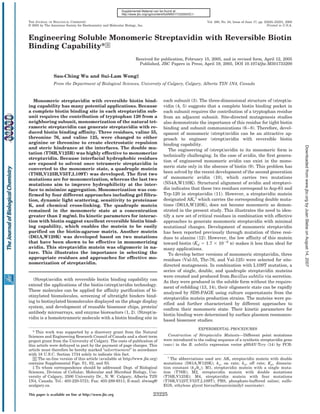
FIG. 1. Structure of tetrameric streptavidin and critical interface residues selected for mutagenesis. A, interfaces of tetrameric
streptavidin. Subunit A forms three subunit contact interfaces with other subunits. These interfaces include A/B, A/C, and A/D. Subunits A, B, C,
and D are highlighted in red, orange, yellow, and green, respectively. B, local environment of Thr-76 in subunit A showing the interfacial interaction
between subunits A and B. Thr-76 and Arg-59 in subunit A are highlighted in red and pink, respectively. Thr-76 and Arg-59 in subunit B are in
dark and bright yellow, respectively. Thr-76 in subunit A is 4 Å from Thr-76 and 3.75 Å from Arg-59 in subunit B. Replacement of Thr-76 by an
arginine would create both electrostatic repulsion (Arg-76 (subunit A)-Arg-76 (subunit B), Arg-76 (subunit A)-Arg-59 (subunit B), and Arg-76
(subunit B)-Arg-59 (subunit A)) and steric hindrance at the interface for subunits A and B. Equivalent effects will also be generated at the interface
between subunits C and D. C, local environment of Val-125 in subunit A showing the interfacial interaction between subunits A and D. Val-125
(red) in subunit A is 3.97 Å from Val-125 (green) in subunit D. It fits into a pocket formed by Val-125 (green) and Thr-123 (green) from subunit D.
A change of Val-125 to arginine will create both charge repulsion and steric hindrance at the A/D subunit interface. The same is true for the B/C
interface interactions. D, local environment of Val-55 in subunit A. Val-55 (red) in subunit A is 3.97 Å from Arg-59 (yellow) in subunit B.
residues should be located on the -strands rather than in the SDS-polyacrylamide gel even in the presence of biotin. In con-
loop regions. Furthermore the selected residue in one subunit trast, V55T mutation had the lowest impact with the majority
should be located very close to the equivalent residue or a of molecules in the tetrameric state even in the absence of
charged residue in another subunit at the interface. Examina- added biotin. Presence of biotin shifts the majority of the three
tion of interfacial residues (Fig. 1, B–D) shows that Thr-76, remaining muteins (V125R, V125T, and V55R) to the tet-
Val-125, and Val-55 meet the criteria. Hence they were selected rameric state. As expected, changing valine to arginine exerted
for mutagenesis. greater impact than changing it to threonine. This is true for
Effects of Single Mutations on Monomerization of Streptavi- both Val-125 and Val-55.
din—Streptavidin muteins carrying a single amino acid change Effects of Multiple Mutations on Monomerization of Strepta-
at the selected site were produced in their soluble form by B. vidin—To develop idealized monomeric streptavidin muteins
subtilis via secretion. Analysis of non-boiled culture superna- that are more likely to remain in the monomeric state at high
tants by SDS-PAGE offers a quick screen for the mutation streptavidin concentrations and have excellent reversible bio-
effect (12). Weaker subunit interaction would result in a higher tin binding capability, two more muteins were created. M2 is
percentage of the sample in the monomeric state on the SDS- the double mutant carrying both the T76R and V125R muta-
polyacrylamide gel. Because biotin can strengthen subunit in- tions. M4 is a quadruple mutant carrying T76R, V125R, V55T,
teraction, samples were analyzed in the presence or absence of and L109T mutations. In this combination, the three interfa-
biotin (9, 26). The impact of the mutation on weakening of the cial hydrophobic residues Val-125, Val-55, and Leu-109 were
subunit interaction followed the order: T76R Ͼ V125R Ͼ changed to hydrophilic ones. The last construct is AK, a double
V125T Ϸ V55R Ͼ V55T (Fig. 2 and Table II). The T76R mutein mutant (D61A,W120K) carrying two mutations (equivalent to
(designated M1) existed 100% in the monomeric state on the those performed in avidin) that have been shown to convert
4. 23228 Engineering of Monomeric Streptavidin
Determination of Apparent Molecular Mass of Streptavidin
Muteins by Dynamic Light Scattering—Because the apparent
molecular mass of wild-type streptavidin in the absence of
biotin is 10 kDa less than expected (56 instead of 66 kDa) as
determined by gel filtration, dynamic light scattering (29) was
used as a second method to estimate the apparent molecular
masses. The apparent molecular mass of wild-type streptavidin
obtained in this way (69 kDa) was closer to that expected (66
kDa) (Table III). The apparent molecular masses for both M2
and M4 in the absence of biotin indicated that they were in the
monomeric state. Addition of biotin caused only a slight in-
crease in their apparent molecular masses. The AK mutein
again was found to be oligomeric independent of the presence
or absence of biotin.
Proteinase K Sensitivity of Streptavidin Muteins—Mono-
FIG. 2. Western blot analysis of culture supernatants from meric streptavidin is expected to be more susceptible to pro-
B. subtilis strains producing streptavidin muteins carrying a teinase K digestion (10). Therefore, wild-type streptavidin and
single mutation. 15 l of non-boiled sample of culture supernatant its muteins were treated with proteinase K (Fig. 5A). Wild-type
was loaded to each lane. Samples in the left set were collected from
B. subtilis strains cultured in super-rich medium without added biotin.
streptavidin was converted to the core form independent of the
Samples in the right set were from culture grown in the presence of presence or absence of biotin. Under the condition used, the
biotin (20 M). The blot was probed with polyclonal antibodies against core streptavidin was resistant to further degradation by pro-
Downloaded from www.jbc.org by Juan Slebe on August 14, 2007
streptavidin. M, molecular weight markers; wt, wild-type streptavidin; teinase K. In contrast, all three muteins including AK, M2, and
Ϫve control, culture supernatant from WB800(pWB705HM) (34) that
did not produce any streptavidin.
M4 were much more susceptible to proteinase K digestion.
Sensitivity to proteinase K is more apparent for M2 and M4,
which were completely digested independent of the presence or
tetrameric avidin to the monomeric state (10). As shown in Fig.
absence of biotin. This property is consistent with the mono-
3 and Table II, just like M1, all these muteins existed in
meric nature of these muteins. The AK mutein behaved differ-
monomeric state on the SDS-polyacrylamide gel even in the
ently. Although most of it was digested by proteinase K in the
presence of biotin.
absence of biotin, it became much more resistant to proteinase
Purification of Streptavidin Muteins—Purification of M4 was
K when biotin was present.
used as an example to illustrate the process (Fig. 4). Proteins
Cross-linking of Streptavidin and Its Muteins—To
partially purified by ion exchange chromatography (lane 2)
strengthen the idea that both M2 and M4 are monomeric
were applied to a biotin-agarose column. M4 could be readily
whereas AK is oligomeric in nature, protein cross-linking was
eluted off from the column using biotin-containing buffer as the
carried out using sulfo-EGS as the cross-linking agent. Sulfo-
eluant (lanes 5–7). Pure streptavidin mutein obtained by this
EGS reacts with both the accessible ␣-amino groups at the N
simple procedure, after removal of biotin by dialysis, could be
termini and the surface-exposed ⑀-amino groups of the lysine
used for biochemical characterizations. To demonstrate that
dialysis could effectively remove any bound biotin from the M4 side chains in proteins. Secreted wild-type streptavidin has
mutein, the dialyzed sample was reloaded to the biotin-agarose eight lysine residues in each subunit. The three-dimensional
matrix. Over 95% of the sample could be retained on the col- structural model of streptavidin suggests that lysine 121 in
umn and eluted off from the column using biotin (data not subunit A is 14.1 Å from lysine 121 in subunit D. As the spacer
shown). Of all the muteins, M1 tended to have a long trailing arm in sulfo-EGS is 16.1 Å, subunits A and D (same for sub-
tail during elution. This indicates that M1 may not have the units B and C) should be easily cross-linked by sulfo-EGS. Also
desirable reversible biotin binding property. Therefore, it was it is possible to have cross-linking between subunits A and B as
not characterized further. the N-terminal region from subunit A, which contains two
Determination of Apparent Molecular Mass of Streptavidin lysine residues, is likely to be positioned close to lysine 80 in
Muteins by Gel Filtration—Observation of 100% monomeriza- subunit B. The same is true for subunits C and D. Therefore,
tion of the streptavidin mutein using a non-boiled sample for one should be able to differentiate tetrameric streptavidin from
SDS-PAGE does not always truly reflect its existence in the the monomeric form with the observation of cross-linked tet-
monomeric state in solution because SDS can promote subunit rameric streptavidin using sulfo-EGS. Lysozyme, well known
dissociation (27, 28). The apparent molecular masses of the to be monomeric in solution (30, 31), served as the negative
purified wild-type streptavidin and the three muteins (M2, M4, control. Fig. 5B shows that the amount of dimeric lysozyme
and AK) were estimated by gel filtration (Supplemental Fig. 2A increased slightly in the presence of the cross-linking agent.
and Table III). The expected molecular mass of monomeric This helped set the upper limit of the concentration of sulfo-
streptavidin is 16.5 kDa. M2 and M4 in the absence of biotin EGS to be used under the experimental condition. The wild-
showed the apparent molecular masses of 19.95 and 21.87 kDa, type streptavidin subunit had an apparent molecular mass of
respectively. These masses increased slightly in the presence of 19 kDa on the SDS gel. After treatment with sulfo-EGS, most
biotin. These data suggest that the muteins are monomeric in of these subunits were cross-linked to dimers and higher oli-
nature because their masses are less than that for the strepta- gomers with small amounts remaining in the monomeric state.
vidin dimer (33 kDa). In contrast, the AK mutein showed an M2 and M4 muteins behaved very similarly (data for M2 are
apparent molecular mass of 45.66 kDa even in the absence of not shown). The majority of the M2 and M4 muteins after the
biotin. This indicates the oligomeric nature of this mutein. cross-linking treatment migrated as monomers with small
Supplemental Fig. 2B shows the elution profile of purified M4 amounts in the dimeric form. These dimers may represent
(in the absence of biotin) from the gel filtration column. The cross-linked monomeric subunits that were artificially gener-
sample (loaded at 2 mg/ml) was eluted as a single peak. There ated in the same manner as with lysozyme. AK showed a
is no evidence for the presence of tetrameric streptavidin, cross-linking profile very similar to that of the wild-type
which would be eluted at 30.5 min. streptavidin. These data strongly support the idea that M2 and
5. Engineering of Monomeric Streptavidin 23229
TABLE II
Summary of the mutagenic effects on weakening of subunit interactions in streptavidin muteins as reflected by the degree of monomerization of
streptavidin muteins after SDS-PAGE
Estimation of the percentage of streptavidin in monomeric and tetrameric states is based on the blots showing the migration pattern of
non-boiled samples in Figs. 2 and 3.
No additional biotin With additional biotin
Streptavidin mutein
Monomer Tetramer Monomer Tetramer
%
M1 (T76R) 100 0 100 0
V125R 92 8 12 88
V125T 25 75 0 100
V55R 20 80 10 90
V55T 1 99 0 100
M2 (T76R,V125R) 100 0 100 0
M4 (T76R,V125R,V55T,L109T) 100 0 100 0
AK 100 0 100 0
TABLE III
Molecular mass determination of wild-type (wt) streptavidin and its
muteins by gel filtration and dynamic light scattering
In dynamic light scattering, the estimated molecular mass (M) was
calculated from the measured hydrodynamic radius (RH) using a pro-
tein calibration curve. The peaks have a polydispersity below 15%.
Downloaded from www.jbc.org by Juan Slebe on August 14, 2007
Theoretical molecular mass is estimated from the amino acid composi-
tion of the mature protein (17).
Gel Dynamic light
Sample filtration scattering Theoretical M
M RH M
kDa nm kDa kDa
wt (no biotin) 56.23 3.69 Ϯ 0.19 69 66.0 (tetramer)
FIG. 3. Analysis of culture supernatants from B. subtilis AK (no biotin) 45.66 3.20 Ϯ 0.23 50
strains producing streptavidin muteins carrying different com- AK (ϩbiotin) 50.12 3.54 Ϯ 0.38 63
binations of mutations. All these strains were cultivated in super- M2 (no biotin) 19.95 1.98 Ϯ 0.24 17 16.5 (monomer)
rich medium supplemented with biotin (20 M). 15 l of non-boiled M2 (ϩbiotin) 23.44 2.09 Ϯ 0.29 18
sample of culture supernatant was loaded. A, Coomassie Blue-stained M4 (no biotin) 21.87 2.08 Ϯ 0.31 18.1 16.5 (monomer)
SDS-polyacrylamide gel. Bands corresponding to tetrameric and mono- M4 (ϩbiotin) 24.54 2.20 Ϯ 0.14 21.5
meric streptavidin are marked by an asterisk and an arrowhead, re-
spectively. B, Western blot probed with polyclonal antibodies against
streptavidin. M, molecular mass markers; wt, wild-type streptavidin; C, DISCUSSION
negative control.
Although streptavidin and avidin have similar three-dimen-
sional structures and biotin binding properties, development of
monomeric streptavidin is much more challenging for two rea-
sons. First, streptavidin has stronger subunit interfacial inter-
actions than avidin (27, 28). More potent mutations are re-
quired to weaken this strong interface interaction. Second,
monomerization of streptavidin may result in the surface ex-
posure of hydrophobic residues that normally would be buried
at the interface in tetrameric streptavidin. This can potentially
affect the solubility of the monomeric streptavidin and lead to
reassociation of the monomers. The problem can be less dra-
FIG. 4. Purification of the M4 streptavidin mutein using bio- matic for avidin, which is a glycosylated protein with a carbo-
tin-agarose. M4 mutein partially purified by Macro S column chroma- hydrate chain in each of the avidin subunits.
tography (PPF) was loaded on a biotin-agarose column. M, molecular Despite the challenge, our study illustrates that, by selecting
mass markers; FT, column flow-through; W, pooled washing fractions; a critical residue located on a rigid surface for mutagenic study
EF1–EF3, eluted fractions.
and the introduction of charge repulsion and steric hindrance
at the interface, a single mutation (T76R) can be greatly effec-
M4 muteins are monomeric, whereas the AK mutein is oligo- tive in developing monomeric streptavidin. Besides the sugges-
meric in solution. tion from SDS-PAGE analysis, gel filtration study of the M1
Reversible Interaction between Streptavidin Muteins and mutein also indicated that the majority of M1 was eluted at a
Biotin—The on rate and off rate of the interactions between position corresponding to the monomeric form (data not
streptavidin muteins and biotin were determined by surface plas- shown). The main drawback for this mutein is its elution be-
mon resonance-based BIAcore biosensor (12). As shown in Table havior on the biotin-agarose column. The elution profile had a
IV (graphical plots for M4 are shown in Supplemental Fig. S3), typical long trailing tail. Furthermore more M1 could be recov-
M2, M4, and AK had their dissociation constant (Kd) in the range ered by soaking the column overnight with buffer containing
of 10Ϫ7 M. The off rates (kd) for these muteins were almost the biotin. This suggests that some of the M1 population have
same, whereas the on rate (ka) for the AK mutein was slightly higher affinity to the matrix.
lower than the rest. One of the factors affecting the on rate is the To increase the efficiency of streptavidin monomerization,
diffusion coefficient (or molecular mass) of the streptavidin mol- M2 mutein was developed by combining two potent mutations
ecule. Because AK is oligomeric in nature, this may account for (T76R,V125R). Data from gel filtration study, dynamic light
the lower on rate for this mutein-biotin interaction. scattering, sensitivity to proteinase K, and cross-linking reac-
6. 23230 Engineering of Monomeric Streptavidin
FIG. 5. Determination of the monomeric or oligomeric states of wild-type streptavidin and its muteins. Pictures show the Coomassie
Blue-stained SDS-polyacrylamide gel. A, proteinase K digestion. B, cross-linking study using sulfo-EGS as the cross-linker. All samples were boiled
prior to loading. M, molecular weight markers; wt, wild-type streptavidin; L, lysozyme. Numbering represents streptavidin molecules in monomeric
(1), dimeric (2), and oligomeric (3–5) states, respectively.
TABLE IV gation of the mutein. Another attractive feature is that M4 has
Kinetic parameters for the interactions between streptavidin a remarkably sharp elution profile with its purification using
muteins and biotin biotin-agarose. Over 95% of the mutein could be readily eluted
Protein ka kd Kd off from the column using just 2 column volumes of the eluant,
Ϫ1 leading to a high rate of protein recovery.
M sϪ1 sϪ1 M
Downloaded from www.jbc.org by Juan Slebe on August 14, 2007
In the site-directed mutagenesis study, it is not difficult to
M2 1.88 Ϯ 0.07 ϫ 104 3.22 Ϯ 0.01 ϫ 10Ϫ3 1.71 Ϯ 0.07 ϫ 10Ϫ7
M4 1.80 Ϯ 0.04 ϫ 104 3.39 Ϯ 0.02 ϫ 10Ϫ3 1.87 Ϯ 0.06 ϫ 10Ϫ7 understand the impact of the mutations in the following order:
AK 1.46 Ϯ 0.05 ϫ 104 3.59 Ϯ 0.03 ϫ 10Ϫ3 2.46 Ϯ 0.10 ϫ 10Ϫ7 T76R Ͼ V125R Ͼ V55R. Analysis of the solvent accessibility of
individual amino acid residues with tetrameric streptavidin
indicates that the solvent-accessible area of Thr-76 in subunit
tion confirmed the monomeric state of M2. The elution profile A is zero. Its close distances to both Thr-76 and Arg-59 in
of this mutein from the biotin-agarose matrix had a consider- subunit B and location on a rigid surface of a -barrel structure
able improvement over that of M1. When a 30-min on-column make it an ideal residue to be changed to arginine to achieve
incubation period was allowed for the eluant in between frac- the maximal electrostatic repulsion and steric hindrance ef-
tion collection, about 90% of M2 could be recovered within 3 fects at the subunit interface (Fig. 1B). The surface-accessible
column volumes of the eluant. area of Val-125 in subunit A is 1.78%. It has extensive inter-
To ensure that the streptavidin mutein will stably remain in actions with Leu-109, Trp-120, Thr-123, and Val-125 in sub-
the monomeric state, more mutations were introduced to M2. unit D (Fig. 1C); Leu-109 in subunit B; and Gln-107 in sub-
The exposure of the AB (or CD) interface will expose three unit C. Its conversion to arginine results in charge repulsion
hydrophobic residues: Val-55, Leu-109, and Val-125. In the M2 in subunit D and potential steric hindrance for subunits B, C,
mutein, Val-125 has been changed to arginine. We chose to and D. As for Val-55 in subunit A, its surface accessible area
further convert Val-55 and Leu-109 to threonine, a more hy- is 29.7%; and it is only close to Arg-59 in subunit B at the
drophilic residue, to develop the M4 mutein. Conversion to interface (Fig. 1D). Thus, V55R has the least impact on
threonine instead of arginine is preferred because proteins monomerization.
with high pI are known to have nonspecific interactions via Although AK mutein shows reversible biotin binding prop-
electrostatic interactions (32, 33). The calculated pI of M2 is erty and monomeric behavior on the SDS-polyacrylamide gel, it
8.1. If both Val-55 and Leu-109 are converted to arginine, the clearly exists in the oligomeric state in solution as suggested by
resulting mutein will have a pI of 9.2. This may increase the gel filtration studies, dynamic light scattering, and cross-link-
chance of charge-related nonspecific interactions. The conver- ing pattern. This study illustrates the importance of selecting
sion of these residues to negatively charged residues was not critical residues and effective approaches to achieve the maxi-
considered because both Val-55 and Leu-109 are located on the mal monomerization effect on tetrameric streptavidin.
-strands, and negatively charged residues are relatively poor
-strand formers. REFERENCES
M4 mutein shares with M2 mutein many desirable features 1. Morag, E., Bayer, E. A., and Wilchek, M. (1996) Anal. Biochem. 243, 257–263
2. Balass, M., Morag, E., Bayer, E. A., Fuchs, S., Wilchek, M., and Katchal-
of an idealized monomeric streptavidin. They both exist in the skikatzir, E. (1996) Anal. Biochem. 243, 264 –269
monomeric state at a reasonably high protein concentration (2 3. Green, N. M. (1990) Methods Enzymol. 184, 51– 67
mg/ml or more as used in the dynamic light scattering study). 4. Weber, P. C., Ohlendorf, D. H., Wendoloski, J. J., and Salemme, F. R. (1989)
Science 243, 85– 88
Both have excellent reversible biotin binding capability as re- 5. Hendrickson, W. A., Pahler, A., Smith, J. L., Satow, Y., Merritt, E. A., and
flected by their on rate and off rate for biotin interaction. Both Phizackerley, R. P. (1989) Proc. Natl. Acad. Sci. U. S. A. 86, 2190 –2194
6. Freitag, S., Le Trong, I., Chilkoti, A., Klumb, L. A., Stayton, P. S., and
have a moderate pI value of 8.1 so that charge-related nonspe- Stenkamp, R. E. (1998) J. Mol. Biol. 279, 211–221
cific interactions will be minimal. In addition, M4 has two 7. Sano, T., and Cantor, C. R. (1995) Proc. Natl. Acad. Sci. U. S. A. 92, 3180 –3184
features that make it even more attractive in practice. Mole- 8. Laitinen, O. H., Airenne, K. J., Marttila, A. T., Kulik, T., Porkka, E., Bayer,
E. A., Wilchek, M., and Kulomaa, M. S. (1999) FEBS Lett. 461, 52–58
cules of M2 mutein tend to aggregate in solution. Filtration of 9. Laitinen, O. H., Marttila, A. T., Airenne, K. J., Kulik, T., Livnah, O., Bayer,
M2 through a 0.02-m filter was essential for obtaining a good E. A., Wilchek, M., and Kulomaa, M. S. (2001) J. Biol. Chem. 276,
8219 – 8224
signal of the mutein for dynamic light scattering studies be- 10. Laitinen, O. H., Nordlund, H. R., Hytonen, V. P., Uotila, S. T., Marttila, A. T.,
cause of poor signal detection caused by the presence of small Savolainen, J., Airenne, K. J., Livnah, O., Bayer, E. A., Wilchek, M., and
amounts of large aggregates in an unfiltered sample. On the Kulomaa, M. S. (2003) J. Biol. Chem. 278, 4010 – 4014
11. Livnah, O., Bayer, E. A., Wilchek, M., and Sussman, J. L. (1993) Proc. Natl.
other hand, a decent signal could at least be obtained with an Acad. Sci. U. S. A. 90, 5076 –5080
unfiltered sample of similarly prepared M4. Thus, conversion 12. Qureshi, M. H., Yeung, J. C., Wu, S.-C., and Wong, S.-L. (2001) J. Biol. Chem.
276, 46422– 46428
of the two hydrophobic residues (Val-55 and Leu-109) to the 13. Nagarajan, V., Ramaley, R., Albertson, H., and Chen, M. (1993) Appl. Environ.
more hydrophilic threonine residue did help minimize aggre- Microbiol. 59, 3894 –3898
7. Engineering of Monomeric Streptavidin 23231
14. Wu, S.-C., Qureshi, M. H., and Wong, S.-L. (2002) Protein Expr. Purif. 24, 24. Atwell, S., Ultsch, M., De Vos, A. M., and Wells, J. A. (1997) Science 278,
348 –356 1125–1128
15. Halling, S. M., Sanchez-Anzaldo, F. J., Fukuda, R., Doi, R. H., and Meares, 25. Swint-Kruse, L., Elam, C. R., Lin, J. W., Wycuff, D. R., and Shive, M. K. (2001)
C. F. (1977) Biochemistry 16, 2880 –2884 Protein Sci. 10, 262–276
16. Gill, S. C., and Von Hippel, P. H. (1989) Anal. Biochem. 182, 319 –326 26. Gonzalez, M., Argarana, C. E., and Fidelio, G. D. (1999) Biomol. Eng. 16, 67–72
17. Gasteiger, E., Gattiker, A., Hoogland, C., Ivanyi, I., Appel, R. D., and Bairoch, 27. Bayer, E. A., Ehrlichrogozinski, S., and Wilchek, M. (1996) Electrophoresis 17,
A. (2003) Nucleic Acids Res. 31, 3784 –3788 1319 –1324
18. Ellison, D., Hinton, J., Hubbard, S. J., and Beynon, R. J. (1995) Protein Sci. 4, 28. Waner, M. J., Navrotskaya, I., Bain, A., Oldham, E. D., and Mascotti, D. P.
1337–1345 (2004) Biophys. J. 87, 2701–2713
19. Guex, N., and Peitsch, M. C. (1997) Electrophoresis 18, 2714 –2723 29. Schurr, J. M. (1977) CRC Crit. Rev. Biochem. 4, 371– 431
20. Freitag, S., Le Trong, I., Klumb, L., Stayton, P. S., and Stenkamp, R. E. (1997) 30. Blake, C. C., Koenig, D. F., Mair, G. A., North, A. C., Phillips, D. C., and
Protein Sci. 6, 1157–1166 Sarma, V. R. (1965) Nature 206, 757–761
21. Jones, S., and Thornton, J. M. (1995) Prog. Biophys. Mol. Biol. 63, 31– 65 31. Schwalbe, H., Grimshaw, S. B., Spencer, A., Buck, M., Boyd, J., Dobson, C. M.,
22. Neshich, G., Togawa, R. C., Mancini, A. L., Kuser, P. R., Yamagishi, M. E., Redfield, C., and Smith, L. J. (2001) Protein Sci. 10, 677– 688
Pappas, G., Jr., Torres, W. V., Fonseca e Campos, T., Ferreira, L. L., Luna, 32. Marttila, A. T., Airenne, K. J., Laitinen, O. H., Kulik, T., Bayer, E. A., Wilchek,
F. M., Oliveira, A. G., Miura, R. T., Inoue, M. K., Horita, L. G., de Souza, M., and Kulomaa, M. S. (1998) FEBS Lett. 441, 313–317
D. F., Dominiquini, F., Alvaro, A., Lima, C. S., Ogawa, F. O., Gomes, G. B., 33. Marttila, A. T., Laitinen, O. H., Airenne, K. J., Kulik, T., Bayer, E. A., Wilchek,
Palandrani, J. F., dos Santos, G. F., de Freitas, E. M., Mattiuz, A. R., Costa, M., and Kulomaa, M. S. (2000) FEBS Lett. 467, 31–36
I. C., de Almeida, C. L., Souza, S., Baudet, C., and Higa, R. H. (2003) Nucleic 34. Szarka, S., Sihota, E., Habibi, H. R., and Wong, S.-L. (1999) Appl. Environ.
Acids Res. 31, 3386 –3392 Microbiol. 65, 506 –513
23. Vetter, I. R., Baase, W. A., Heinz, D. W., Xiong, J. P., Snow, S., and Matthews, 35. Wu, S.-C., Ye, R., Wu, X.-C., Ng, S.-C., and Wong, S.-L. (1998) J. Bacteriol. 180,
B. W. (1996) Protein Sci. 5, 2399 –2415 2830 –2835
Downloaded from www.jbc.org by Juan Slebe on August 14, 2007