
Recommandé
Recommandé
Contenu connexe
Similaire à Anatomy related to obstetrics (U G).pptx
Similaire à Anatomy related to obstetrics (U G).pptx (20)
Dernier
Dernier (20)
Anatomy related to obstetrics (U G).pptx
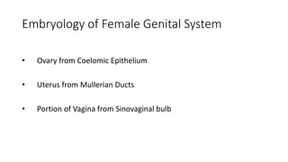
- 1. Embryology of Female Genital System • Ovary from Coelomic Epithelium • Uterus from Mullerian Ducts • Portion of Vagina from Sinovaginal bulb
- 2. APPLIED ANATOMY -IMPERFORATE HYMEN
- 8. MANAGEMENT
- 9. ISTHMUS- Narrow part between cervix & corpus of uterus. Same histological structures as corpus of uterus incorporated into cavity of uterus during pregnancy – isthmus opens up, so anatomical internal os ceases to exist, only histological os remains.
- 11. Hegar’s sign- At 6-10 wks. Seen in 2/3rd of cases Bimanual palpation Fingers of both hands approximate ,there is softening of isthmus with gestational sac in the upper cavity and empty lower cavity
- 12. Symphysio fundal height ( SFH ) in cm, after 24 weeks upto 36 weeks corresponds to number of weeks SFH can be used to clinically calculate the estimated fetal weight using Johnson’s formula Fetal weight = (SFH – n) x 155 n=12 if vertex is above ischial spine, n=11 is vertex is below ischial spine
- 14. Arrangement of muscle fibres- 1.Outer longitudinal: hoodlike layer ,arches over fundus and extends into various ligaments. 2. Middle layer: thickest and strongest, arranged in criss cross fashion. Apposition of 2 double curve muscle fibres – fig of 8 form which form the “ living ligatures”. 3. Inner circular: sphincter like fibres around fallopian tube orifices and internal os of cervix.
- 15. • Muscle fibre undergoes elongation, apposition of 2 double curves make figure of 8 that contract to occlude the blood vessels running in between them. • CLINICAL SIGNIFICANCE : occlusion of vessels during contractions helps in reducing the amount of blood loss postpartum