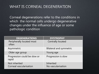
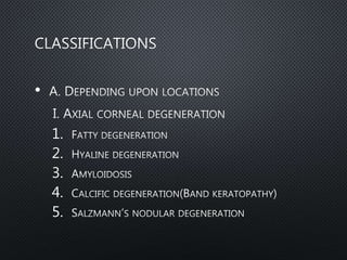
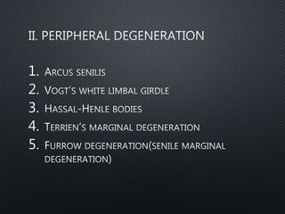
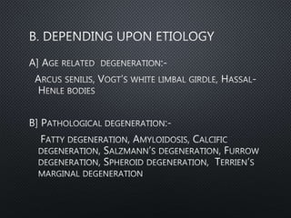
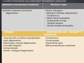
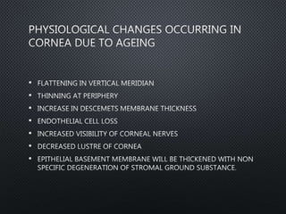
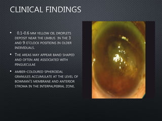
CORNEAL
DEGENERATIONS
MODERATOR-DR UMA
PRESENTER-DR AKSHAY NAYAK
DEGENERATIONS DYSTROPHY
Peripherally located most
often
Centrally located
Asymmetric Bilateral and symmetric
Older age group Young age
Progression could be slow or
rapid
Progression is slow
Not inherited
Corneal vascularisation
Inherited
No vascularisation
Corneal degenerations refer to the conditions in
which the normal cells undergo degenerative
changes under the influence of age or some
pathologic condition
•
1.
2.
3.
4.
5.
1.
2.
3.
4.
5.
Epithelium and basement membrane Bowman’s Layer
Epithelial basement membrane
degeneration
- Pannus ,Pterygium
- Salzmann’s Nodular degeneration
- Corneal arcus
- Calcific Band keratopathy
- Limbal girdle of Vogt
- Nodular amyloid
- Climatic droplet keratopathy
-
STROMA ENDOTHELIUM AND DESCEMET’S
MEMBRANE
-Scarring with or without vascularisation
-Lipid degeneration
-Polymorphic amyloid degeneration
-Crocodile shagreen
-Cornea farinata
-Terrien ‘s Marginal degeneration
-Corneal arcus
-Cornea guttae
-Hassal henle warts
-Retrocorneal fibrous membrane
CLASSIFICATION BASED ON LAYER MOST PROMINENTLY INVOLVED
•
•
•
•
•
•
•
•
•
•
•
•
•
•
•
•
•
•
CORONARY ARTERY DISEASE
•
•
•
•
BOWMAN’S MEMBRANE
•
•
•
•
•
•
•
•
•
•
•
•
•
•
•
•
•
1.
2.
3.
•
•
•
•
•
•
•
•
•
1.
2.
•
•
•
•
•
•
•
•
•
•
•
•
•
•
•
•
•
•
•
•
•
•
•
•
•
•
•
•
•
•
•
•
•
•
•
•
•
•
•
•
•
•
•
•
•
•
•
•
•
•
•
•
•
•
•
•
•
•
•
•
•
•
•
•
•
•
•
•
•
•
•
•
•
•
•
•
•
•
•
•
•
•
•
•
•
•
1.
2.
3.
•
•
•
•
•
•
•
•
•
•
•
•
•
•
•
•
•
•
•
•
•
•
•
•
•
•
•
•
•
•
•
•
•
•
•
•
•
•
•
•
• CLINICALLY, AMYLOID DEPOSITS USUALLY OCCUR AS RAISED, YELLOW-PINK
NODULAR MASSES IN THE CORNEA.
•
•
•
•
•
•
•
•
•
•
•
•
•

Corneal degenerations