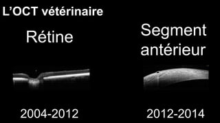
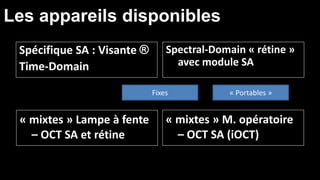
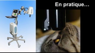
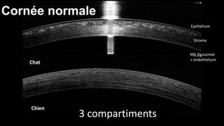
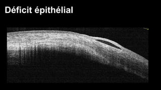
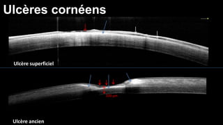
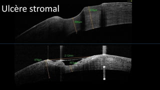
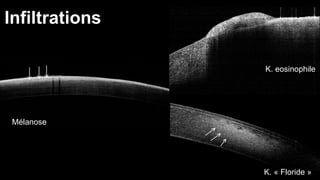
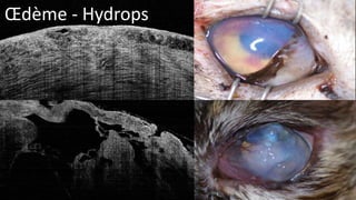
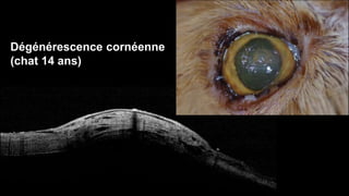
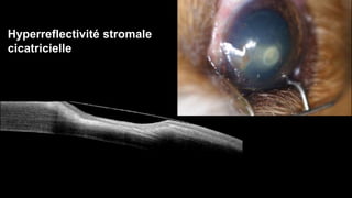
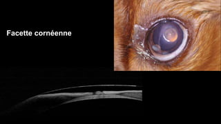
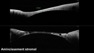
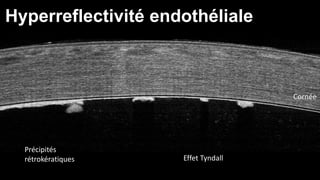
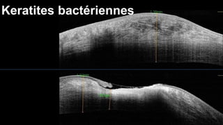
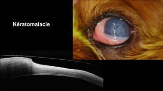
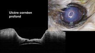
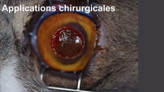
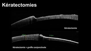
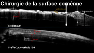
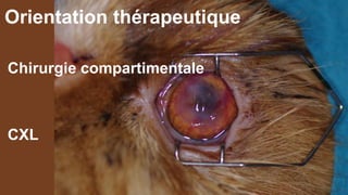
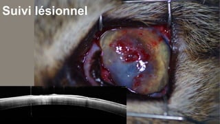
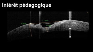
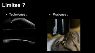
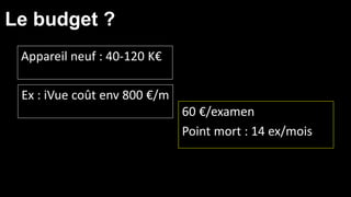
OCT 
INTERET DE L’OCT DANS 
LE DIAGNOSTIC DES 
AFFECTIONS CORNEENNES 
Dr Frank FAMOSE – Toulouse 
frankfamose@gmail.com 
* 
* Optical Coherence Tomography
Infrarouges 
840 – 1310 nm 
Time-Domain (TD) 
Spectral- Domain (SD) 
Analyse de réflectivité
L’histoire… 
1991 
1999 
2010 (SD) 
Rétine 
2004 (SD) 
2007 (SD) 
Segment antérieur 
2005 (TD)
L’OCT vétérinaire 
Rétine Segment 
antérieur 
2004-2012 2012-2014
Les appareils disponibles 
Spécifique SA : Visante ® 
Time-Domain 
Spectral-Domain « rétine » 
avec module SA 
« mixtes » Lampe à fente 
– OCT SA et rétine 
Fixes « Portables » 
« mixtes » M. opératoire 
– OCT SA (iOCT)
• Image conf Trieste 
En pratique…
Cornée normale 
Chat 
Chien 
Epithélium 
Stroma 
Mb Descemet 
+ endothelium 
3 compartiments
Interprétation des images OCT 
• Absence d’un compartiment 
• Modifications de l’épaisseur d’un 
compartiment 
• Augmentation partielle ou totale de 
réflectivité 
• Diminution partielle ou totale de réflectivité
Kératites bactériennes
Etat des lieux d’une kératite 
Lésions spécifiques 
Facteurs favorisants/aggravants 
Identification des germes en cause
Les moyens du diagnostic 
Examen clinique –Test de Schirmer, 
Fluorescéine- rose bengale 
Lampe à fente 
Instrumentation « lourde » : OCT, 
microscopie confocale, UBM
Déficit épithélial
Ulcères cornéens 
Ulcère superficiel 
Ulcère ancien 
320 μm
Ulcère stromal
Infiltrations 
Mélanose 
K. eosinophile 
K. « Floride »
OEdème - Hydrops
Dégénérescence cornéenne 
(chat 14 ans)
Hyperreflectivité stromale 
cicatricielle
Facette cornéenne
Amincissement stromal
Cornée 
Hyperreflectivité endothéliale 
Précipités 
rétrokératiques Effet Tyndall
Keratites bactériennes
Kératomalacie
Ulcère cornéen 
profond
Applications chirurgicales
Kératectomies 
Kératectomie 
Kératectomie + greffe conjonctivale
Chirurgie de la surface cornénne 
324 μm 
Vetbiosis J0 
Greffe Conjonctivale J 30 
Vetbiosis sheets 
OEdème cornéen 
Conjunctiva 
Corneal 
Stroma 
150 μm 
320 μm
Greffe de biomatériau (ACell-Vet)
Greffe ACell-vet 
(8 jours post-op)
J30 J60 J90 
Ectasie cornéenne post-opératoire
Orientation pathogénique 
complémentaire 
Cicatrisation épithéliale ? 
Epaisseur du stroma résiduel ? 
Implication endoculaire ?
Orientation thérapeutique 
Chirurgie compartimentale 
CXL
Suivi lésionnel
Intérêt pédagogique
Limites ? 
• Techniques : • Pratiques :
Le budget ? 
Appareil neuf : 40-120 K€ 
60 €/examen 
Point mort : 14 ex/mois 
Ex : iVue coût env 800 €/m
2 questions 
Faut-il investir dans un OCT ? 
? 
Peut-on devenir OCT-addict ? OUI

Oct sferov-2014

Notes de l'éditeur

  • #14 In chronic superficial ulcerations, the thickness of the epithelium is increased and we observe zones of detachement and an increased density of the proximal stroma. These observations are similar to those collected with traditionnal histology. When the ulceration is deeper, we oberve an increased density of the stroma around the ulceration. The depth of stromal involvement can be measured, and we can see a localized corneal fibrosis made visible by the increase of the corneal density with a strong absorption of the IR beam.
  • #22 A normal anterior chamber does not appear in OCT. However, in pathological conditions, we can identify the presence of blood or fibrin clots, and in this case of anterior uveitis, Tyndall effects and retrokeratic precipitates.
  • #28 We have also evaluated OCT imaging in surgical conditions. As you can see, we can measure residual stromal thickness after a superficial keratectomy : this helps in the choice of the surgical technique. We can also follow the healing process after surgeries like conjonctival or vetbiosis grafts.