SlideShare une entreprise Scribd logo
1  sur  34
Essential idea: Information transferred from DNA to mRNA is
translated into an amino acid sequence.
7.3 Translation
Section of Titin, our largest known protein
http://circ.ahajournals.org/content/124/8/876/F2.large.jpg
Understandings
Statement Guidance
7.3 U.1 Initiation of translation involves assembly of the
components that carry out the process.
Examples of start and stop codons are
not required.
7.3 U.2 Synthesis of the polypeptide involves a repeated cycle of
events.
7.3 U.3 Disassembly of the components follows termination of
translation.
Names of the tRNA binding sites are
expected as well as their roles.
7.3 U.4 Free ribosomes synthesize proteins for use primarily within
the cell.
7.3 U.5 Bound ribosomes synthesize proteins primarily for
secretion or for use in lysosomes.
7.3 U.6 Translation can occur immediately after transcription in
prokaryotes due to the absence of a nuclear membrane.
7.3 U.7 The sequence and number of amino acids in the
polypeptide is the primary structure.
7.3 U.8 The secondary structure is the formation of alpha helices
and beta pleated sheets stabilized by hydrogen bonding.
7.3 U.9 The tertiary structure is the further folding of the
polypeptide stabilized by interactions between R groups.
Polar and non-polar amino acids are
relevant to the bonds formed between
R groups.
7.3 U.10 The quaternary structure exists in proteins with more than
one polypeptide chain.
Quaternary structure may involve the
binding of a prosthetic group to form a
conjugated protein.
Applications and Skills
Statement Utilization
7.2 A.1 tRNA-activating enzymes illustrate enzyme–substrate
specificity and the role of phosphorylation.
7.3 S.1 Identification of polysomes in electron micrographs of
prokaryotes and eukaryotes.
7.3 S.2 The use of molecular visualization software to analyze the
structure of eukaryotic ribosomes and a tRNA molecule.
Components of Translation
1. mRNA = message
2. tRNA = interpreter
3. Ribosome = site of translation
7.3 U.1 Initiation of translation involves assembly of the components
that carry out the process.
tRNA
• Transcribed in nucleus
• Specific to each amino acid
• Transfer AA to ribosomes
• Anticodon: pairs with
complementary mRNA codon
• Base-pairing rules between 3rd
base of codon & anticodon are
not as strict. This is called
wobble.
7.3 U.1 Initiation of translation involves assembly of the components
that carry out the process.
Ribosomes
• Ribosome = rRNA + proteins
• made in nucleolus
• 2 subunits
7.3 U.1 Initiation of translation involves assembly of the components
that carry out the process.
The role of RNA in Protein Synthesis
• 3 Types of RNA molecules in the steps from gene to
protein:
1. Messenger RNA (mRNA), Complementary copy of DNA
2. Transfer RNA (tRNA) carries amino acid to the site of
synthesis.
3. Ribosomal RNA (rRNA), stabilizes the site of synthesis
7.3 U.1 Initiation of translation involves assembly of the components
that carry out the process.
Translation stages:
Initiation, Elongation and Termination
• Translation occurs in the 5' to 3' direction along the mRNA
A. Initiation begins with the attachment of the ribosome to the mRNA
B. Elongation begins at the mRNA start codon AUG and continues
with the addition of amino acids to the polypeptide.
C. Termination occurs at the STOP codon (UGA, UAG or UAA).
7.3 U.1 Initiation of translation involves assembly of the components
that carry out the process.
Ribosomes
Active sites:
• A site: holds AA to be
added
• P site: holds growing
polypeptide chain
• E site: exit site for
tRNA
7.3 U.1 Initiation of translation involves assembly of the components
that carry out the process.
7.3 U.2 Synthesis of the polypeptide involves a repeated cycle of events.
• While the first tRNA is still
attached, a second tRNA
attaches to the mRNA at
the A site on the ribosome,
carrying the amino acid
that corresponds to the
mRNA codon.
• The methionine amino
acid (Met) is the start code
for all amino acids, is the
first tRNA to arrive at the P
site binds to the amino
acid carried by the second
tRNA located at the A site.
7.3 A.1 tRNA-activating enzymes illustrate enzyme–substrate specificity
and the role of phosphorylation.
• Each tRNA binds with a
specific amino acid in the
cytoplasm in a
reaction catalyzed by a
specific tRNA-activating
enzyme.
• Each specific amino acid binds
covalently to the 3'- terminal
nucleotide (CCA) at the end of
the tRNA molecule.
• The binding of the specific
amino acid to the tRNA
requires energy from ATP.
Bioninja
7.3 A.1 tRNA-activating enzymes illustrate enzyme–substrate specificity
and the role of phosphorylation.
http://www.phschool.com/science/biology_place/biocoach/translation/addani.html
7.3 U.2 Synthesis of the polypeptide involves a repeated cycle of events.
• The two amino acids are
joined together through
a condensation
reaction that creates
a peptide bond between
the two amino acids.
• The ribosome moves along
the mRNA one codon
shifting the tRNA that was
attached to methionine to
the E site.
• The tRNA is released back
into the cytoplasm from the
E site, allowing it to pick up
another amino acid
(methionine) to build
another polypeptide.
7.3 U.2 Synthesis of the polypeptide involves a repeated cycle of events.
• Another tRNA moves into the
empty A site bringing the next
amino acid corresponding to
themRNA codon.
• Again, the amino acid is attached
to the polypeptide forming a
peptide bond, the ribosome slides
across one codon and tRNA at the
P site moves into the E site
releasing it back into the
cytoplasm.
• The ribosome continues to move
along the mRNA adding amino
acids to the polypeptide chain.
• This process continues until a stop
codon is reached.
7.3 U.3 Disassembly of the components follows termination of
translation.
• Termination begins
when 1 of the 3 stop
codons
 UAA
 UGA
 UAG
moves into the A site.
• These tRNA have no
attached amino acids.
• When the stop codon
is reached
the ribosome
dissociates and the
polypeptide is
released.
http://highered.mheducation.com/sites/0072507470/st
udent_view0/chapter3/animation__how_translation_w
orks.html
http://www.stolaf.edu/people/giannini/flashanimat/molgen
etics/translation.swf
Watch these animations about the process of translation. Can you narrate?
7.3 U.4 Free ribosomes synthesize proteins for use primarily within the
cell.
https://www.studyblue.com/notes/note/n/molecular-exam-3/deck/2630328
• Free ribosomes in the
cytoplasm synthesize
proteins that will be
used inside the cell in
the cytoplasm,
mitochondria and
chloroplasts (in
autotrophs)
7.3 U.5 Bound ribosomes synthesize proteins primarily for secretion or
for use in lysosomes.
• Ribosomes attached to ER create
proteins that are secreted from the
cell by exocytosis or are used in
lysosomes.
• Proteins that are destined to be
used in lysosomes, ER, Golgi
Apparatus, the plasma membrane
or secreted by the cell are made by
ribosomes bound by the
endoplasmic reticulum
• Ribosomes that become bound to
the ER are directed here by a signal
sequence that is part of that
specific polypeptide
• This signal sequence on the
polypeptide binds to a signal
recognition protein (SRP)
• The SRP guides the polypeptide and
ribosome to the ER where it binds
to an SRP receptor http://herbmitchell.info/Figure.4-8-Synthesissecretoryprotein.jpg
7.3 U.6 Translation can occur immediately after transcription in
prokaryotes due to the absence of a nuclear membrane.
• Since prokaryotic DNA is not compartmentalized into a nucleus, once transcription
begins creating a strand of mRNA, translation can begin immediately as the mRNA
strand is created
• In eukaryotes, the completed mRNA has to be transported from the nucleus,
through the nuclear pore to the ribosome on the ER or in the cytosol
http://www.mun.ca/biology/scarr/iGen3_05-09_Figure-L.jpg
Prokaryotes vs. Eukaryotes
Prokaryotes Eukaryotes
• Transcription and
translation both in
cytoplasm
• DNA/RNA in cytoplasm
• RNA poly binds directly to
promoter
• Transcription makes mRNA
(not processed)
• No introns
• Transcription in nucleus;
translation in cytoplasm
• DNA in nucleus, RNA travels
in/out nucleus
• RNA poly binds to TATA box
& transcription factors
• Transcription makes pre-
mRNA  RNA processing 
final mRNA
• Exons, introns (cut out)
7.3 U.6 Translation can occur immediately after transcription in
prokaryotes due to the absence of a nuclear membrane.
Structure of Proteins
The complex structure of
proteins is explained by
referring to 4 levels of
organization
A. Primary
B. Secondary
C. Tertiary
D. Quaternary
http://upload.wikimedia.org/wikipedia/commons/2/26/225_Peptide_Bond-01.jpg
Structure of Proteins
Primary structure:
• The order/ number of amino acids in a polypeptide chain.
• Linear shape (no internal bonding)
7.3 U.7 The sequence and number of amino acids in the polypeptide is
the primary structure.
7.3 U.8 The secondary structure is the formation of alpha helices and
beta pleated sheets stabilized by hydrogen bonding
Secondary Structure:
Hydrogen bonding causes
The primary structure of the
polypeptide to fold and coil
Into some characteristic
ways:
• Alpha Helix
• Beta pleated sheets
Beta-pleated sheets:
• Flat, zig-zag structure
• A number of chains which are hydrogen bonded together
• Forms a sheet
Example: Fibers in in silk
7.3 U.8 The secondary structure is the formation of alpha helices and
beta pleated sheets stabilized by hydrogen bonding
Changes to the primary and secondary
structure comes from additional bonds
Folding in the primary structure is
caused by charged groups on the
amino acid chain.
These charged groups include:
» Hydrogen bonds
» Ionic bonds
» Covalent bonds.
(disulphide bridge)
7.3 U.9 The tertiary structure is the further folding of the polypeptide
stabilized by interactions between R groups.
• Tertiary structure is the three-dimensional conformation
of a polypeptide.
• The polypeptide folds just after it is formed in
translation.
• The shape is maintained by intermolecular bonds
7.3 U.9 The tertiary structure is the further folding of the polypeptide
stabilized by interactions between R groups.
http://cnx.org/resources/36c08f3ac1c144763610fa69fbb9e278/Figure_03_04_08.jpg
7.3 U.10 The quaternary structure exists in proteins with more than one
polypeptide chain.
• Quaternary structure is the linking together of two or more
polypeptides to form a single protein.
• The protein structure below has 4 different polypeptide chains.
http://www.topsan.org/@api/deki/files/6029/=EK5976M_Fig3Comparisons.png
1. Fibrous Proteins
– Insoluble in Water
– Structural (support/strength)
Example
–Collagen (tissue strengthening)
–Keratin (hair/nails)
–Elastin (skin)
Two major types of quaternary Proteins
Each color represents an alpha helix
7.3 U.10 The quaternary structure exists in proteins with more than one
polypeptide chain.
2. Globular Proteins
• Can be soluble in water
• Functional (enzymes and antibodies)
Examples
• Amylase (digestion of starch)
• Insulin (blood sugar regulation)
• Hemoglobin (carry O2)
• Immunoglobulin (antibodies)
7.3 U.10 The quaternary structure exists in proteins with more than one
polypeptide chain.
Ribosomes effect in translation
• Ribosome are found in Prokaryotes (70's) and Eukaryotes (80's).
Including P and A sites. START codons and STOP codons begin and
termination translation.
Polyribosome= Polysomes
• Multiple ribosomes on the same mRNA at the same time.
• All ribosome move 5' to 3' in sequence.
• In protein synthesis polyribosomes increase the quantity of
polypeptide synthesized.
7.3 S.1 Identification of polysomes in electron micrographs of
prokaryotes and eukaryotes.
7.3 S.1 Identification of polysomes in electron micrographs of
prokaryotes and eukaryotes.
• In prokaryotes,
several ribosomes
can attach
themselves to the
growing mRNA
chains to form a
polysome while
the mRNA chains
are still attached
to the DNA
7.3 S.1 Identification of polysomes in electron micrographs of
prokaryotes and eukaryotes.
• In eukaryotes, the mRNA
detaches from the DNA
and is then transported
through pores in the
nuclear envelope to the
ribosomes in the
cytoplasm. Once in the
cytosol, eukaryote mRNA
can also form polysomes
Conserved sequence: a
homologous sequence of DNA
that is identical across all
members of a species.
Bioinformatics: uses computer
databases to store and analyze
gene & protein sequences from
large amounts of data collected
from sequencing genes of
various organisms
Faster, more powerful computers allow scientist to identify
conserved sequences & genes by looking for patterns and
homologous sequences within organisms’ genome. If a
sequence is homologous across species or individuals of a
species, it usually has a functional role. Eg. It codes for a
protein (a gene).
7.3 S.2 The use of molecular visualization software to analyze the
structure of eukaryotic ribosomes and a tRNA molecule.

Contenu connexe

Tendances

IB Biology 3.1 genes
IB Biology 3.1 genesIB Biology 3.1 genes
IB Biology 3.1 genes
Bob Smullen
 

Tendances (20)

IB Biology 2.4 & 7.3 Slides: Proteins
IB Biology 2.4 & 7.3 Slides: ProteinsIB Biology 2.4 & 7.3 Slides: Proteins
IB Biology 2.4 & 7.3 Slides: Proteins
 
IB Biology 2.7 Slides: Transcription & Translation
IB Biology 2.7 Slides: Transcription & TranslationIB Biology 2.7 Slides: Transcription & Translation
IB Biology 2.7 Slides: Transcription & Translation
 
9.4 plant reproduction
9.4 plant reproduction9.4 plant reproduction
9.4 plant reproduction
 
IB Biology 2.1 Slides: Molecules to Metabolism
IB Biology 2.1 Slides: Molecules to MetabolismIB Biology 2.1 Slides: Molecules to Metabolism
IB Biology 2.1 Slides: Molecules to Metabolism
 
2.8 cellular respiration
2.8 cellular respiration 2.8 cellular respiration
2.8 cellular respiration
 
IB Biology 3.1 genes
IB Biology 3.1 genesIB Biology 3.1 genes
IB Biology 3.1 genes
 
IB Biology Genetics 2015
IB Biology Genetics 2015IB Biology Genetics 2015
IB Biology Genetics 2015
 
IB Biology 2.9 Slides: Photosynthesis
IB Biology 2.9 Slides: PhotosynthesisIB Biology 2.9 Slides: Photosynthesis
IB Biology 2.9 Slides: Photosynthesis
 
IB Biology 3.1 Slides: Genes
IB Biology 3.1 Slides: GenesIB Biology 3.1 Slides: Genes
IB Biology 3.1 Slides: Genes
 
2.1 molecules to metabolism me
2.1 molecules to metabolism me2.1 molecules to metabolism me
2.1 molecules to metabolism me
 
2.5 enzymes
2.5 enzymes 2.5 enzymes
2.5 enzymes
 
6.1 digestion HL Year One
6.1 digestion HL Year One6.1 digestion HL Year One
6.1 digestion HL Year One
 
concept of gene and protein synthesis
concept of gene and protein synthesisconcept of gene and protein synthesis
concept of gene and protein synthesis
 
IB Biology 2.8 Slides: Respiration
IB Biology 2.8 Slides: RespirationIB Biology 2.8 Slides: Respiration
IB Biology 2.8 Slides: Respiration
 
DNA Replication
DNA ReplicationDNA Replication
DNA Replication
 
IB Biology 2.5 Slides: Enzymes
IB Biology 2.5 Slides: EnzymesIB Biology 2.5 Slides: Enzymes
IB Biology 2.5 Slides: Enzymes
 
Transcription process
Transcription processTranscription process
Transcription process
 
Transcription of DNA
Transcription of DNATranscription of DNA
Transcription of DNA
 
2.4 proteins
2.4 proteins2.4 proteins
2.4 proteins
 
Ch 11: Cell Communication
Ch 11: Cell CommunicationCh 11: Cell Communication
Ch 11: Cell Communication
 

Similaire à 7.3 translation

• Define transcription• Define translation• What are the 3 steps.pdf
• Define transcription• Define translation• What are the 3 steps.pdf• Define transcription• Define translation• What are the 3 steps.pdf
• Define transcription• Define translation• What are the 3 steps.pdf
arihantelehyb
 
27 28 105 fa13 transcription and translation skel
27 28 105 fa13 transcription and translation skel27 28 105 fa13 transcription and translation skel
27 28 105 fa13 transcription and translation skel
Afton Chase
 
6 - Protein Synthesis and Tracking
6 - Protein Synthesis and Tracking 6 - Protein Synthesis and Tracking
6 - Protein Synthesis and Tracking
tchubb
 

Similaire à 7.3 translation (20)

Biosynthesis of protein in eukariotes
Biosynthesis of protein in eukariotesBiosynthesis of protein in eukariotes
Biosynthesis of protein in eukariotes
 
7.3 translation
7.3 translation7.3 translation
7.3 translation
 
Gene expression
Gene expressionGene expression
Gene expression
 
• Define transcription• Define translation• What are the 3 steps.pdf
• Define transcription• Define translation• What are the 3 steps.pdf• Define transcription• Define translation• What are the 3 steps.pdf
• Define transcription• Define translation• What are the 3 steps.pdf
 
Translation
TranslationTranslation
Translation
 
2_2018_03_19!02_55_44_PM.pdf
2_2018_03_19!02_55_44_PM.pdf2_2018_03_19!02_55_44_PM.pdf
2_2018_03_19!02_55_44_PM.pdf
 
protien synthesis (1).pptx
protien synthesis (1).pptxprotien synthesis (1).pptx
protien synthesis (1).pptx
 
DNA- Transcription and Tranlation, RNA, Ribosomes and membrane proteins.pptx
DNA- Transcription and Tranlation, RNA, Ribosomes and membrane proteins.pptxDNA- Transcription and Tranlation, RNA, Ribosomes and membrane proteins.pptx
DNA- Transcription and Tranlation, RNA, Ribosomes and membrane proteins.pptx
 
TRANSLATION
TRANSLATIONTRANSLATION
TRANSLATION
 
27 28 105 fa13 transcription and translation skel
27 28 105 fa13 transcription and translation skel27 28 105 fa13 transcription and translation skel
27 28 105 fa13 transcription and translation skel
 
Protein synthesis.
Protein synthesis.Protein synthesis.
Protein synthesis.
 
Transcription
TranscriptionTranscription
Transcription
 
PROTEIN SYNTHESIS IN EUKARYOTES.pptx
PROTEIN SYNTHESIS IN EUKARYOTES.pptxPROTEIN SYNTHESIS IN EUKARYOTES.pptx
PROTEIN SYNTHESIS IN EUKARYOTES.pptx
 
Divakaran Molecular level of Eukaryotic translation
Divakaran Molecular level of Eukaryotic translationDivakaran Molecular level of Eukaryotic translation
Divakaran Molecular level of Eukaryotic translation
 
6 - Protein Synthesis and Tracking
6 - Protein Synthesis and Tracking 6 - Protein Synthesis and Tracking
6 - Protein Synthesis and Tracking
 
Biomol-Translation Eukaryote.pptx
Biomol-Translation Eukaryote.pptxBiomol-Translation Eukaryote.pptx
Biomol-Translation Eukaryote.pptx
 
post transcriptional modifications
post transcriptional modificationspost transcriptional modifications
post transcriptional modifications
 
protein synthesis
protein synthesisprotein synthesis
protein synthesis
 
AP Bio Ch 10 Power Point
AP Bio Ch 10 Power PointAP Bio Ch 10 Power Point
AP Bio Ch 10 Power Point
 
Subin cology
Subin cologySubin cology
Subin cology
 

Plus de Bob Smullen

Topic 8: Ecology Option C Part 2
Topic 8: Ecology Option C Part 2Topic 8: Ecology Option C Part 2
Topic 8: Ecology Option C Part 2
Bob Smullen
 

Plus de Bob Smullen (20)

1. complete stats notes
1. complete stats notes1. complete stats notes
1. complete stats notes
 
Topic One: Cell
Topic One: CellTopic One: Cell
Topic One: Cell
 
Endocrine System
Endocrine SystemEndocrine System
Endocrine System
 
IB Biology Human Reproduction
IB Biology Human Reproduction IB Biology Human Reproduction
IB Biology Human Reproduction
 
Topic 8: Ecology Option C Part 2
Topic 8: Ecology Option C Part 2Topic 8: Ecology Option C Part 2
Topic 8: Ecology Option C Part 2
 
Topic 8: Ecology Option C Part 1
Topic 8: Ecology Option C Part 1Topic 8: Ecology Option C Part 1
Topic 8: Ecology Option C Part 1
 
Topic 7: Plant Science
Topic 7: Plant ScienceTopic 7: Plant Science
Topic 7: Plant Science
 
Topic 6: Evolution
Topic 6: Evolution Topic 6: Evolution
Topic 6: Evolution
 
Topic Five: Genetics
Topic Five: GeneticsTopic Five: Genetics
Topic Five: Genetics
 
Topic 3: Nucleic Acid
Topic 3: Nucleic AcidTopic 3: Nucleic Acid
Topic 3: Nucleic Acid
 
Topic 2: Biochemistry
Topic 2: Biochemistry Topic 2: Biochemistry
Topic 2: Biochemistry
 
11.3 kidney
11.3 kidney11.3 kidney
11.3 kidney
 
11.2 Movement
11.2 Movement 11.2 Movement
11.2 Movement
 
6.5 neurons and synapses
6.5 neurons and synapses 6.5 neurons and synapses
6.5 neurons and synapses
 
Immune system hl
Immune system hlImmune system hl
Immune system hl
 
Ia headings
Ia headingsIa headings
Ia headings
 
6.2 and 6.4 transport and respiration
6.2 and 6.4  transport and respiration6.2 and 6.4  transport and respiration
6.2 and 6.4 transport and respiration
 
Ecology Optional Topic Student Notes
Ecology Optional Topic Student NotesEcology Optional Topic Student Notes
Ecology Optional Topic Student Notes
 
C.6 nitrogen cycle .pptx
C.6 nitrogen cycle .pptxC.6 nitrogen cycle .pptx
C.6 nitrogen cycle .pptx
 
C.5 population.pptx
C.5 population.pptxC.5 population.pptx
C.5 population.pptx
 

7.3 translation

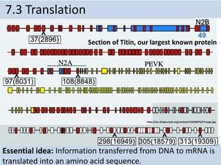
  • 1. Essential idea: Information transferred from DNA to mRNA is translated into an amino acid sequence. 7.3 Translation Section of Titin, our largest known protein http://circ.ahajournals.org/content/124/8/876/F2.large.jpg
  • 2. Understandings Statement Guidance 7.3 U.1 Initiation of translation involves assembly of the components that carry out the process. Examples of start and stop codons are not required. 7.3 U.2 Synthesis of the polypeptide involves a repeated cycle of events. 7.3 U.3 Disassembly of the components follows termination of translation. Names of the tRNA binding sites are expected as well as their roles. 7.3 U.4 Free ribosomes synthesize proteins for use primarily within the cell. 7.3 U.5 Bound ribosomes synthesize proteins primarily for secretion or for use in lysosomes. 7.3 U.6 Translation can occur immediately after transcription in prokaryotes due to the absence of a nuclear membrane. 7.3 U.7 The sequence and number of amino acids in the polypeptide is the primary structure. 7.3 U.8 The secondary structure is the formation of alpha helices and beta pleated sheets stabilized by hydrogen bonding. 7.3 U.9 The tertiary structure is the further folding of the polypeptide stabilized by interactions between R groups. Polar and non-polar amino acids are relevant to the bonds formed between R groups. 7.3 U.10 The quaternary structure exists in proteins with more than one polypeptide chain. Quaternary structure may involve the binding of a prosthetic group to form a conjugated protein.
  • 3. Applications and Skills Statement Utilization 7.2 A.1 tRNA-activating enzymes illustrate enzyme–substrate specificity and the role of phosphorylation. 7.3 S.1 Identification of polysomes in electron micrographs of prokaryotes and eukaryotes. 7.3 S.2 The use of molecular visualization software to analyze the structure of eukaryotic ribosomes and a tRNA molecule.
  • 4. Components of Translation 1. mRNA = message 2. tRNA = interpreter 3. Ribosome = site of translation 7.3 U.1 Initiation of translation involves assembly of the components that carry out the process.
  • 5. tRNA • Transcribed in nucleus • Specific to each amino acid • Transfer AA to ribosomes • Anticodon: pairs with complementary mRNA codon • Base-pairing rules between 3rd base of codon & anticodon are not as strict. This is called wobble. 7.3 U.1 Initiation of translation involves assembly of the components that carry out the process.
  • 6. Ribosomes • Ribosome = rRNA + proteins • made in nucleolus • 2 subunits 7.3 U.1 Initiation of translation involves assembly of the components that carry out the process.
  • 7. The role of RNA in Protein Synthesis • 3 Types of RNA molecules in the steps from gene to protein: 1. Messenger RNA (mRNA), Complementary copy of DNA 2. Transfer RNA (tRNA) carries amino acid to the site of synthesis. 3. Ribosomal RNA (rRNA), stabilizes the site of synthesis 7.3 U.1 Initiation of translation involves assembly of the components that carry out the process.
  • 8. Translation stages: Initiation, Elongation and Termination • Translation occurs in the 5' to 3' direction along the mRNA A. Initiation begins with the attachment of the ribosome to the mRNA B. Elongation begins at the mRNA start codon AUG and continues with the addition of amino acids to the polypeptide. C. Termination occurs at the STOP codon (UGA, UAG or UAA). 7.3 U.1 Initiation of translation involves assembly of the components that carry out the process.
  • 9.
  • 10. Ribosomes Active sites: • A site: holds AA to be added • P site: holds growing polypeptide chain • E site: exit site for tRNA 7.3 U.1 Initiation of translation involves assembly of the components that carry out the process.
  • 11. 7.3 U.2 Synthesis of the polypeptide involves a repeated cycle of events. • While the first tRNA is still attached, a second tRNA attaches to the mRNA at the A site on the ribosome, carrying the amino acid that corresponds to the mRNA codon. • The methionine amino acid (Met) is the start code for all amino acids, is the first tRNA to arrive at the P site binds to the amino acid carried by the second tRNA located at the A site.
  • 12. 7.3 A.1 tRNA-activating enzymes illustrate enzyme–substrate specificity and the role of phosphorylation. • Each tRNA binds with a specific amino acid in the cytoplasm in a reaction catalyzed by a specific tRNA-activating enzyme. • Each specific amino acid binds covalently to the 3'- terminal nucleotide (CCA) at the end of the tRNA molecule. • The binding of the specific amino acid to the tRNA requires energy from ATP. Bioninja
  • 13. 7.3 A.1 tRNA-activating enzymes illustrate enzyme–substrate specificity and the role of phosphorylation. http://www.phschool.com/science/biology_place/biocoach/translation/addani.html
  • 14. 7.3 U.2 Synthesis of the polypeptide involves a repeated cycle of events. • The two amino acids are joined together through a condensation reaction that creates a peptide bond between the two amino acids. • The ribosome moves along the mRNA one codon shifting the tRNA that was attached to methionine to the E site. • The tRNA is released back into the cytoplasm from the E site, allowing it to pick up another amino acid (methionine) to build another polypeptide.
  • 15. 7.3 U.2 Synthesis of the polypeptide involves a repeated cycle of events. • Another tRNA moves into the empty A site bringing the next amino acid corresponding to themRNA codon. • Again, the amino acid is attached to the polypeptide forming a peptide bond, the ribosome slides across one codon and tRNA at the P site moves into the E site releasing it back into the cytoplasm. • The ribosome continues to move along the mRNA adding amino acids to the polypeptide chain. • This process continues until a stop codon is reached.
  • 16. 7.3 U.3 Disassembly of the components follows termination of translation. • Termination begins when 1 of the 3 stop codons  UAA  UGA  UAG moves into the A site. • These tRNA have no attached amino acids. • When the stop codon is reached the ribosome dissociates and the polypeptide is released.
  • 18. 7.3 U.4 Free ribosomes synthesize proteins for use primarily within the cell. https://www.studyblue.com/notes/note/n/molecular-exam-3/deck/2630328 • Free ribosomes in the cytoplasm synthesize proteins that will be used inside the cell in the cytoplasm, mitochondria and chloroplasts (in autotrophs)
  • 19. 7.3 U.5 Bound ribosomes synthesize proteins primarily for secretion or for use in lysosomes. • Ribosomes attached to ER create proteins that are secreted from the cell by exocytosis or are used in lysosomes. • Proteins that are destined to be used in lysosomes, ER, Golgi Apparatus, the plasma membrane or secreted by the cell are made by ribosomes bound by the endoplasmic reticulum • Ribosomes that become bound to the ER are directed here by a signal sequence that is part of that specific polypeptide • This signal sequence on the polypeptide binds to a signal recognition protein (SRP) • The SRP guides the polypeptide and ribosome to the ER where it binds to an SRP receptor http://herbmitchell.info/Figure.4-8-Synthesissecretoryprotein.jpg
  • 20. 7.3 U.6 Translation can occur immediately after transcription in prokaryotes due to the absence of a nuclear membrane. • Since prokaryotic DNA is not compartmentalized into a nucleus, once transcription begins creating a strand of mRNA, translation can begin immediately as the mRNA strand is created • In eukaryotes, the completed mRNA has to be transported from the nucleus, through the nuclear pore to the ribosome on the ER or in the cytosol http://www.mun.ca/biology/scarr/iGen3_05-09_Figure-L.jpg
  • 21. Prokaryotes vs. Eukaryotes Prokaryotes Eukaryotes • Transcription and translation both in cytoplasm • DNA/RNA in cytoplasm • RNA poly binds directly to promoter • Transcription makes mRNA (not processed) • No introns • Transcription in nucleus; translation in cytoplasm • DNA in nucleus, RNA travels in/out nucleus • RNA poly binds to TATA box & transcription factors • Transcription makes pre- mRNA  RNA processing  final mRNA • Exons, introns (cut out) 7.3 U.6 Translation can occur immediately after transcription in prokaryotes due to the absence of a nuclear membrane.
  • 22. Structure of Proteins The complex structure of proteins is explained by referring to 4 levels of organization A. Primary B. Secondary C. Tertiary D. Quaternary http://upload.wikimedia.org/wikipedia/commons/2/26/225_Peptide_Bond-01.jpg
  • 23. Structure of Proteins Primary structure: • The order/ number of amino acids in a polypeptide chain. • Linear shape (no internal bonding) 7.3 U.7 The sequence and number of amino acids in the polypeptide is the primary structure.
  • 24. 7.3 U.8 The secondary structure is the formation of alpha helices and beta pleated sheets stabilized by hydrogen bonding Secondary Structure: Hydrogen bonding causes The primary structure of the polypeptide to fold and coil Into some characteristic ways: • Alpha Helix • Beta pleated sheets
  • 25. Beta-pleated sheets: • Flat, zig-zag structure • A number of chains which are hydrogen bonded together • Forms a sheet Example: Fibers in in silk 7.3 U.8 The secondary structure is the formation of alpha helices and beta pleated sheets stabilized by hydrogen bonding
  • 26. Changes to the primary and secondary structure comes from additional bonds Folding in the primary structure is caused by charged groups on the amino acid chain. These charged groups include: » Hydrogen bonds » Ionic bonds » Covalent bonds. (disulphide bridge) 7.3 U.9 The tertiary structure is the further folding of the polypeptide stabilized by interactions between R groups.
  • 27. • Tertiary structure is the three-dimensional conformation of a polypeptide. • The polypeptide folds just after it is formed in translation. • The shape is maintained by intermolecular bonds 7.3 U.9 The tertiary structure is the further folding of the polypeptide stabilized by interactions between R groups. http://cnx.org/resources/36c08f3ac1c144763610fa69fbb9e278/Figure_03_04_08.jpg
  • 28. 7.3 U.10 The quaternary structure exists in proteins with more than one polypeptide chain. • Quaternary structure is the linking together of two or more polypeptides to form a single protein. • The protein structure below has 4 different polypeptide chains. http://www.topsan.org/@api/deki/files/6029/=EK5976M_Fig3Comparisons.png
  • 29. 1. Fibrous Proteins – Insoluble in Water – Structural (support/strength) Example –Collagen (tissue strengthening) –Keratin (hair/nails) –Elastin (skin) Two major types of quaternary Proteins Each color represents an alpha helix 7.3 U.10 The quaternary structure exists in proteins with more than one polypeptide chain.
  • 30. 2. Globular Proteins • Can be soluble in water • Functional (enzymes and antibodies) Examples • Amylase (digestion of starch) • Insulin (blood sugar regulation) • Hemoglobin (carry O2) • Immunoglobulin (antibodies) 7.3 U.10 The quaternary structure exists in proteins with more than one polypeptide chain.
  • 31. Ribosomes effect in translation • Ribosome are found in Prokaryotes (70's) and Eukaryotes (80's). Including P and A sites. START codons and STOP codons begin and termination translation. Polyribosome= Polysomes • Multiple ribosomes on the same mRNA at the same time. • All ribosome move 5' to 3' in sequence. • In protein synthesis polyribosomes increase the quantity of polypeptide synthesized. 7.3 S.1 Identification of polysomes in electron micrographs of prokaryotes and eukaryotes.
  • 32. 7.3 S.1 Identification of polysomes in electron micrographs of prokaryotes and eukaryotes. • In prokaryotes, several ribosomes can attach themselves to the growing mRNA chains to form a polysome while the mRNA chains are still attached to the DNA
  • 33. 7.3 S.1 Identification of polysomes in electron micrographs of prokaryotes and eukaryotes. • In eukaryotes, the mRNA detaches from the DNA and is then transported through pores in the nuclear envelope to the ribosomes in the cytoplasm. Once in the cytosol, eukaryote mRNA can also form polysomes
  • 34. Conserved sequence: a homologous sequence of DNA that is identical across all members of a species. Bioinformatics: uses computer databases to store and analyze gene & protein sequences from large amounts of data collected from sequencing genes of various organisms Faster, more powerful computers allow scientist to identify conserved sequences & genes by looking for patterns and homologous sequences within organisms’ genome. If a sequence is homologous across species or individuals of a species, it usually has a functional role. Eg. It codes for a protein (a gene). 7.3 S.2 The use of molecular visualization software to analyze the structure of eukaryotic ribosomes and a tRNA molecule.