
Recommandé
Recommandé
Contenu connexe
Tendances
Tendances (19)
Similaire à Fundamentals of Mass Spectrometry
Similaire à Fundamentals of Mass Spectrometry (20)
Dernier
Dernier (20)
Fundamentals of Mass Spectrometry
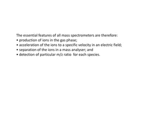
- 1. The essential features of all mass spectrometers are therefore: • production of ions in the gas phase; • acceleration of the ions to a specific velocity in an electric field; • separation of the ions in a mass analyser; and • detection of particular m/z ratio for each species.
- 2. Basic Design of a Mass Spectrometer
- 3. Components of a mass spectrometer All mass spectrometers are basically similar. They consist of the following: • A high vacuum system : A high vacuum system is the very first requirement to produce high level of vacuum inside the mass spectrometer. Under this high vacuum ions don’t collide with air molecules and, their path length and life in increased. • A sample inlet: Through sample inlet, sample is introduce in the mass spectrometer. Sample or analyte must be introduced in gaseous form. Sample inlet may be a sample or target plate; a high-performance liquid chromatography (HPLC), gas chromatography (GC) or capillary electrophoresis system; solids probe; electron impact chaber direct chemical ionization chamber. • An ion source: Ion source convert analyte molecules into ion. There are many methods of Ionization that can be used to produce ions, such as; Electron impact ionization, chemical Ionization methods, field ionization, fast atom bombardment (FAB); matrix desorption ionization or Electro spray ionization. • A mass filter/analyser: Mass filter separates the ions on the basis of m/z of ions. A mass filter separate the ion by any of the following types: Magnetic sector or Electric sector, Quadrupole , ion trap or Time of Flight (TOF). • A detector: After the ions are separated by mass analyzer, the detector detects these ions and tell about their mass. A detector can be a conversion dynode, electron multiplier, microchannel plate or array detector.
- 4. Vacuum Pump: Purpose- All mass analysers operate under vacuum in order to minimise collisions between Ions and air molecules. Without a high vacuum, the ions produced in the source will be lost and will not reach the detector. At atmospheric pressure, the mean free path of a typical ion is around 52 nm; at 1 mtorr, it is 40 mm; and at 1 µtorr, it is 40 m. In most instruments, two vacuum pump types are used (i) a rotary vane pump(to produce the main reduction in pressure, which reduces the pressure from 760 torr to 1 torr) followed by (ii) a turbomolecular pump or diffusion pump (to produce the high vacuum of 1 micro torr to 1 nano torr) Ionization : Ionization is a process of converting neutral analyte molecules into ions. Ions may be produced from a neutral molecule by either (i) removal of an electron that produce a positively charged cation, or (ii) by adding an electron so that a negatively charged anion is formed. For the purpose of mass spectroscopy each analyte molecule is needed to be converted into ionic form. There are many types of ionization methods and any one of them is used in a mass spectrometers.
- 5. Types of Ionization methods 1. Electron Impact Ionization 2. Chemical ionization 3. Field ionization 4. Field desorption ionization 5. Fast Atom Bombardment ionization 6. Electro spray ionization 7. Matrix assisted desorption ionization Note: 1 and 2 methods are strong or hard ionization methods while from 3-7 are soft Ionization method. Note: 1, 2 and 3 need vapour phase of sample while 4 -7 can use liquid or gaseous form of sample molecules. Note: Soft ionzation methods specially no. 6 and 7 are used for study of biomolecules Such as DNA, RNA and proteins
- 6. Electron Impact ionization (EI): Electron impact ionisation (EI) is widely used for the analysis of metabolites, pollutants and pharmaceutical compounds, and in drug testing programmes. In EI a beam of electron is produced by heated metal filaments and this electron beam is accelerated to 70 eV potential. Sample is introduced in gaseous form into ionization chamber and ionisation occurs when the high speed electrons Interact with the sample molecules. After collision electrons may be released from the substance (to produce a cation) or electron can be captured (to produce an anion). EI can be used only with substance or molecules having molecular weight below 400 Da, because large molecules can not be easily converted ion gaseous state. Accelerated electrons have very high energy, which cause breaking of bonds. Due to breaking of bonds the resulting fragment (ions) is left with single unpaired electron called Radical ion (M.+/- ) which can be either positively or negatively charged. A positive charged cation is represented as M+. and a negatively charged anion is represented as M-. . This (+ or -) sign indicates the ionic state and the (.) a radical. Such radical ions Having (.) are termed “molecular ions, parent ions or precursor ions”. These precursor ions are very unstable, due to having high energy. These precursor ion disintegrating into a number of smaller ‘fragment ions’ or ‘daughter ions’ or ‘product ion’ that may be relatively unstable and further fragmentation may occur. This gives rise to a series of daughter ions or product ions, which are recorded as the mass spectrum.
- 7. When a precursor ion breaks, it splits into two parts one part carries the charge (ion) and other part carries an unpaired electron(radical). The ions are true ions and not radical ions. The radicals are neutral in nature and therefore do not take any further part in the mass spectrometry and are pumped away by the vacuum system. Structure of Electron impact Ionization chamber Example of disintegration of Precursor ion into daughter ion or fragment ion
- 8. Chemical Ionization (CI): Chemical ionisation (CI) is similar to EI. The difference is that it cause lesser fragmentation than EI and produces high intensity molecular ions, due to less fragmentation the spectra obtained by CI is more clean than EI. Structure of CI is same as EI the only difference is that ionization chamber contains a suitable reagent gas such as methane (CH4) or ammonia (NH3). Accelerated electrons collide with reagent gas and reagent gas ions NH3+ and CH4+ are formed. Some of these gas ions react with analyte moelcules to produces analyte ions. The peak obtained from CI shows the addition of CH4+ or NH3+ in the spectra. Structure of Chemical Ionization chamber NH3 or CH4 NH3+ or CH4+ Field Ionization: in this type of ionization method, a very high electric filed 107-108 volt/ Cm is applied and Sample is kept in this electric field. Under this high electric field outer bonding Electrons are released and molecules becomes positively charged.
- 9. Ion desorption methods: These methods don’t require the sample to be converted into gaseous phase. They can use solid or liquid form of the sample. (i) Field Desorption: Field desorption (FD) source is difficult to produce and source is destroyed each time after use. The filament of source is to be prepared fresh and in advance. A tungeston wire 10um in diameter is coated with an appropriate organic material, from this wire under suitable conditions carbon filaments or dendrites are grown. A solution of sample is carefully coated on these dendrites and allowed to dry. This make a solid coat on dendrites and the filament is carefully placed inside ionization chamber. Ionization chamber in evacuated and high voltage 107 -108 volt is applied. Under high voltage ions are formed. (ii) Plasma desorption Plasma is group of atomic nuclei from which all the electrons are removed. Source of plasma Ii californium Cf 252. from this Cf two nuclei Ba 20+ and Tc18+ are ejected in opposite direction these nuclei have very high energy and when the collide with biological molecules they ionize them. This method was the old one and now is not much used. Fig: Tungeston filament showing growth of carbon filament or dendrites on it.
- 10. (iii) Fast Atom Bombardment (FAB): It is a soft ionization technique and was developed in early 1980s. The important advantage of this technique is that it produces ions with low internal energies, due to which ions don’t go further fragmentation. This allows the analysis of biomolecules in solution form without prior derivatisation. The sample is mixed with a relatively involatile, viscous matrix such as glycerol, thioglycerol or m-nitrobenzyl alcohol. The mixture, placed on a probe, is introduced into the ionization chamber and bombarded with an ionising beam of neutral atoms (such as Ar, He, Xe) of high velocity. The new version of this ionization method uses beam of caesium (Cs+) ions and this type of spectroscopy is known as ‘liquid secondary ion mass spectrometry (LSIMS). FAB produces Pseudomolecular ion (M + H)+ and (M - H)_. Pseudomolecular ions are the mass of the ion formed from a substance of a given mass by the gain or loss of one or more protons instead of electrons. Diagram of FAB Diagram of FAB
- 11. (iv) Matrix Assisted Laser Desorption ionization (MALDI): This method of ionization uses Laser beams in stead of electrons (EI), or fast moving atom (FAB). Laser beam contains enough energy to cause ionization and desorption of the sample coated on suitable probe surface. Matrix function as energy sink which absorb the laser energy and transfer it to the sample for ionization. It also protect the sample from destructive effect of laser by covering the sample. The matrix is a conjugated organic compound (normally a weak organic acid). Commonly used matrix in MALDI is α-Cyano Hydroxy Cinnamic Acid (CHCN). Instead of laser beams other radiations, such as UV/ visible/ Infrared can also be used, with suitable matrix. The advantage of MALDI is that it produces large mass ions with high sensitivity and produces molecular ions which are stable and usually don’t undergo fragmentation. For obtaining Mass spectra by MALDI, sample is properly mixed with matrix solution and placed on grid of stainless steel MALDI sample plate (made up of stainless steel) in form of spot. The Matrix+sample spot is allowed to dry so that the matrix get crystallized. After the matrix is properly crystallized, a laser is fired on dried matrix+ sample spot on MALDI plate and ions are generated. These ions are extracted by using a high voltage pulse to aviod time lag focusing (TLF) or time delayed extraction (TDE). These ions are then transferred to mass analyzer. The reflectron in used in MALDI-TOF-MS to avoid post source decay (PSD)
- 13. MALDI–TOF instrument components. (1) Sample mixed with matrix is dried on the target plate which is introduced into high-vacuum chamber. (2) The camera allows viewing of the position of the laser beam which can be tracked to optimise the signal. (3) The sample/matrix is irradiated with laser pulses. (4) The clock is started to measure time-of-flight. (5) Ions are accelerated by the electric field to the same kinetic energy and are separated according to mass as they fly through the flight tube. (6) Ions strike the detector either in linear (dashed arrow) or reflectron (full arrows) mode at different times, depending on their m/z ratio. (7) A data system controls instrument parameters, acquires signal versus time and processes the data.
- 14. Ion Evaporation methods: (i) Thermospray ionization: here the thermal effect is used to convert droplets in vapour form . Due to use of heat this method is not suitable for thermolabile . compound (i) Ion spray ionization: Ion spray use a gas (mostly nitrogen) to cause nebulization of sample solution to form spray. This is also called pneumatic assisted ionization. (i) Electro Spray Ionization : This is the most commonly used methods than above two. This involves the production of ions by spraying a solution of the analyte into an electrical field. The electrospray (ES) creates very small droplets of solvent-containing analyte. The essential principle in ES is that a spray of charged liquid droplets is produced by atomisation or nebulisation. Solvent (typically 50 : 50 water and organic solvent) is removed as the droplets enter the mass spectrometer. ESI is the result of the strong electric field (around 4 keV at the end of the capillary and 1 keV at the counter electrode) acting on the surface of the sample solution. As the solvent evaporates in the high-vacuum region, the droplet size decreases and eventually charged analyte (free of solvent) remains. Ionisation can occur at atmospheric pressure and this method is also sometimes referred to as atmospheric pressure ionisation (API). The flow rate into the source is normally around a few mm3 min-1 and m/z limit of measurement of ESI-MS is normally 2000- 3000Da. A curtain or sheath gas (usually nitrogen) around the spray needle at a slow flow rate may be used to assist evaporation of the solvent at or below room temperature. This may be an advantage for thermally labile compounds.
- 15. Electrospray ionisation source. The ESI creates very small droplets of solvent-containing analyte by atomisation or nebulisation as the sample is introduced into the source through the fine glass (or other material) hollow needle capillary. The solvent evaporates in the high-vacuum region as the spray of droplets enters the source. As the result of the strong electric field acting on the surface of the sample droplets, and electrostatic repulsion, their size decreases and eventually single species of charged analyte (free of solvent) remain. These may have multiple charges depending on the availability of ionisable groups.
- 16. Mass Analyzers: Once ions are created and leave the ion source, they pass into a mass analyser, the function of which is to separate the ions and to measure their masses. (Remember, what is really measured is the mass-to-charge ratio (m/z) for each ion.) At any given moment, ions of a particular mass are allowed to pass through the analyser where they are counted by the detector. Subsequently, ions of a different mass are allowed to pass through the analyser and again the detector counts the number of ions. In this way, the analyser scans through a large range of masses. In the majority of instruments, a particular type of ionisation is coupled to a particular mass analyser that operates by a particular principle. That is, EI, CI and FAB are combined with magnetic sector instruments; ESI and its derivatives with quadrupole (or its variant ion-trap) and MALDI is coupled to TOF detection.
- 17. Magnetic sector analysis: In magnetic sector analyser ions are first accelerated by an electric field. The electric sector acts as a kinetic energy filter and separates the ions on the basis of their velocity, irrespective of the m/z. A given ion with the appropriate velocity then enters the magnetic sector analyser. Under magnetic sector ion will travel in a curved trajectory and radius of this trajectory depends upon m/z and velocity of the ion (the latter has already been selected). Thus only ions of a particular m/z will be detected at a particular magnetic field strength. The trajectory of the ions is through a sector of the circular poles of the magnet, hence the term magnetic sector.
- 18. Quadrupole: Quadrupole analyser consists of four parallel cylindrical rods . A direct current (DC) voltage and a superimposed radio frequency (RF) voltage are applied to each rod, creating a continuously varying electric field along the length of the analyser. Inside the quadruple ions are accelerated towards the detector. By controlling electric field ions of one particular mass-to-charge ratio (m/z) can be selected and retained inside quadrupole. Ions with different m/z will impact on the quadrupole rods and will not be detected. By changing the electric field (scanning), the ions of different m/z successively arrive at the detector. Quadrupoles can routinely analyse up to m/z 3000. Note that hexapole and octapole devices are also used, to direct a beam into the next section of a triple quadrupole or into the ion trap for example, but the principle is the same. Quadrupole analyser. The fixed (DC) and oscillating (RF)fields cause the ions to undergo complicated trajectories through the quadrupole filter. For a given set of fields, only certain trajectories are stable, which only allows ions of specific m/z to travel through to the detector.
- 19. Ion- trap: Ion trap mass spectrometers is used with ESI. All ions are are transferred into and subsequently measured almost simultaneously (within milliseconds) in an ion trap. The trap must then be refilled with the ions that are arriving from the source. Ion trap has higher sensitivity relative to quadrupole where at any given moment only ions of one particular m/z are detected. ESI–ion trap mass spectrometers have found wide application for analysis of peptides and small biomolecules such as in protein identification by tandem MS; liquid chromatography/mass spectrometry (LC/MS); combinatorial libraries and rapid analysis in drug discovery and drug development. Ion trap
- 20. Principle of time-of-flight (TOF): is a method of mass spectrometry in which an ion's mass-to-charge ratio is determined via a time measurement. Its principle is that ‘If the ions are accelerated with the same potential at a fixed point and a fixed initial time, the ions will separate according to their mass to charge ratios. This time of flight can be converted to mass. Ions are accelerated by an electric field of and this acceleration results in an ion having the same kinetic energy. The velocity of the ion depends on the mass-to-charge ratio. The time that it subsequently takes for the particle to reach a detector at a known distance is measured and this time is known as time of flight (TOF). This time will depend on the mass-to-charge ratio of the particle (heavier particles reach lower speeds). From this time and the known experimental parameters one can find the mass-to-charge ratio of the ion. Time of flight (TOF)
- 21. Detectors: Following types of detectors are used in mass spectrometry •Faraday cup: •Conversion dynode •Electron Multiplier •Array detectors