2. ISSN 2231 – 2250 Inflammatory Dentigerous Cyst Associated With... 50
interface was flat. Underlying fibrous
connective tissue wall showed loosely
arranged collagen fibers with islands of
odontogenic epithelium and severe chronic
inflammatory cells infiltration (Fig 1c).
According to WHO criteria, radiographic
feature and histopathologic findings the
diagnosis was inflamed dentigerous cyst
associated with the unerupted first premolar.
The patient remained under follow-up for
one year and no complications were
observed.
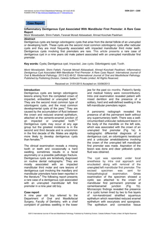
Figure 1: The sectional panoramic radiograph showing a well-circumscribed, unilocular
radiolucent lesion in the body of the mandible, on the left side in relation to the crown of
unerupted mandibular left first premolar (a). The gross specimen showing the relation of the cyst
to the crown of first premolar (b), however the hematoxylin and eosin stained photomicrograph
under low power view showing the lining epithelium which is thin and non-keratinized with
exosytosis and spongiosis along with inflammation in the cyst wall (c).
Discussion
Dentigerous cysts are developmental cysts
that enclose crown of an unerupted tooth at
cemento-enamel junction and thus they are
also named “tooth containing cysts”.9
These
cysts are the second most common type of
odontogenic cysts and make up an
estimated 14% to 24% of all jaw cysts.2,10
The exact histopathogenesis of these cysts
is unknown. They are believed to be caused
by expansion of dental follicles resulting
from accumulation of fluid in the space
between the tooth crown and epithelial
components.11
The highest incidence of
dentigerous cysts occurs during the second
and third decades and they are rarely seen
during childhood.2,12
Shear has estimated
that about 9% of dentigerous cysts occur in
the first decade of life.13
Our patient was only
nine years old at the time of presentation.
However in the only reported case by Muthu
kumar et al the age of the patient was 62
years. The sex predilection of dentigerous
cyst is 1.6:1 in favor of male2,15
, and the
prevalence is higher for whites than for
blacks.14
Dentigerous cyst most commonly develops
around the crown of the third mandibular
molar tooth.14
But in this case, mandibular
first premolar was found to be the cause of
the dentigerous cyst. In our review of the
literature, we have found only one case of
dentigerous cyst associated with mandibular
first premolar (Fig 4).15
Dentigerous cysts are
typically asymptomatic and may be large,
destructive, expansile lesions of bone.16
In
present case, the cyst was painless and was
3. 51 Monir Moradzadeh, et al. ISSN 2231 - 2250
discovered during investigation of
asymptomatic swellings.
On radiographic examination, dentigerous
cysts appear as unilocular radiolucent area,
with well-defined and often sclerotic borders,
associated with the crown of an unerupted
tooth. The borders may be ill-defined when
infected.5,14
Diagnosis should not be made
on radiographic evidence alone but should
include both macroscopic and microscopic
examination of the lesion. The
histopathological examination of cyst in the
present case showed thin and non-
keratinized lining epithelium, resembling
reduced enamel epithelium overlying a
fibrous connective tissue capsule in most of
the areas.5
In presence of inflammation the
capsule shows foci of chronic inflammatory
cells14
and the lining epithelium
corresponding to these areas shows an
increase in thickness.2,14
So this case was
diagnosed as an inflammatory dentigerous
cyst.
Treatment of choice for dentigerous cysts is
removal of the associated tooth and
enucleation of the soft tissue component.2
Complete removal of the cyst is extremely
important because ameloblastoma,
squamous cell carcinoma and
mucoepidermoid carcinoma have been
reported as potential complications of
untreated dentigerous cysts.17
Conclusion
Dentigerous cyst is most commonly seen in
association with impacted third molars. In
this case report we have presented a rare
case of inflammatory dentigerous cyst
associated with mandibular first premolar.
The case was successfully treated. Post
operatively there was no complication.
Acknowledgement
We would like to acknowledge all the staff
members for their support and guidance.
Author Affiliations
1.Dr.Monir Moradzadeh, Associate Professor,
2.Dr.Shirin Fattahi, Assistant Professor,
3.Dr.Faranak Moradi Abbasabadi, Post-graduate
Student, 4.Dr.Ahmad Koochaki Pasikhani, Post-
graduate Student, Department of Oral and
Maxillofacial Pathology, Faculty of Dentistry,
Tabriz University of Medical Sciences, Tabriz,
Iran.
References
1. Larsen PE. Odontogenesis and
odontogenic cysts and tumors. In:
Cummings CC (ed). Otolaryngology
head and neck surgery. 2nd. St Louis;
Mo: Mosby- Year Book Inc; 1993.
2. Regezi AJ, Sciubba JJ, Jordan RCK.
Oral Pathology: Clinical Pathologic
Correlations. 5th
ed. St. Louis: Saunders;
2008. 242-4p.
3. Sumita M, Vineet R, Karen B, Thomas
G. Non-syndromic bilateral dentigerous
cysts of mandibular premolars: a rare
case and review of literature. HongKong
Dental Journal 2006;3:129-33.
4. Shah N, Thuau H, Beale T.
Spontaneous regression of bilateral
dentigerous cysts associated with
impacted mandibular third molars. Br
Dent J 2002;192:75-6.
5. Shun Y. Dentigerous cyst associated
with an impacted anterior maxillary
supernumerary tooth. J Dent Child
(Chic) 2008;75:104-7.
6. Shetty R, Sandler PJ. Keeping your eye
on the ball. Dental Update 2004;31398-
402.
7. McCrea S. Adjacent dentigerous cysts
with the ectopic displacement of a third
mandibular molar and supernumerary
(forth) molar: a rare occurrence. Oral
Surg Oral Med Oral Pathol Oral Radiol
Endod 2009;107:e15-20.
8. Miyakawi S, Hyomoto M, Kirita J,
Sugimura M. Eruption speed and rate of
angulation change of a cyst associated
mandibular second premolar after
marsupialization of a dentigerous cyst.
Am J Ortho Dentofac Orthop
1999;116:578-84.
9. González AE, Maestre PM, Fernandez
CD, Vilchez I, Egea SJJ, Perez GJL.
Dentigerous cyst associated with a
formocresol pulpotomized deciduous
molar. J Endod 2007;33:488-92.
10. Rubin DM, Vendrenne D, Portnof JE.
Orthodontically guided eruption of
mandibular second premolar following
enucleation of an inflammatory cyst:
case report. J Clin Pediatr Dent
2002;27:19-24.
11. Edamatsu M, Kumamoto H, Ooya K,
Echigo S. Apoptosis related factors in
the epithelial components of dental
follicles and dentigerous cysts
associated with impacted third molars of
the mandible. Oral Surg Oral Med Oral
Pathol Oral Radiol Endod 2005;99:17-
23.
12. Trimble LD, West RA, McNeill RW.
Cleidocranial dysplasia. Comprehensive
treatment of dentofacial abnormalities, J
Am Dent Assoc 1982;5:661-6.
4. ISSN 2231 – 2250 Inflammatory Dentigerous Cyst Associated With... 52
13. Shear M. Developmental odontogenic
cysts. An update. J Oral Pathol Med
1994;23:1-11.
14. Neville BW, Damm DD, Allen CM,
Bouquot JE. Oral and Maxillofacial
Pathology. 3rd
ed. St. Louis: Saunders;
2008. 679-82p.
15. Muthukumar SR, Parthiban SV,
Alagappan M, Arunkumar S.
Dentigerous Cyst Associated with
Mandibular First Premolar: A Rare Case
Report. Indian Journal of
Multidisciplinary Dentistry 2011:298-8.
16. McDonald RE, Avery DR, Dean JA.
Tumors of the oral soft tissues and cysts
and tumors of the bone. Dentistry for the
Child and Adolescent. 8th
ed. St. Louis:
Mosby; 2004. 159-161p.
17. Shafer WG, Hine MK, Levy BM. A
textbook of oral pathology. 4th
ed. India:
Reed Elsevier India Private Ltd; 2006.
260–5p.
Corresponding Author
Dr.Shirin Fattahi,
Department of Pathology,
Dentistry Faculty,
Daneshgah Street, Tabriz,
Eastern Azerbaijan, Iran.
Fax: +984113307953,
Mob: +989143150165,
E-mail: Shirin_Fattahi@yahoo.com
Source of Support: Nil, Conflict of Interest: None Declared.