Case record...Vertebral hemangioma with multisegmental retromedullary lipoma
•
2 j'aime•2,176 vues

Case record...Vertebral hemangioma with multisegmental retromedullary lipoma

Recommandé
Recommandé
Contenu connexe
Tendances
Tendances (20)
En vedette
En vedette (9)
Similaire à Case record...Vertebral hemangioma with multisegmental retromedullary lipoma
Similaire à Case record...Vertebral hemangioma with multisegmental retromedullary lipoma (20)
Plus de Professor Yasser Metwally
Plus de Professor Yasser Metwally (20)
Dernier
Dernier (20)
Case record...Vertebral hemangioma with multisegmental retromedullary lipoma
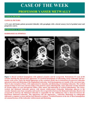
- 1. CASE OF THE WEEK PROFESSOR YASSER METWALLY CLINICAL PICTURE CLINICAL PICTURE: A 51 years old female patient presented clinically with paraplegia with a dorsal sensory level of gradual onset and progressive course. RADIOLOGICAL FINDINGS RADIOLOGICAL FINDINGS: Figure 1. Dorsal vertebral hemangioma with epidural extension and fat overgrowth. Postcontrast CT scan of D4 vertebra showing the characteristic appearance of vertebral hemangioma of alternating coarse trabeculae and low- density cystic areas. Notice the dotted appearance of the vertebral, mostly due to destruction of transverse trabeculae and preservation and thickening of the longitudinal trabeculae. Such haemangiomas are usually of the cavernous type. There is also resorption of bone, with replacement by sinusoids, and thickening of remaining trabeculae. The cortical margin is intact in most cases and may bulge at the posterior aspect. Histologically, many abnormal vascular channels of varying calibre are seen interspersed within a fatty matrix and thickening of vertical trabiculations. The coarse, vertical, and thickened trabecular pattern, with osseous reinforcement (trabecular thickening) adjacent to the vascular channels cause bone resorption and reactive fat overgrowth in between the thickened trabeculae. Reactive fat overgrowth in between the thickened trabeculae is responsible for the radiolucency that is observed between the hyperdense thickened trabeculae. This appearance (the dot appearance.... trabecular thickening) on radiographs represents a response to stress and has been likened to corduroy. Vertebral fractures at the site of these hemangiomas are unusual because of this trabecular reinforcement and thickening.
- 2. Figure 2. Post intravenous contrast CT scan image (A) and CT myelography (B,C). Notice the epidural extension of the hemangioma that showed contrast enhancement (A). The remnant of dye in the subarachnoid spaces (B,C) showed that the spinal cord is actually pushed anteriorly by AN epidural mass of fat density (epidural lipoma) Figure 3. Sagittal reconstruction images showing the vertebral bony hemangioma with enhanced epidural extension and the multisegmental retromedullary lipoma pushing the spinal cord anteriorly. The retromedullary lipoma is probably reactive to the epidural hemangioma rather than a true neoplasm. Operative finding, however, revealed an epidural angiolipoma.
- 3. Figure 4. CT myelography showing the vertebral hemangioma and the multisegmental retromedullary lipoma pushing the spinal cord anteriorly. The retromedullary lipoma is probably reactive to the epidural hemangioma rather than a true neoplasm. Operative finding, however, revealed an epidural angiolipoma. Figure 5. A, Plain CT scan, B, CT scan with intravenous contrast showing a vertebral bony hemangioma extending into the epidural spaces. quot;vertebral hemangiomas are rarely implicated in symptoms formation (unless proved otherwise by radiological studies)quot;. Notice the epidural retromedullary lipoma pushing the spinal cord anteriorly and apparently is the cause of compressive myelopathy in this case. The retromedullary lipoma is probably reactive to the epidural hemangioma rather than a true neoplasm. Operative finding, however, revealed an epidural angiolipoma.
- 4. Figure 6. Plain x ray showing a vertebral body haemangioma. The vertebral bodies appear dotted in cross-section due to destruction of transverse striation and preservation of the longitudinal striation. These lesions are well demonstrated by plain CT scan and do not enhance after i.v. contrast injection. The vertebral hemangioma in this patient was not causely related to the paraplegic state. The cause of paraplegia was an epidural retromedullary lipoma pushing the spinal cord anteriorly, which when debulked surgically the patient much improved. On plain radiographs and CT, there is exaggeration of the vertebral trabeculae of the vertebral body (also called quot;accordionquot; or quot;honeycombquot; vertebrae). This appearance may still be preserved after vertebral collapse. The cortical margin is intact in most cases and may bulge at the posterior aspect Figure 7. A, Plain CT scan, B, CT myelography showing a vertebral body haemangioma associated with an extradural, retromedullary lipoma pushing the spinal cord anteriorly. On plain radiographs and CT, there is exaggeration of the vertebral trabeculae of the vertebral body (also called quot;accordionquot; or quot;honeycombquot; vertebrae). This appearance may still be preserved after vertebral collapse. The cortical margin is intact in most cases and may bulge at the posterior aspect Figure 8. Operative findings revealed an epidural angiolipoma, following debulking of this lipoma the patient much improved.
- 5. Box 1. Characteristics of vertebral hemangiomas Histologically, many abnormal vascular channels of varying calibre are seen interspersed within a fatty matrix and thickening of vertical trabiculations. The coarse, vertical, trabecular pattern, with osseous reinforcement (trabecular thickening) adjacent to the vascular channels that have caused bone resorption. This appearance on radiographs represents a response to stress and has been likened to corduroy. Vertebral fractures at the site of these hemangiomas are unusual because of this trabecular reinforcement. Fatty overgrowth are probably reactive in nature secondary to the hemangioma and probably it represents a response to stress. The vascular channels frequently causes bone resorption that progress to destruction of horizontal trabeculae. Thickening of the vertical trabeculae, with osseous reinforcement represents a response to stress. Trabecular thickening causes vertebral reinforcement thus making vertebral fractures at the site of these hemangiomas unusual. The dotted appearance of CT scan imaging is due to the thickened vertical trabeculae. Trabeculae thickening occurs through intramembranous bone formation adjacent to the angiomatous channel. The radiolucency interspersed between the hyperdense dots is due to increased fat content in between the thickened vertical trabeculae. Bone resorption and cystic changes probably play a part in this radiolucency but to much lesser extent, and that is why vertebral hemangiomas appear hyperintense on non-contrast MRI T1 images due to increased fat content. The multisegmental epidural lipoma observed in this case is probably reactive to the epidural extension of the hemangioma rather than true neoplasm. Lipomas are neither true tumours nor they are hamartomas, they are simply normal adipose tissues in abnormal sites. The epidural lipoma, whatever its aetiology and pathogenesis, has apparently reached a large size enough to exert significant spinal cord compression and myelopathy. Although epidural, extraosseous extension of the vertebral hemangioma is observed in this case, however it was not causing any spinal cord compression or displacement as CT myelography demonstrated that the spinal cord is pushed anteriorly by the retromedullary lipoma rather than posteriorly by the epidural hemangioma. One can not, however, negates the possible role of a vertebral hemangioma in symptom formation by causing bleeding with epidural hematoma or vascular steal with spinal cord ischemia. DIAGNOSIS: DIAGNOSIS: VERTEBRAL HEMANGIOMA WITH MULTISEGMENTAL EXTRADURAL RETROMEDULLARY LIPOMA DISCUSSION DISCUSSION: Vertebral hemangioma Haemangioma of the vertebra is a benign, slow growing tumour of the blood vessels. It makes up 30% of all haemangiomas of bone and is found in approximately 10% of autopsies. It is commonly seen in women over 40 and has a predilection for the lower thoracic or upper lumbar spine. Most vertebral haemangiomas are asymptomatic and are of no clinical significance. However vertebral collapse or extension into the spinal canal may result in pain, spinal cord compression and/or paraplegia. Histologically, many abnormal vascular channels of varying calibre are seen interspersed within a fatty matrix. The haemangioma is usually of the cavernous type. There is also resorption of bone, with replacement by sinusoids, and thickening of remaining trabeculae.
- 6. Hemangioma of bone histologically resembles cavernous hemangiomas (cavernomas). Radiographically it has a pathognomonic appearance of alternating coarse trabeculae and low-density cystic areas. The alternating pattern is particularly well seen on CT. Hemangiomas typically arise in the vertebral body, unlike osteoid osteoma, osteoblastoma, and aneurysmal bone cyst, which arise in the posterior neural arch. Although most hemangiomas of the spine are discovered as incidental findings, they have been reported to cause vertebral canal compromise. They can grow over time and have been associated with hematoma in cases of trauma. On MRI the lesion is hyperintense on the precontrast MRI T1 studies, and On MRI T2 studies the lesion appears of heterogenous signal intensity, probably due to the presence of blood products. The author did not observe enhancement of these lesions following contrast injection. Figure 9. Asymptomatic spinal hemangioma at T-6 in a 67-year-old man. A Lateral conventional tomogram shows typical trabecular thickening (arrows) at the site of the hemangioma. (B and C) Photographs of the macerated dry bone (B) and gross specimens sectioned coronally (C) demonstrate the trabecular thickening (arrowheads). (D) Photomicrograph (original magnification, approximately X5; hematoxylin-eosin stain) also shows trabecular thickening (solid arrows), angiomatous components (open arrows), and fat overgrowth (arrowheads), compared with the normal, more cellular marrow (*) of adjacent vertebrae. Trabeculae thickening occurs through intramembranous bone formation adjacent to the angiomatous channel. These tumours are commonly found in the vertebral bodies and laminae, cavernous hemangiomas are slowly growing, small benign lesions that have been demonstrated in 11% of spines at autopsy, but are only rarely symptomatic. They occur in the body of the vertebra, although the posterior elements can be affected. Rarely, compression of neural
- 7. elements by dilated epidural and paraspinal draining veins can occur in association with hypervascular hemangiomas. They occur most commonly in the thoracic spine. Radiologically, these are very distinct lesions of the bony casing of the spinal cord that, in time, will expand into the epidural space and produce a compressive myelopathy. In the author experience, vertebral hemangiomas are rarely implicated in symptoms formation (unless proved otherwise by radiological studies) and even when discovered in a paraplegic patient a search for anther cause of paraplegia must be made. See figs. 12,13 On plain radiographs and CT, there is exaggeration of the vertebral trabeculae of the vertebral body (also called quot;accordionquot; or quot;honeycombquot; vertebrae). This appearance may still be preserved after vertebral collapse. The cortical margin is intact in most cases and may bulge at the posterior aspect. The vertebral haemangioma may have extraosseous extension and surround the cord several levels above the bone lesion. Cord compression usually occurs in middle aged adults, sparing children and the older age group. Paravertebral soft tissue extensions are occasionally seen. The vertebral bodies appear dotted in cross-section due to destruction of transverse striation and preservation of the longitudinal striation. These lesions are well demonstrated by plain CT scan and do not enhance after i.v. contrast injection. A characteristic appearance is usually seen on MRI. A mottled pattern with increased signal intensity is seen on T1 weighted images due to fatty tissue interspersed with the thickened trabeculae. Extraosseous tumour, if present, is better evaluated with MRI than with any other imaging modality. Appearance of exaggerated vertical trabeculae as described above may also be seen in secondary osteoporosis, multiple myeloma, lymphoma, metastases, Paget's disease or blood dyscrasia. Hence, any symptomatic lesion of the vertebrae with features of a haemangioma must be carefully evaluated. Figure 10. Plain x ray showing a vertebral body haemangioma, B, A characteristic appearance is usually seen on MRI. A mottled pattern with increased signal intensity is seen on T1 weighted images due to fatty tissue interspersed with the thickened trabeculae. Vertebral hemangiomas are the most common primary spinal neoplasm and are present in 10 to 12% of the population, based on autopsy studies performed in adults.[4,7,29,31,37] Usually incidental, asymptomatic, and solitary, these are benign vascular lesions that can give rise to symptoms in rare circumstances. Symptoms include radicular pain in most cases, and neurological compromise can occur in up to 40% of symptomatic cases. Symptoms are thought to develop by the following mechanisms: 1) vascular expansion of a vertebral body, pedicle, lamina, or facet leading to direct compression of nerve roots and/or the spinal canal; 2) subperiosteal extension of the hemangioma resulting in an extradural mass compressing the spinal cord; and 3) compression fractures and vertebral collapse secondary to replacement of bone with hemangioma. Rarely, a vertebral hemangioma may cause bleeding with epidural hematoma or vascular steal with spinal cord ischemia. Vertebral hemangiomas exhibit a classic radiographic appearance of coarse vertical striations, referred to as a
- 8. honeycomb pattern, on plain spine x-ray films or CT scans. Conventional MR imaging is less definitive for hemangioma, but the high blood content of the lesion demonstrates high T2-weighted signal intensity on fat- suppression sequences. Angiograms can also aid in diagnosis and provide a road map for treatment (that is, embolization). The vascular supply to vertebral hemangiomas usually originates from small branches of the intercostal or lumbar arteries that arise proximal to the radicular branches. Certain radiographic features such as neural arch involvement, complete vertebral body involvement, expanded osseous cortex with indistinct margins, an irregular honeycomb pattern of bone, an epidural mass, and fatty stroma are associated with the development of symptoms,[22] which most pregnancy-related cases demonstrate. The histological appearance of hemangiomas consists of benign vascular proliferation with normal capillary and venous structure.[8,29,35] Two types have been described: cavernous or capillary. The most common type of vertebral hemangioma is the cavernous type, which is characterized by large sinusoidal spaces lined by a single layer of epithelium. The capillary type of vertebral hemangioma differs from the cavernous type only by having smaller vascular channels. Whereas asymptomatic hemangiomas occur primarily in the thoracolumbar spine, symptomatic hemangiomas most commonly occur at the thoracic level.[27] Lumbar or cervical locations for symptomatic hemangiomas are exceedingly rare. Epidural lipoma These tumours are situated in the dorsal region in the extradural retromedullary spaces. They usually extend for more than one vertebral segment and are well demonstrated by plain CT scan as regions of fat density. No significant enhancement occurs after i.v. contrast injection. Lipomas are occasionally associated with bony haemangiomas and are considered reactive to it. The larger the degree of fat overgrowth in the stroma between thickened trabeculae (seen on CT images as low attenuation between thickened trabeculae and as areas of high intensity on T1-weighted images and intermediate intensity on T2-weighted images), the less likely these lesions will be symptomatic (inactive hemangioma) as shown by Laredo and coworkers [50]. Lipomas originate from the focal premature dysjunction of neural ectoderm from cutaneous ectoderm that allows migration of periaxial mesoderm into the developing neural tube. This pluripotent mesoderm primarily develops into fat, but may also develop into other tissues, including blood vessels. So the combined lesion might be explained by the focal maturation of this pluripotent mesoderm, which migrates between the two ectodermal layers and primarily matures as a lipoma in the primitive vascular plexus. Subsequent failure to form the capillary component in this plexus might lead to the formation of a combined lesion (vascular lesion combined with epidural lipoma) [39,40]. Overgrowth of adipose tissue is most frequently associated with hemangiomas and are considered reactive to it, and this characteristic led some authors in the past to refer to these lesions as angiolipomas. However, fat overgrowth should be considered a reactive phenomenon as opposed to a true neoplastic component; therefore, the term angiolipoma is not appropriate for the vast majority of these musculoskeletal vascular lesions [41,42,43]. True angiolipomas are rare lesions. Operative debulking of excessive epidural fat (whither it is considered reactive to the hemangioma or a true neoplasm) is probably indicated and might be useful to the patient especially when it exerts significant spinal cord displacement and compression. Box 2. Radiographic features that is associated with development of symptoms in vertebral hemangiomas 1. Neural arch involvement 2. Complete vertebral body involvement 3. Expanded osseous cortex with indistinct margins 4. An irregular honeycomb pattern of bone 5. An epidural mass, and fatty stroma
- 9. Figure 11. Hemangioma of L3 in a 65-year-old man. Nearly the entire body is occupied by this predominantly lytic lesion, which has multiple areas of distinct high density representing thickened trabeculae. The demonstrated radiolucency is due to increased fat content in between the thickened trabeculae. Figure 12. Epidural lipoma associated with multiple spinal haemangiomas. The vertebral hemangiomas are an incidental discovery in this patient and was not causely related to the paraplegic state of the patient. Spinal cord compression by the epidural lipoma was the cause of paraplegia in this patient.
- 10. Figure 13. A MRI precontrast T1 (A) AND MRI T2 (B,C,D,E) studies showing a case of low dorsal cord compression. MRI showed multiple vertebral hemangiomas (the lesions are hyperintense on the precontrast MRI T1 image [A] and of heterogenous signal intensity on the MRI T2 images). The vertebral hemangiomas in this patient were not causely related to the paraplegic state. The cause of paraplegia was a heavily calcified dorsal disc herniation compressing the spinal cord (C,D,E), which when removed surgically the patient much improved.
- 11. Figure 14. Asymptomatic hemangioma at L-3 in a 30-year-old man. A Axial T1-weighted MR image shows low-signal- intensity thickened trabeculae (arrowheads) in a background of fat overgrowth (arrows). B Sagittal conventional spin- echo T2-weighted MR image reveals high signal intensity in the vascular portions of the lesion (arrows), although the majority of the lesion appears similar to adipose tissue because of the large amount of fat overgrowth. SUMMARY SUMMARY Osseous Hemangioma The majority of hemangiomas that involve bone are discovered incidentally in asymptomatic patients. Men are affected twice as often as women, and lesions are usually discovered in the 4th 5th decades of life. Soft-tissue components may also be associated with these lesions. Osseous hemangioma is particularly common in the spine and calvaria and less frequently affects long bones such as the tibia, femur, and humerus. Vertebral hemangioma is extraordinarily common, seen in 11% of the cases in one large autopsy series [45]. It accounts for 28% of all skeletal hemangiomas [46]. These lesions can involve only a portion of or the entire vertebral body and are multiple in one-third of the cases [47]. The thoracic spine is the most common location for vertebral hemangiomas. [44,45,46,47]. At radiography, vertebral hemangiomas classically have a coarse, vertical, trabecular pattern, with osseous reinforcement (trabecular thickening) adjacent to the vascular channels that have caused bone resorption [48]. This appearance on radiographs represents a response to stress and has been likened to corduroy. Vertebral fractures at the site of these hemangiomas are unusual because of this trabecular reinforcement. At CT, the thickened trabeculae are seen in cross section as small punctate areas of sclerosis, often called the polka-dot appearance. At MR imaging, areas of trabecular thickening have low signal intensity, regardless of the pulse sequence used. On T1-weighted MR images, the signal intensity of vertebral hemangiomas varies from low to high, depending on the degree of adipose tissue present. T2-weighted MR images usually show areas of very high intensity corresponding to the vascular components [49]. CT or MR images obtained after intravenous administration of contrast material demonstrate lesion
- 12. enhancement. Vertebral hemangiomas occasionally cause neurologic symptoms from spinal cord compression, particularly if these lesions extend into the posterior elements or surrounding soft tissues, expand and fracture bones. [44,45,46,47]. The larger the degree of fat overgrowth in the stroma between thickened trabeculae (seen on CT images as low attenuation between thickened trabeculae and as areas of high intensity on T1-weighted images and intermediate intensity on T2- weighted images), the less likely these lesions will be symptomatic (inactive hemangioma) as shown by Laredo and coworkers [50]. Calvarial hemangiomas account for 20% of all hemangiomas and are most frequent in the frontal or parietal region [46]. These lesions arise in the diploic space and cause expansion that often involves the outer table to a greater extent. At radiography and CT, a calvarial hemangioma commonly appears as a lytic lesion with a pattern of radiating, weblike or spoke-wheel, trabecular thickening [44,45,46,47]. This characteristic appearance, as in vertebral lesions, is caused by preexisting trabeculae that have become thickened through intramembranous bone formation adjacent to the angiomatous channels. Recognition of the pattern should alert the radiologist to the vascular nature of the lesion. Osseous hemangiomas in other locations may also have radiating trabecular thickening on radiographs. Another common pattern is a bubbly bone lysis that creates a honeycomb, latticelike, or quot;hole-within-holequot; appearance. These lytic areas are invariably multifocal and usually metaphyseal or epiphyseal. Bone lysis can have linear and circular components on radiographs, suggestive of a vascular lesion, with linear and circular elements representing vascular channels seen longitudinally and en face, respectively. However, these serpentine vascular channels are recognized more easily with CT and MR imaging. Characteristically, these channels have low signal intensity on T1-weighted images and very high signal intensity on T2-weighted images because of slow blood flow. In arteriovenous lesions with faster blood flow, low signal intensity may persist with all MR imaging pulse sequences. The appearance of osseous hemangiomas at bone and red blood cell labeled scintigraphy is variable, from photopenia to moderate increased activity [51,52]. Periosteal or cortical hemangiomas occur most frequently in the anterior tibial diaphysis. These lytic cortical lesions may also show the characteristic multifocal vascular channels or be seen as a larger, nonspecific region of bone destruction. Cortical hemangiomas may predispose the bone to fracture, and periosteal reaction may accompany these lesions. Addendum A new version of this PDF file (with a new case) is uploaded in my web site every week (every Saturday and remains available till Friday.) To download the current version follow the link quot;http://pdf.yassermetwally.com/case.pdfquot;. You can also download the current version from my web site at quot;http://yassermetwally.comquot;. To download the software version of the publication (crow.exe) follow the link: http://neurology.yassermetwally.com/crow.zip The case is also presented as a short case in PDF format, to download the short case follow the link: http://pdf.yassermetwally.com/short.pdf At the end of each year, all the publications are compiled on a single CD-ROM, please contact the author to know more details. Screen resolution is better set at 1024*768 pixel screen area for optimum display. For an archive of the previously reported cases go to www.yassermetwally.net, then under pages in the right panel, scroll down and click on the text entry quot;downloadable case records in PDF formatquot; REFERENCES References 1. Acquaviva R, Thevenot C: [Recurrent spinal cord compression caused by vertebral hemangioma; determinant role of pregnancy.] Maroc Med 36:942–944, 1957 (Fre) 2. Askenasy H, Behmoaram A: Neurological manifestations in haemangioma of the vertebrae. J Neurochem
- 13. 20:276–284, 1957 3. Baker ND, Klein MJ, Greenspan A, et al: Symptomatic vertebral hemangiomas: a report of four cases. Skelet Radiol 15: 458–463, 1986 4. Bandiera S, Gasbarrini A, De Iure F, et al: Symptomatic vertebral hemangioma: the treatment of 23 cases and a review of the literature. Chir Organi Mov 87:1–15, 2002 5. Bouchez B, Gozet G, Le Coutour X, et al: [Medullary compression due to vertebral angioma in pregnancy. A case treated by embolization.] J Gynecol Obstet Biol Reprod (Paris) 13: 885–888, 1984 (Fre) 6. Bouchez B, Gozet G, Lecoutour X, et al: [Spinal cord compression caused by vertebral angioma during pregnancy. A case treated by embolization.] Presse Med 13:1696–1697, 1984 (Fre) 7. Bremnes RM, Hauge HN, Sagsveen R: Radiotherapy in the treatment of symptomatic vertebral hemangiomas: technical case report. Neurosurgery 39:1054–1058, 1996 8. Castel E, Lazennec JY, Chiras J, et al: Acute spinal cord compression due to intraspinal bleeding from a vertebral hemangioma: two case-reports. Eur Spine J 8:244–248, 1999 9. David M, Constans-Lamarch JP: Compression medullaire recidivante par hemangiome extradural. Role determinant des grossesses sur les rechutes. Rev Neurol (Paris) 87:638–644, 1952 10. Doppman JL, Oldfield EH, Heiss JD: Symptomatic vertebral hemangiomas: treatment by means of direct intralesional injection of ethanol. Radiology 214:341–348, 2000 11. Esparza J, Castro S, Portillo JM, et al: Vertebral hemangiomas: spinal angiography and preoperative embolization. Surg Neurol 10:171–173, 1978 12. Faria SL, Schlupp WR, Chiminazzo H Jr: Radiotherapy in the treatment of vertebral hemangiomas. Int J Radiat Oncol Biol Phys 11:387–390, 1985 13. Feydy A, Cognard C, Miaux Y, et al: Acrylic vertebroplasty in symptomatic cervical vertebral haemangiomas: report of 2 cases. Neuroradiology 38:389–391, 1996 14. Fields WS, Jones JR: Spinal epidural hemangioma in pregnancy. Neurology 7:825–828, 1957 15. Gabal AM: Percutaneous technique for sclerotherapy of vertebral hemangioma compressing spinal cord. Cardiovasc Intervent Radiol 25:494–500, 2002 16. Gangi A, Guth S, Imbert JP, et al: Percutaneous vertebroplasty: indications, technique, and Results. Radiographics 23:e10, 2003 17. Gerszten PC, Ozhasoglu C, Burton SA, et al: CyberKnife frameless single-fraction stereotactic radiosurgery for benign tumors of the spine. Neurosurg Focus 14(5):E16, 2003 18. Goyal M, Mishra NK, Sharma A, et al: Alcohol ablation of symptomatic vertebral hemangiomas. AJNR Am J Neuroradiol 20:1091–1096, 1999 19. Guthkelch A: Hemangiomas involving the spinal epidural space. J Neurol Neurosurg Psychiatry 11:199–210, 1948 20. Jayakumar PN, Vasudev MK, Srikanth SG: Symptomatic vertebral haemangioma: endovascular treatment of 12 patients. Spinal Cord 35:624–628, 1997 21. Lam RL, Roulhac GE, Erwin HJ: Hemangioma of the spinal canal and pregnancy. J Neurosurg 8:668–671, 1951 22. Laredo JD, Reizine D, Bard M, et al: Vertebral hemangiomas: radiologic evaluation. Radiology 161:183–189, 1986
- 14. 23. Lavi E, Jamieson DG, Granat M: Epidural haemangiomas during pregnancy. J Neurol Neurosurg Psychiatry 49:709–712, 1986 24. Liu CL, Yang DJ: Paraplegia due to vertebral hemangioma during pregnancy. A case report. Spine 13:107–108, 1988 25. Nelson DA: Spinal cord compression due to vertebral angiomas during pregnancy. Arch Neurol 11:408–413, 1964 26. Newman MJ: Spinal angioma with symptoms in pregnancy. J Neurochem 21:38–41, 1958 27. Nguyen JP, Djindjian M, Badiane S: [Vertebral hemangioma with neurologic signs. Clinical presentation, Results of a survey by the French Society of Neurosurgery.] Neurochirurgie 35: 270–274, 305–308, 1989 (Fre) 28. Nguyen JP, Djindjian M, Pavlovitch JM, et al: Vertebral hemangioma with neurologic signs. Therapeutic Results. Survey of the French Society of Neurosurgery. Neurochirurgie 35: 299–303, 305–308, 1989 (Fre) 29. Pastushyn AI, Slin'ko EI, Mirzoyeva GM: Vertebral hemangiomas: diagnosis, management, natural history and clinicopathological correlates in 86 patients. Surg Neurol 50:535–547, 1998 30. Pavlovitch JM, Nguyen JP, Djindjian M, et al: [Radiotherapy of vertebral hemangioma with neurologic complications.] Neurochirurgie 35:296–298, 305–308, 1989 (Fre) 31. Poungvarin N, Bhoopat W: Symptomatic vertebral haemangiomas: report of two cases. J Med Assoc Thai 74:363–368, 1991 32. Rami PM, McGraw JK, Heatwole EV, et al: Percutaneous vertebroplasty in the treatment of vertebral body compression fracture secondary to osteogenesis imperfecta. Skelet Radiol 31: 162–165, 2002 33. Redekop GJ, Del Maestro RF: Vertebral hemangioma causing spinal cord compression during pregnancy. Surg Neurol 38: 210–215, 1992 34. Schwartz DA, Nair S, Hershey B, et al: Vertebral arch hemangioma producing spinal cord compression in pregnancy. Diagnosis by magnetic resonance imaging. Spine 14:888–890, 1989 35. Schwartz TH, Hibshoosh H, Riedel CJ: Estrogen and progesterone receptor-negative T11 vertebral hemangioma presenting as a postpartum compression fracture: case report and management. Neurosurgery 46:218–221, 2000 36. Shapiro GS, Millett PJ, DiCarlo EF, et al: Spinal epidural hemangioma related to pregnancy. Skelet Radiol 30:290–294, 2001 37. Tekkok IH, Acikgoz B, Saglam S, et al: Vertebral hemangioma symptomatic during pregnancy—report of a case and review of the literature. Neurosurgery 32:302–306, 1993 38. Templin CR, Stambough JB, Stambough JL: Acute spinal cord compression caused by vertebral hemangioma. Spine J 4: 595–600, 2004 39. Metwally, MYM: Value of CT scan in the evaluation of the spinal cord and vertebral column diseases. MD thesis, Ain shams university, department of neurology, Cairo, Egypt. 1991 40. Metwally, MYM: Textbook of neuroimaging, A CD-ROM publication, (Metwally, MYM editor) WEB-CD agency for electronic publication, version 9.2a April 2008 41. Enzinger FM, Weiss SW. Benign tumors and tumor like lesions of blood vessels. In: Enzinger FM, Weiss SW, eds. Soft tissue tumors. 3rd ed. St Louis, Mo: Mosby, 1995; 579-626. 42. Pribyl C, Burke SW, Roberts JM, et al. Infiltrating angiolipoma or intramuscular hemangioma: a report of five cases. J Pediatr Orthop 1986; 6:172-176. 43. Lin JJ, Lin F. Two entities in angiolipoma: a study of 459 cases of lipoma with review of the literature on
- 15. infiltrating angiolipoma. Cancer 1974; 34:720-727. 44. Resnick D, Kyriakos M, Greenway GD. Tumors and tumor-like lesions of bone: imaging and pathology of specific lesions. In: Resnick D, ed. Diagnosis of bone and joint disorders. 3rd ed. Philadelphia, Pa: Saunders, 1995; 3821-3840. 45. Huvos AG. Hemangioma, lymphangioma, angiomatosis/lymphangiomatosis, glomus tumor. In: Huvos AG, ed. Bone tumors: diagnosis, treatment and prognosis. 2nd ed. Philadelphia, Pa: Saunders, 1991; 553-578. 46. Mirra JM. Vascular tumors. In: Mirra JM, ed. Bone tumors: clinical, radiologic, and pathologic considerations. Philadelphia, Pa: Lea & Febiger, 1989; 1340-1478. 47. Karlin CA, Brower AC. Multiple primary hemangiomas of bone. AJR 1977; 129:162-164. 48. Laredo JP, Reizine D, Bard M, Merland JJ. Vertebral hemangiomas: radiologic evaluation. Radiology 1986; 161:183-189. 49. Ross JS, Masaryk TJ, Modic MT, Carter JR, Mapstone T, Dengel FH. Vertebral hemangioma: MR imaging. Radiology 1987; 165:165-169. 50. Laredo JD, Assouline E, Gelbert F, Wybier M, Merland JJ, Tubiana JM. Vertebral hemangiomas: fat content as a sign of aggressiveness. Radiology 1990; 177:467-472. 51. Makhija M, Bofill ER. Hemangioma, a rare cause of photopenic lesion on skeletal imaging. Clin Nucl Med 1988; 13:661-662. 52. Moreno AJ, Reeves TA, Turnbull GL. Hemangioma of bone. Clin Nucl Med 1988; 13:668-669.