Genome mosaicism
•Télécharger en tant que PPTX, PDF•
17 j'aime•12,269 vues

This presentation is fetures the basic introduction to Genome mosaicism in humans and nature, with some examples of its harmful effects on humans, with

Recommandé
Contenu connexe
Tendances
Tendances (20)
En vedette
En vedette (20)
Similaire à Genome mosaicism
Similaire à Genome mosaicism (20)
Plus de Surender Rawat
Dernier
Dernier (20)
Genome mosaicism
- 1. Surender Rawat M. Sc. Microbial Biotechnology Mahrishi Dayanand University Rohtak, Haryana
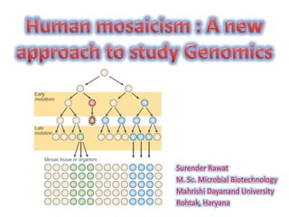
- 3. • GENOME - The genome is the genetic material of an organism. • MOSAICISM - Mosaicism is a condition in which cells within the same person have a different genetic makeup. • The phenomenon was discovered by Curt Stern. • In 1936, he demonstrated that recombination, normal in meiosis, can also take place in mitosis. • When it does, it results in somatic (body) mosaics. These are organisms which contain two or more genetically distinct types of tissue.
- 5. • Named after the intricate images created by the craftsman from small pieces of coloured tiles. • The coexistence of two or more genetically distinct cell populations derived origina lly from a single zygote. • Mosaics may arise at any stage of development, from the two- cell stage onward, or in any tissue which actively proliferates thereafter. • The phenomenon is commonly observed in many species of animals and plants and may be caused by somatic mutation or chromosomal nondisjunction. • An individual animal or plant may exhibit mosaicism, or it may occur in a culture of a single cell- or tissue-type obtained from an individual.
- 6. When eye colours vary between the two eyes, or within one or both eyes, the condition is called heterochromia iridis (= 'different coloured iris'). Changes may take place in the dividing cells leading up to iris formation in the embryo.
- 7. • These females are heterozygous for the X-linked colour genes: the genes for their coat colours are carried on the X chromosome. • X-inactivation causes groups of cells to carry either one or the other X- chromosome in an active state.
- 8. FEMALE MALE MOSAIC GYNANDROMORPH • A gynandromorph is an organism that contains both male and female characteristics. • While the organism is only a few cells large, one of the dividing cells does not split its sex chromosomes typically. • This leads to one of the two cells having sex chromosomes that cause male development and the other cell having chromosomes that cause female development.
- 9. • Use of aCGH and SNP chips has allowed detection of events that were missed by karyotyping • Scientists analyzed the genomes of a bunch of iPSC lines and then compared them to the “parental” fibroblast cells from which they had been made CNV arose specifically during the iPSC production process CNVs varied from one cell to another even though all the cells came from a single person. • Mosaicism for small for small intragenic CNV was detected in 10% of 30 molecularly diagonised • Subjects and extensive genomic CNVs have been found in clonal isolates of embryonic stem cell. • In addition, varying levels of mosaicism have reported in somatic human tissues including the skin, Brain, blood and induced pluripotent stem cells
- 12. • Mosaicism may result from: – Unusual events in cell division (mitosis). – A gene mutation during development • De novo pre zygotic mutation • De novo post zygotic mutation – A chromosomal mutation during development – X-inactivation: one X chromosome is randomly switched off in cells of a female mammal – Numerical or structural abnormalities of chromosomes • Deletion • Aneuploidy
- 13. • Chimerism — the presence of two or more cell lines that are derived from different zygotes in an organism . • A 52-year-old woman from Boston was told that she was not the biological mother of her children, after years of raising them. • The woman who claimed to have given birth to her children had a set of genes completely different from those of two of her three children. but the DNA of the children matched their father’s DNA. • After working for nearly two years on the case, doctors came up with an answer to the problem: the woman was a “human chimera.”
- 14. MOSAICS • Cells in the organism have same genetic origins • Nearly all loci are identical in the different cell populations as all cells are derived from the same zygotic genotype • Somatic mutations make us all mosaic CHIMERA • A single organism composed of genetically distinct cells. • In chimerism there are divergent genotypes all across the genome. • Chimeras can be artificially produced: organ transplants, chimera of diffferent animals
- 15. • Cancer • Heterochromia iridis • Lines of Blaschko • Neurofibromatosis type 1 • Proteus syndrome • Osteogenesis imperfecta type II
- 16. • The non-germ cells of the body are heterogeneous: some have a mutation and some do not. • The person may or may not be affected by the disorder caused by that mutation. • Individuals will express the phenotype depending on how many and which cells are affected. • Typically, individuals with somatic mosaicism exhibit a milder phenotype since only a proportion of cells contain the mutation and/or because the mutation is confined to a finite segment of the body. • Clinical Information on Diseases: – Cancer – Down Syndrome - Lines of Blaschko
- 17. • A classic example of somatic mosaicism. • Cancer arises when cells grow uncontrollably, which can be caused by mutations in genes that prevent the cell from properly functioning within the cell cycle or prevent it from cell death at the proper time. • Most cancers occur sporadically, meaning they are not due to an inherited predisposition. • Some inherited cancer syndromes include breast and ovarian cancer (BRCA1/2 genes), colon cancer, retinoblastoma, and thyroid cancer. • Since these mutations are only present in the cancer cells and not the rest of the cells in the body, the individual has somatic mosaicism for these mutations.
- 18. • Blaschko lines or the lines of Blaschko are thought to represent pathways of epidermal cell migration and proliferation during the development of the fetus. • These lines are invisible but many inherited and acquired diseases of skin manifest themselves according to these patterns creating the visual appearance of these lines.
- 19. • The diploid germ cell precursors in the gonad are heterogeneous: some have a mutation and some do not. • A mosaic germline mutation is significant because it can be passed to offspring. • Germline mosaicism can be observed with any inheritance pattern, but it is most commonly seen with autosomal dominant and X-linked disorders. • Most individuals are unaware they possess a germline mutation until they have children that are affected. • Clinical Information on Diseases: – Osteogenesis imperfecta – Ovarian dysgenesis
- 21. • Sometimes known as brittle bone disease • Bone disorder characterized by brittle bones that are prone to fracture. • People with OI are born with defective connective tissue, or without the ability to make it, usually because of a deficiency of Type-I collagen. • Eight types of OI can be distinguished. Most cases are caused by mutations in the COL1A1 and COL1A2 genes. • Severe and usually lethal in the perinatal period • Most cases die within the first year of life due to respiratory failure or intracerebral hemorrhage
- 22. • A combination of germline and somatic mosaicism • Example is Neurofibromatosis type 2 (NF2) is an autosomal dominant cancer syndrome • Symptoms are dizziness; headache; facial weakness, numbness, or pain; ringing in the ears and progressive hearing loss. • This disease is caused by inactivating mutations in the NF2 tumour-suppressor gene, located in 22q12, that normally gives rise to a product called Merlin. • This peptide is thought to have a tumor-suppressive function • Merlin's deficiency can result in unmediated progression through the cell cycle due to the lack of contact-mediated tumour suppression, sufficient to result in the tumors characteristic of NF II.
- 23. • Recently, a range of disorders has been shown to be caused by mosaicism for point mutations, primarily using NGS technologies • Non-overgrowth mosaic disorders like Benign keratinocytic epidermal nevi, which have been shown to be caused by mutations in fibroblast growth factor receptor 3 (FGFR3), phosphatidylinositol-4, - 5-bisphosphate 3-kinase and different RAS family members. • A series of mosaic overgrowth disorders was molecularly delineated, beginning with Proteus syndrome, which was shown to be due to AKT1 mutations
- 24. • Causes skin overgrowth and a typical bone development, often accompanied by tumours over half the body. • Proteus syndrome results from a mutation in the AKT1 gene. • The AKT1 gene helps regulate cell growth and division (proliferation) and cell death. • A mutation in this gene disrupts a cell's ability to regulate its own growth, allowing it to grow and divide abnormally. • Increased cell proliferation in various tissues and organs leads to the abnormal growth characteristic of Proteus syndrome. • Studies suggest that an AKT1 gene mutation is more common in groups of cells that experience overgrowth than in the parts of the body that grow normally
- 26. •Chromosome nondisjunction is probably the principal cause of chromosomal aberration, which in turn may lead to the d evelopment of mosaicism. •Chromosomal alterations (such as whole-chromosome aneuploidy, segmental aneuploidy (that is, aneuploidy of parts of chromosomes) and structural alterations) have been historically identified by cytogenetic analyses
- 27. • Mosaicism can result when that genetic lesion is completely or partially reverted, perhaps driven by selective pressure, in a subset of cells in the multicellular organism. • For example, the phenomenon of revertant mosaicism has recently been described, in which a mutation was spontaneously directly corrected in a subset of cells in an affected organ. • This phenomenon has been demonstrated in blood, muscle, liver and skin, of which skin is most dramatic, as the normal patches can be directly visualized among the abnormal patches.
- 28. • Recent technological advances have provided us with tools to assess mosaicism on a much broader scale, in many disorders and in diverse tissues, with increasing resolution to detect smaller-scale mutations • Different approaches used to study mosaicism include – Cytogenetics – Microarray – DNA sequencing
- 29. • Cytogenetic analysis by study of banded metaphase chromosomes or by fluorescence in situ hybridization (FISH) is carried out in a cell-by-cell manner, and recognition of mosaicism is therefore fairly straightforward. • Mosaicism is recognized when cells within an individual are found to have divergent chromosome contents, such as monosomic or trisomic chromosomes in the karyotype of one cell and a normal karyotype in another. • The cytogenetic detection of low-level mosaicism is challenging, as a sufficient number of cells must be counted
- 30. • Beginning in 2005, microarray-based techniques began to replace cytogenetic testing, with the introduction first of array-based comparative genomic hybridization (aCGH), which can analyse genomic copy number variants (CNVs). • It is followed by genome-wide SNP arrays, which can analyse both SNPs and CNVs. • The advantages of array-based testing (which typically analyses DNA extracted from whole blood) for mosaicism detection include: – many cells are analysed simultaneously; – different cell types are analysed; – cells of all cell cycle phases are analysed; – samples do not require culturing, which might itself cause mutations • SNP arrays are much more sensitive than aCGH for mosaicism detection.
- 31. DNA sequencing • Sanger-based sequencing can be extremely effective for many applications, but it is a low throughput technique. • NGS is a high-throughput technique that is massively used nowadays. • These digital data are more amenable to statistical approaches to distinguish mosaicism from sequencing errors. • It also does not require genetic mapping data to identify causative genetic variants. • For this reason, subtractive informatic approaches — in which two samples are informatically processed to identify only those positions in the genome that differ — can be coupled to NGS to identify mosaic alterations.
- 32. • The recognition of mosaic disorders is rapidly increasing. • It can affect somatic tissues, the germ line or both. • It can arise at various stages of development or adult life and can be caused by mutation from a normal genotype to a variant genotype or from a variant genotype to a normal genotype. • Recent advances in genomic technologies have enhanced our ability to study the widespread nature of mosaicism in both healthy individuals and in patients with a wide range of disorders. • Recent insights include: – Identification of disease genes for disorders that are seen only as mosaics as they are lethal when constitutional – the identification of chimerism by whole-genome tools – recognition of the changing rates of mosaicism with increasing age represents a window on the ageing process.
- 33. • Abyzov, A. et al. Somatic copy number mosaicism in human skin revealed by induced pluripotent stem cells. Nature 492, 438–442 (2012). • Biesecker and Nancy . A genomic view of mosaicism and human disease , Nature, 2013 • Youssoufian & Pyeritz MECHANISMS AND CONSEQUENCES OF SOMATIC MOSAICISM IN HUMANS, Nature, 2002 • Mardis, E. R. The impact of next-generation sequencing technology on genetics. Trends Genet. 24, 133–141 (2008). • Happle, R. Mosaicism in human skin. Understanding the patterns and mechanisms. Arch. Dermatol. 129, 1460–1470 (1993). • Lindhurst, M. J. et al. A mosaic activating mutation in AKT1 associated with the Proteus syndrome. New Engl. J. Med. 365, 611–619 (2011). • Callum, P. et al. Gonosomal mosaicism for an NF1 deletion in a sperm donor: evidence of the need for coordinated, long-term communication of health information among relevant parties. Hum. Reprod. 27, 1223–1226 (2012). • Maertens, O. et al. Molecular dissection of isolated disease features in mosaic neurofibromatosis type 1. Am. J. Hum. Genet. 81, 243–251 (2007). • Conlin, L. K. et al. Mechanisms of mosaicism, chimerism and uniparental disomy identified by single nucleotide polymorphism array analysis. Hum. Mol. Genet. 19, 1263–1275 (2010). • Hook, E. B. Exclusion of chromosomal mosaicism: tables of 90%, 95% and 99% confidence limits and comments • Shin, S. Y., Yoo, H. W., Lee, B. H., Kim, K. S. & Seo, E. J. Identification of the mechanism underlying a human chimera by SNP array analysis. Am. J. Med. Genet. A 158, 2119–2123 (2012). • Conlin, L. K. et al. Utility of SNP arrays in detecting, quantifying, and determining meiotic origin of tetrasomy 12p in blood from individuals with Pallister–Killian syndrome. Am. J. Med. Genet. A 158, 3046– 3053 (2012).
Notes de l'éditeur
- Curt Stern (30 August 1902 – 23 October 1981) was a German-American geneticist.[1] Born in Hamburg, Germany, he studied zoology at the University of Berlin, receiving his PhD in 1923 at the age of 21. He was selected for a fellowship to study at Columbia University, then the site of Thomas Hunt Morgan's famous drosophila fly room. In 1936, he demonstrated that recombination can also take place in mitosis resulting in somatic mosaics, organisms that contain two or more genetically distinct types of tissues.[2] He later demonstrated that there were multiple genes on the Drosophila Y chromosome, and described the mechanism of dosage compensation.
- Greek "gyne" female and "andro" male, For example, an XY cell undergoing mitosis duplicates its chromosomes, becoming XXYY. Usually this cell would divide into two XY cells, but in rare occasions the cell may divide into an X cell and an XYY cell. If this happens early in development, then a large portion of the cells are X and a large portion are XYY. Since X and XYY dictate different sexes, the organism has tissue that is female and tissue that is male
- “geep” chimera comprised of goat and sheep cells
- Approximately 2-4% of individuals with Down syndrome are mosaic for cells that have three copies of chromosome 21. Neurofibromatosis, in its mosaic form, is termed segmental NF. This means that only one part of the body is affected. This is most likely due to a mutation in the NF gene that arose very early on in development and led to a proportion of affected cells in the same area of the body.
- Typically, a person with only germline mosaicism will not be affected with the disorder caused by the mutation because the mutation is not in the other cells of the body. Genetic testing using blood or tissue samples (other than germline tissue) from individuals who only have a germline mutation will be negative for the mutation.
- In a normal cell, the concentrations of active (dephosphorylated) merlin are controlled by processes such as cell adhesion (which would indicate the need to restrain cell division).
- Proteus syndrome causes an overgrowth of skin, bones, muscles, fatty tissues, and blood and lymphatic vessels.
- These include its limited throughput and the fact that both alleles of an autosomal locus are sequenced concurrently and are displayed as analogue electropherograms, from which the often subtle contribution from mosaic alleles cannot be accurately determined (