

Recommandé
Contenu connexe
Tendances
Tendances (20)
Similaire à Penicillium
Similaire à Penicillium (20)
Plus de SyedaFari2
Dernier
Dernier (20)
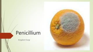
Penicillium
- 2. Taxonomic position Kingdom : Fungi Division : Ascomycota Class : Eurotiomycetes Order : Eurotiales Family :Trichocomaceae
- 3. Occurrence They are • Common to occur • Cosmopolitan genus • Commonly called as green mold • Found in variety of habitat citrus fruits, jellies , foot stuffs , old leather , paper , soil etc. • Conidia of Penicillium are present everywhere in air and soil • It was accidently discovered by Alexander Fleming , who was working on bacteria and his culture got contaminated by Penicillium notatum
- 4. Plant body - Mycelium It is well developed Profusely branched Composed of colorless , slender , tubular, branched and septate hyphae Overall hyphae spreads on surface but some hyphae penetrate inside the substratum to absorb nourishment The mycelium becomes colored due to production of colored conidia
- 5. Cell structure Hyphae is septate Each cell is uninucleate The cell wall is microfibrillar , and in Penicillium notatum , it is of 3 layers The outer layer is made up of glucan The next layer is made up of proteins the 3rd layer is made up if chitin fibrils embedded in granular matrix The innermost layer is made up of pectic and hemicellulose The plasma membrane surrounds the cytoplasm that contains organelles like mitochondria , ribosomes and endoplasmic reticulum in embedded form The reserve food is in form of oil globules The adjacent cells are connected by a pore present in septa of their walls
- 7. Reproduction It reproduces by Vegetative reproduction Asexual reproduction Sexual reproduction
- 8. Vegetative reproduction • It takes place by fragmentation • During fragmentation the hyphae breaks into short fragments • That grows by repeated division into new mycelium
- 9. Asexual reproduction It takes place by formation of conidia Conidia are produced on special hyphae called conidiophore The conidia are erect , brown and broom like in fashion Each branch bears short branch at its tip , called metulae at each metula , there are bottle shaped sterigmata Conidia are produced in chains at these sterigmata Whole group of metulae and sterigmata are called -- pencillus
- 10. Structure of conidia Each conidia is tiny, uninucleate, spore like structure, May be globose and ovoid in shape The spore wall is pigmented and it is made up of two layers The outer layer is thick, pigmented is called exine The inner layer smooth, and thin called as intine Inside the spore wall plasma membrane is present , which encloses the mitochondria , ribosomes in embedded form The reserve food is in oil globules
- 11. Development of conidia Each sterigmata is uninucleate During reproduction the nucleus conidia divided into two One daughter nucleus along with some cytoplasm migrate into the slightly swollen tip of sterigmata The portion is then cut off from the rest of the sterigmata by formation of cross walls The protoplast then contract slightly and secretes its on wall The process repeats several times and chain of conidia are formed Each newly formed conidia is uninucleate but later own its nucleus divided several times , making conidia multinucleated
- 12. Germination of conidium The conidia germinates by producing hyphae directly on falling on suitable substrate
- 14. Oidia formation Penicillium hyphae when grown in sugar medium , it divides into small, uninucleate segments which become rounded and separate into thin walled spore like structure called oidia or oidiospore These increase in number by budding , this is called torula stage , this is important in bringing the fermentation of sugar into alcohol The oidia when transferred to solid medium , germinates to form new mycelium
- 15. Torula stage
- 17. Sexual reproduction It is the perfect state of Penicillium All the species are homothallic The sexual reproduction is oogamous Male sex organ is called antheridia Female sex organ is called ascogonia
- 18. Ascogonium It’s a long , erect, multinucleate , tubular structure , with curved upper end It arises from a uninucleate , septate hypha as a finger – like lateral outgrowth which elongates in to an ascogonium The nucleus of the ascogonium divides many times mitotically to produce 32 to 64 nuclei.
- 19. Antheridium While the ascogonium is developing , A slender uninucleate branch originates from a cell of the same hypha adjacent to the developing ascogonium , or from neighboring hypha . This is called antheridial branch It grows up and coils around the ascogonium The tip of the antheridial branch swells up and cut off from rest of the branch to form a uninucleate antheridium
- 20. Fertilization The tip of the antheridia comes into contact with the wall of the ascogonium and the wall of contact between the two dissolves to form a pore The protoplast of the gametangia come in contact with each other through this pore The antheridial and ascogonial nuclei arrange themselves in pairs . Each pair is called a dikaryon
- 21. Cleistothecium development As soon as the septation of ascogonium and development of ascogenous hyphae starts The sterile hyphae present at the base of ascogonium grow upwards and surround the sexual apparatus These hyphae become interwoven to form a hollow , ball like structure , the peridium wall of ascocarp The ascocarp continues to grow even after the maturation of ascospores The ascocarp is without an opening , therefore it is called Cleistothecium
- 23. Dispersal and germination of ascospores The asci are evanescent - - dissolve soon after the formation of ascospores, leaving the ascospores free in the ascocarp. The ascospores are released by the decay of the wall of the ascocarp Ascospores germinate by the formation of a germ tube to form a new mycelium