
Recommandé
Recommandé
Contenu connexe
Tendances
Tendances (20)
Similaire à Cardiac Amyloidosis - Dr. Akif
Similaire à Cardiac Amyloidosis - Dr. Akif (20)
Plus de akifab93
Plus de akifab93 (20)
Dernier
Dernier (20)
Cardiac Amyloidosis - Dr. Akif
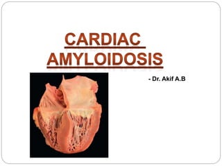
- 1. - Dr. Akif A.B
- 3. AMYLOIDOSIS Group of protein-folding disorders in which >1 organ is infiltrated by proteinaceous deposits known as amyloid The deposits are derived from 1 of several amyloidogenic precursor proteins, and the prognosis of the disease is determined both by the organ(s) involved and the type of amyloid Amyloid involvement of the heart (cardiac amyloidosis) carries the worst prognosis of any involved organ, and light-chain (AL) amyloidosis is the most serious form of the disease
- 4. EPIDEMIOLOGY There is a slight male predominance of AL cardiac amyloidosis Disease generally presents from the fifth to seventh decade, although it may occur at all ages from the fourth decade onward Clinical evidence of cardiac involvement is seen in 22% to 34% of AL patients
- 5. AMYLOIDOSIS IN HEART Transthyretin amyloidosis (ATTR) AL amyloidosis Secondary Amyloidosis
- 7. Light-Chain Amyloidosis (AL or Primary) Caused by the proliferation of an abnormal clone of plasma cells that overproduce lambda, or less commonly, kappa immunoglobulin light-chain usually associated with plasma cell dyscrasia, but usually not associated with multiple myeloma Cardiac involvement in AL amyloidosis almost always occurs in the setting of another significant organ involvement, and that isolated cardiac disease is present in <5% of cases Heart disease occurs in one-third to half of AL patients, and heart failure tends to progress rapidly once the heart is affected and has a very poor prognosis
- 9. Organ Involvements in AL- Amyloidosis It most commonly affects the kidney, resulting in nephrotic syndrome, with clinical cardiac involvement being the second most common presenting manifestation It also affects liver, peripheral/autonomic nerves, soft tissue, and the gastrointestinal system Cerebral involvement does not occur in AL amyloidosis
- 10. Amyloidosis Vs Multiple Myeloma In contrast to myeloma, most patients with AL amyloidosis have <20% of plasma cells in the marrow, and the manifestations of the disease are due to the formation of amyloid from the abnormal circulating free light chains Approximately 5% to 10% of patients with AL amyloidosis will have evidence of overt multiple myeloma, and vice vers
- 11. Transthyretin-associated amyloidoses (ATTR) TTR is a transport protein of thyroid hormone and retinol (vitamin A); hence, the name, transthyretin These include 2 types of amyloidosis: Wild type transthyretin Mutated type of transthyretin-associated amyloidosis (ATTRm)
- 12. Wild-Type Transthyretin (ATTR) Also known as senile systemic amyloidosis (SSA) The main involved organ is the heart This type is almost exclusively found in elderly men (male: female=20:1~50:1) and the symptoms are slowly progressive
- 13. Mutated Type Transthyretin Also called “familial type of amyloid polyneuropathy (FAP)” This type is caused by mutant transthyretin produced in the liver It affects peripheral/autonomic nerves and the heart Autosomal dominant
- 14. AL Vs ATTR Amyloidosis The pathophysiology of cardiac AL amyloidosis differs from that of ATTR amyloidosis Severity of heart failure in AL amyloidosis is more severe than in TTR amyloidosis, despite higher left ventricular (LV) mass in the latter Lack of change in standard echocardiographic parameters after successful hematologic treatment This led to the suggestion that AL amyloidosis might have both toxic and infiltrative components
- 15. Infiltration of the myocardium causes physical cellular damage and restrictive pathophysiology Circulating cardiotoxic light chains produce cellular damage through various pathways In other forms of cardiac amyloid, such as TTR, the sole mechanism of cardiac
- 16. It appears that there are different deposition patterns in TTR and AL amyloidosis Predominance of diffuse peri-cellular, endocardial, and arterial and/or arteriolar deposits in AL amyloidosis and nodular deposits in ATTR
- 17. Secondary amyloidosis This type of amyloidosis is caused by serum amyloid A and also called AA amyloidosis It is seen in association with juvenile or adult rheumatoid arthritis and other rheumatic disorders such as ankylosing spondylitis, as well as with inflammatory bowel disease Hepatic and renal amyloid deposition dominates the clinical features, and clinical heart disease related to cardiac amyloidosis is rare Treatment is to heal the underlying inflammatory process
- 19. MEDIAN SURVIVAL AL 24months (6months if HF at diagnosis) ATTRwild 57months ATTRmutated 31months
- 21. The nonbranching fibrils of amyloid are composed not only of fragments of the precursor protein, but also contain proteoglycans and serum amyloid P (SAP) They are generally extremely resistant to degradation, although evidence suggests that amyloid does slowly resorb from the body once fibrillogenesis has been halted
- 22. The amyloid deposits expand the extracellular space and stiffen the heart without producing compensatory dilation, which results in a restrictive pathophysiology involving both ventricles Atrial amyloid infiltration is almost always present and frequently causes atrial contractile dysfunction Amyloid deposition also occurs on valves and perivascularly
- 25. The most common early manifestation of cardiac amyloidosis of any type is dyspnea on exertion, which progresses relatively rapidly Dyspnea is due to LV diastolic dysfunction. However, the atria, although they do dilate, are also abnormally stiff, and likely contribute to dyspnea on
- 26. Often followed by peripheral edema and ascites Peripheral edema in AL amyloidosis may also be due to hypoalbuminemia associated with amyloid-related nephrotic syndrome Atrial arrhythmia may be the initial manifestation of the disease
- 27. Atrial infiltration with amyloid deposits results in atrial dysfunction, and thrombus formation may occur, even in this setting of sinus rhythm Thromboembolism may thus be an early manifestation of the disease
- 28. MICROVASCULAR DYSFUNCTION Coronary microvascular dysfunction is ubiquitous in AL and ATTR CA Manifest as angina in the absence of obstructive coronary artery disease Microvascular dysfunction is not limited to the heart, and occasionally, causes jaw or buttock claudication, particularly in AL amyloidosis Peri-vascular amyloid deposits, as well as autonomic dysfunction , may account for microvascular dysfunction, which can be aggravated by thick walls and high LV filling pressure
- 29. ADVANCE DISEASE MANIFESTATIONS Exertional syncope Most likely due to a low and fixed cardiac output, can occur, and this has a particularly poor prognosis Jaw claudication, leg claudication, and angina may all occur due to small vessel amyloid deposition
- 31. In AL amyloidosis, signs of the noncardiac disease are commonly present Approximately 10% of patients have macroglossia, which may vary from obvious tongue enlargement to subtle tooth indentation of the tongue
- 32. MACROGLOSSIA
- 33. Periorbital bruising in the setting of heart failure is almost pathognomonic of AL amyloidosis Because it may be subtle, the eyelids should be inspected for small bruises
- 35. NAIL DYSTROPHY
- 37. Jugular venous pressure frequently reveals an inspiratory rise (Kussmaul sign), a sign that is shared with patients with constrictive pericarditis Unlike severe heart failure caused by most other etiologies, a third heart sound is uncommon in cardiac amyloidosis, as is a fourth heart sound Valvular dysfunction due to amyloidosis is rarely severe, although on occasion, tricuspid regurgitation can be severe
- 38. Hepatomegaly usually reflects congestion, but an enlarged liver may also be due to amyloid infiltration in patients with AL amyloidosis When hepatic infiltration occurs, the liver is hard and nonpulsatile, and quite distinct from the congested liver of heart failure
- 39. Nephrotic syndrome is a common manifestation of AL amyloidosis It is a clinical clue to the presence of hypoalbuminemia in a patient with anasarca is extensive edema in the absence of an elevated jugular venous pressure
- 40. Blood Pressure The blood pressure in suspected cardiac amyloidosis is frequently low, due to a combination of reduced cardiac output and low peripheral tone It should always be measured both supine and standing, because autonomic nervous system dysfunction may manifest as significant postural hypotension
- 41. PERIPHERAL NEUROPATHY Peripheral neuropathy is also a feature of both AL amyloidosis and familial TTR amyloidosis It is symmetric, predominantly sensory, and may be missed if the neurological examination is performed casually
- 42. Carpel Tunnel Syndrome Numbness and pain in the hands may be due to carpal tunnel syndrome, and a history of surgery for carpal tunnel syndrome may precede the onset of heart failure by 7 or 8 years Commonly associated with ATTR wild type
- 44. When to suspect Amyloidosis ? Non-diabetic nephrotic syndrome Non-ischaemic cardiomyopathy with an echocardiogram showing 'hypertrophy' Hepatomegaly or alkaline phosphatase elevation without imaging abnormality Peripheral neuropathy with MGUS or CIDP with autonomic features Atypical myeloma with monoclonal light chains and modest marrow plasmacytosis
- 45. Laboratory Radiological Serum and urine Immunofixation ECG Serum free light chains ECHO Biopsy (HPE) Cardiac MRI Mass Spectrometry Serum troponin, B-type natriuretic peptide, and beta-2- microglobulin should be performed in all patients as they provide prognostic information.
- 50. Cardiac amyloidosis is a condition in which the extracellular space of the heart is expanded by an amorphous, fibrillar proteinaceous material known as amyloid
- 51. Sulfated Alcian Blue Amyloid deposits are extensive and extracellular, compressing the cardiomyocytes This leads to myocardial dysfunction, due to both stiffening of the extracellular space and direct myocyte damage
- 52. The inset on the right shows amyloid surrounding a small vessel This can result in angina with normal- appearing epicardial coronary arteries seen on coronary angiography
- 53. WHAT IS THE GOLD STANDARD FOR THE DIAGNOSIS OF CA? Congo red staining of amyloid deposits showing apple-green birefringence under polarized light is the definitive way of determining the presence of amyloid in tissue This test does not determine the type of amyloid
- 54. Congo red stained section showing apple green birefringence of amyloid-like material (A-Amyloid material showing apple green birefringence, C-Connective tissue separating overlying epithelium-E)
- 55. Fat Pad Biopsy Sensitivity Type of Amyloidosis Sensitivity AL 84% ATTRwild 15% ATTRmutated 45%
- 56. Once amyloid deposits are found, typing is most accurately characterized (>98% sensitivity) by mass spectrometry
- 57. ECG in Amyloidosis Electrocardiogram showing very low limb lead voltage with indeterminate axis and poor precordial R-wave progression in AL amyloid cardiomyopathy Coronary angiography was normal, and the echocardiogram showed a thick left ventricle and no pericardial effusion. This voltage/left ventricular mass mismatch strongly suggests cardiac amyloidosis
- 58. The QRS is said to be low voltage when: The amplitudes of all the QRS complexes in the limb leads are < 5 mm; or The amplitudes of all the QRS complexes in the precordial leads are < 10 mm
- 60. ECHO in Amyloidosis Thick-walled ventricle Small left ventricular chamber volume Valve thickening Atrial enlargement and signs of elevated filling pressures with a restrictive diastolic filling Interventricular septal thickness of > 12 mm, in the absence of aortic valve disease or systemic hypertension RV wall thickness>7mm, elevated RA Pressure
- 61. Amyloidosis & LV Hypertrophy In either form of cardiac amyloidosis, the dominant imaging finding is the appearance of cardiac "hypertrophy The "hypertrophy" on imaging represents amyloid fibril deposition rather than myocyte hypertrophy/hyperplasia, which explains why ECG voltages are decreased rather than increased
- 67. Tissue Doppler and Speckle Tracking Can assist in differentiation from other causes of wall thickening, including hypertension and hypertrophic cardiomyopathy.
- 68. Two-dimensional (2D) strain mapping shows relative preservation of apical function Early clue to amyloidosis because it gives rise to a ‘bulls-eye’ pattern when the segmental strain is plotted, which is rare in other cardiomyopathies
- 69. Cardiovascular magnetic resonance CMR is typically considered in individuals with echocardiograms that are suspicious but not typical for amyloidosis or Individuals with high clinical suspicion, irrespective of echocardiogram results (gene- positive individuals with symptoms or at the age when disease onset is expected)
- 70. Standard CMR sequences for cardiac amyloidosis include cine images in the standard long-axis and short-axis views to study cardiac structure, systolic function, calculate LV wall thickness and mass late gadolinium enhancement imaging (about 5 min after injection of gadolinium) in axial, short-, and long- axis views optimal myocardial inversion time is assessed about 4 min after injection of gadolinium to identify specific patterns of myocardial nulling in amyloid as opposed to left ventricular myocardial hypertrophy
- 74. Scintigraphy with 123I-Labeled Serum Amyloid P Component Amyloid deposits contain amyloid P component, which is derived from the normal circulating protein SAP In patients with amyloidosis, SAP leaves the circulation and is deposited on the amyloid fibrils, presumably because of its specific calcium-dependent binding affinity for them Scintigraphy with 123I-SAP produces high-quality images because a high proportion of the tracer is deposited in amyloid and retained there for prolonged periods, whereas circulating SAP that has not been deposited is rapidly catabolized and excreted Retention of SAP in amyloid was highly specific and that the quantity of Radio-labeled SAP in each tissue correlated with the amount of amyloid present
- 76. NUCLEAR SCINTIGRAPHY Nuclear Scintigraphy Bone-avid, phosphate- based isotopes, including 99mTc-PYP (pyrophosphate) and 99mTcDPD (3,3- diphosphono-1,2-propanodiacarboxylic acid), have a specific avidity for ATTR amyloid deposits An international consensus document has confirmed the utility of the bone-avid compounds for the accurate identification of ATTR amyloidosis , and differentiation from AL amyloidosis or other wall thickening diseases
- 77. A 99mTc-PYP scan that does not show uptake, or shows mild uptake (Perugini grade 1), is completely consistent with AL amyloidosis Conversely, a bone scintigraphy scan (99mTc-PYP or 99mTc-DPD) with abnormal uptake (grade 2 or 3), in combination with a negative plasma cell dyscrasia evaluation has a specificity and positive predictive value of 100% for diagnosis of ATTR cardiac amyloidosis However, TTR genotyping must still be performed to differentiate wild-type from mutant ATTR cardiac amyloidosis Finally, there is increasing evidence that amyloid specific positron emission tomography (PET) tracers, including 18- F florbetapir , and the 11-C Pittsburgh B compound (PiB) , can identify cardiac amyloidosis, specifically the AL type
- 81. Systolic function Rapid filling Atrial contraction
- 87. Because ATTR amyloidosis is not a malignant process, chemotherapy has no role Although there are no currently approved disease-modifying medications for ATTR amyloidosis, multiple agents have been studied and/or are in late-phase trials These include the following: Diflunisal Tafamidis RNA interference approaches
- 89. Diflunisal This nonsteroidal anti-inflammatory drug that is approved for arthritis pain stabilizes the tetrameric form of transthyretin A randomized trial demonstrated slowing of disease progression among patients with polyneuropathy due to mutant ATTR amyloidosis Because nonsteroidal anti-inflammatory drugs are relatively contraindicated in HF, this is not likely to be a good option for ATTR cardiomyopathy.
- 90. Tafamidis This agent is approved in some parts of the world (Europe and Japan) for mutant ATTR amyloidosis causing polyneuropathy Transthyretin Tetramer stabilisation
- 91. GUIDELINES
- 92. RNA interference approaches Two agents that work via small-interfering RNA or RNA interference (decreasing production of transthyretin by the liver) are in Phase 3 trials for ATTR amyloidosis, including both polyneuropathy and cardiomyopathy forms
- 93. If heart involvement presents beforehand, cardiac amyloidosis may progress despite liver transplantation in some cases
- 97. Treatment of primary amyloidosis (AL) The goal of treatment for AL is suppression of the plasma cell clone responsible for the synthesis of the immunoglobulin light chain Interruption of light chain deposition allows the body to solubilise and eliminate the amyloid deposit This prevents further amyloid deposition, which would result in progressive organ failure
- 104. PROGNOSTIC MARKERS
- 105. Prognosis in amyloidosis is dependent primarily on the degree of cardiac involvement and In AL amyloidosis, on circulating light-chain levels Because chemotherapy options have greatly expanded in recent years, prognosis for AL amyloidosis has markedly improved as well, with the life expectancy of most patients, including many of those with significant cardiac involvement, measured in years rather than months Prognosis in ATTR amyloidosis is generally better than in AL amyloidosis, though both forms of the disease still carry a high annual mortality
- 107. Biomarkers can be used for patients with light chain amyloidosis and transthyretin amyloidosis for staging and prognosis Biomarkers can be used for patients with light chain amyloidosis to determine disease progression and response to therapies