SlideShare une entreprise Scribd logo
1  sur  45
Rhythm Analysis Step 1:	Calculate rate. Step 2:	Determine regularity. Step 3:	Assess the P waves. Step 4:	Determine PR interval. Step 5:	Determine QRS duration.
Step 1: Calculate Rate Option 1 Count the # of R waves in a 6 second rhythm strip, then multiply by 10. Interpretation? 3 sec 3 sec 9 x 10 = 90 bpm
Step 1: Calculate Rate Option 2  Find a R wave that lands on a bold line. Count the # of large boxes to the next R wave. If the second R wave is 1 large box away the rate is 300, 2 boxes - 150, 3 boxes - 100, 4 boxes - 75, etc.  (cont) R wave
Step 1: Calculate Rate Option 2  Interpretation? 300 150 100 75 60 50 Approx. 1 box less than 100 = 95 bpm
What is the heart rate?
Step 2 : Determine Regularity Regular: If the difference between the longest R-R interval in the ECG and the shortest R-R interval is less than 0.12 second Irregular: If the difference between the longest R-R interval in the ECG and the shortest R-R interval is greater than 0.12 second
Step 2: Determine regularity Look at the R-R distances (using a caliper or markings on a pen or paper). Interpretation? R R Regular
Step 3: Assess the P waves Are there P waves? Do the P waves all look alike? Do the P waves occur at a regular rate? Is there one P wave before each QRS? Interpretation? Normal P waves with 1 P wave for every QRS
Step 4: Determine PR interval Normal: 0.12 - 0.20 seconds. 		        (3 - 5 boxes) Interpretation? 0.12 seconds
Step 5: QRS duration Normal: 0.04 - 0.12 seconds.                  (1 - 3 boxes) Interpretation? 0.08 seconds
Rhythm Summary Rate				90-95 bpm	 Regularity			regular P waves				normal PR interval			0.12 s QRS duration			0.08 s Interpretation? Normal Sinus Rhythm
Normal Sinus Rhythm (NSR) The electrical impulse is formed in the SA node and conducted normally. This is the normal rhythm of the heart; other rhythms that do not conduct via the typical pathway are called arrhythmias.
NSR Parameters Rate			60 - 100 bpm	 Regularity		regular P waves			normal PR interval		0.12 - 0.20 s QRS duration		0.04 - 0.12 s
Cardiac Arrhythmias and Their Electrocardiographic Interpretation Abnormal rhythmicity of the pacemaker  Shift of the pacemaker from the sinus node to another place in the heart  Blocks at different points in the spread of the impulse through the heart  Abnormal pathways of impulse transmission through the heart  Spontaneous generation of spurious impulses in almost any part of the heart
Tachycardia: The term "tachycardia" means fast heart rate, usually defined in an adult person as faster than 100 beats per minute This electrocardiogram is normal except that the heart rate The general causes of tachycardia include increased body temperature, stimulation of the heart by the sympathetic nerves, or toxic conditions of the heart Abnormal Sinus Rhythms
130 bpm ,[object Object]
Regularity?regular ,[object Object],normal ,[object Object],0.16 s ,[object Object],0.08 s Interpretation? Sinus Tachycardia
Sinus Tachycardia Deviation from NSR 		- Rate			> 100 bpm
Sinus Tachycardia SA node is depolarizing faster than normal, impulse is conducted normally.
Bradycardia The term "Bradycardia" means a slow heart rate, usually defined as fewer than 60 beats per minute The cause of Bradycardia is Vagal Stimulation
30 bpm ,[object Object]
Regularity?regular ,[object Object],normal ,[object Object],0.12 s ,[object Object],0.10 s Interpretation? Sinus Bradycardia
Sinus Bradycardia Deviation from NSR 		- Rate			< 60 bpm
Sinus Bradycardia SA node is depolarizing slower than normal, impulse is conducted normally (i.e. normal PR and QRS interval).
Atrioventricular Block Delay or blocks conduction from the atria to the ventricles. Causes are ischemia, extreme stimulation of the heart by vagus nerves, inflammation of the AV node or A-V bundle
1st Degree AV Block Prolonged conduction delay in the AV node or Bundle of His.
1st Degree AV Block 60 bpm ,[object Object]
Regularity?regular ,[object Object],normal ,[object Object],0.36 s ,[object Object],0.08 s Interpretation? 1st Degree AV Block
1st Degree AV Block Deviation from NSR PR Interval		> 0.20 s
2nd Degree Block When conduction through the A-V bundle is slowed enough to increase the P-R interval to 0.45 second. There will be P wave but no QRS-T wave There is drop beat of the ventricles
2nd Degree Block 50 bpm ,[object Object]
  Regularity?regularly irregular ,[object Object],nl, but 4th no QRS ,[object Object],lengthens ,[object Object],0.08 s Interpretation? 2nd Degree AV Block, Type I
2nd Degree AV Block Deviation from NSR PR interval progressively lengthens, then the impulse is completely blocked (P wave not followed by QRS).
3rd Degree AV Block Deviation from NSR The P waves are completely blocked in the AV junction; QRS complexes originate independently from below the junction.
3rd Degree AV Block There is complete block of conduction in the AV junction, so the atria and ventricles form impulses independently of each other. Without impulses from the atria, the ventricles own intrinsic pacemaker kicks in at around 30 - 45 beats/minute.
3rd Degree AV Block 40 bpm ,[object Object]
  Regularity?regular ,[object Object],no relation to QRS ,[object Object],none ,[object Object],wide (> 0.12 s) Interpretation? 3rd Degree AV Block
Premature Contractions  A premature contraction is a contraction of the heart before the time that normal contraction would have been expected. This condition is also called extrasystole, premature beat, or ectopic beat The causes are local areas of ischemia, toxic irritation of A-V node, purkinje system caused by drugs or caffeine
Premature Atrial Contractions Deviation from NSR These ectopic beats originate in the atria (but not in the SA node), therefore the contour of the P wave, the PR interval, and the timing are different than a normally generated pulse from the SA node.
Premature Atrial Contraction 70 bpm ,[object Object]
Regularity?occasionally irreg. ,[object Object],2/7 different contour ,[object Object],0.14 s (except 2/7) ,[object Object],0.08 s Interpretation? NSR with Premature Atrial Contractions
Premature Ventricular Contraction Deviation from NSR Ectopic beats originate in the ventricles resulting in wide and bizarre QRS complexes. The QRS complex is usually prolonged. The reason is that the impulse is conducted mainly through slowly conducting muscle of the ventricles
Premature Ventricular Contraction 60 bpm ,[object Object]

Contenu connexe

Tendances

ELectrophysiology basics part4
ELectrophysiology basics part4ELectrophysiology basics part4
ELectrophysiology basics part4salah_atta
 
Electrophysiology study protocol
Electrophysiology study protocolElectrophysiology study protocol
Electrophysiology study protocolSatyam Rajvanshi
 
Basics of ecg interpretation by dr sai
Basics of ecg interpretation by dr saiBasics of ecg interpretation by dr sai
Basics of ecg interpretation by dr saiSainath Hiwrale
 
Cardiac+Electrophysiology
Cardiac+ElectrophysiologyCardiac+Electrophysiology
Cardiac+Electrophysiologydhavalshah4424
 
How to differentiate VT from SVT
How to differentiate VT from SVTHow to differentiate VT from SVT
How to differentiate VT from SVTHaneen Hassan
 
Cardiac pacemakers part ii
Cardiac pacemakers part iiCardiac pacemakers part ii
Cardiac pacemakers part iisalah_atta
 
P wave axis and escape rhythms
P wave axis and escape rhythmsP wave axis and escape rhythms
P wave axis and escape rhythmsEMSMedic79
 
Basic EP study part 2
Basic EP study part 2Basic EP study part 2
Basic EP study part 2SolidaSakhan
 
How to perform and interpret entrainment pacing Basics
How to perform and interpret entrainment pacing BasicsHow to perform and interpret entrainment pacing Basics
How to perform and interpret entrainment pacing BasicsBenjamin Jacob, CEPS (IBHRE)
 
WPW EP evaluation
WPW EP evaluationWPW EP evaluation
WPW EP evaluationRohitWalse2
 
Anatomy & physiology for the EP professional part II 8.4.14
Anatomy & physiology for the EP professional part II 8.4.14Anatomy & physiology for the EP professional part II 8.4.14
Anatomy & physiology for the EP professional part II 8.4.14lpesbens
 
Arrhythmia diagnosis and management
Arrhythmia diagnosis and managementArrhythmia diagnosis and management
Arrhythmia diagnosis and managementMuhammed Arslan
 

Tendances (20)

ELectrophysiology basics part4
ELectrophysiology basics part4ELectrophysiology basics part4
ELectrophysiology basics part4
 
Electrophysiology study protocol
Electrophysiology study protocolElectrophysiology study protocol
Electrophysiology study protocol
 
Basics of ecg interpretation by dr sai
Basics of ecg interpretation by dr saiBasics of ecg interpretation by dr sai
Basics of ecg interpretation by dr sai
 
Avrt vz vt
Avrt vz vtAvrt vz vt
Avrt vz vt
 
Cardiac+Electrophysiology
Cardiac+ElectrophysiologyCardiac+Electrophysiology
Cardiac+Electrophysiology
 
How to differentiate VT from SVT
How to differentiate VT from SVTHow to differentiate VT from SVT
How to differentiate VT from SVT
 
Cardiac pacemakers part ii
Cardiac pacemakers part iiCardiac pacemakers part ii
Cardiac pacemakers part ii
 
Intracardiac Electrograms
Intracardiac ElectrogramsIntracardiac Electrograms
Intracardiac Electrograms
 
P wave abnormalities in ECG
P wave  abnormalities in ECGP wave  abnormalities in ECG
P wave abnormalities in ECG
 
Electrophysiology AVRT
Electrophysiology AVRTElectrophysiology AVRT
Electrophysiology AVRT
 
Arrythmias in ICCU
Arrythmias in ICCUArrythmias in ICCU
Arrythmias in ICCU
 
FRACTIONAL FLOW RESERVE
FRACTIONAL FLOW RESERVEFRACTIONAL FLOW RESERVE
FRACTIONAL FLOW RESERVE
 
Heart block
Heart blockHeart block
Heart block
 
SVT-Alogarythm
SVT-AlogarythmSVT-Alogarythm
SVT-Alogarythm
 
P wave axis and escape rhythms
P wave axis and escape rhythmsP wave axis and escape rhythms
P wave axis and escape rhythms
 
Basic EP study part 2
Basic EP study part 2Basic EP study part 2
Basic EP study part 2
 
How to perform and interpret entrainment pacing Basics
How to perform and interpret entrainment pacing BasicsHow to perform and interpret entrainment pacing Basics
How to perform and interpret entrainment pacing Basics
 
WPW EP evaluation
WPW EP evaluationWPW EP evaluation
WPW EP evaluation
 
Anatomy & physiology for the EP professional part II 8.4.14
Anatomy & physiology for the EP professional part II 8.4.14Anatomy & physiology for the EP professional part II 8.4.14
Anatomy & physiology for the EP professional part II 8.4.14
 
Arrhythmia diagnosis and management
Arrhythmia diagnosis and managementArrhythmia diagnosis and management
Arrhythmia diagnosis and management
 

En vedette

thyriod gd & parathyod
thyriod gd & parathyodthyriod gd & parathyod
thyriod gd & parathyodMBBS IMS MSU
 
Pathology seminar groups
Pathology seminar groupsPathology seminar groups
Pathology seminar groupsMBBS IMS MSU
 
Histo Respiratory2
Histo Respiratory2Histo Respiratory2
Histo Respiratory2MBBS IMS MSU
 
10. triangles of neck, tmj & applied anatomy[1]
10. triangles of neck, tmj & applied anatomy[1]10. triangles of neck, tmj & applied anatomy[1]
10. triangles of neck, tmj & applied anatomy[1]MBBS IMS MSU
 
Lower Limb 2 Gluteal Area
Lower Limb 2 Gluteal AreaLower Limb 2 Gluteal Area
Lower Limb 2 Gluteal AreaMBBS IMS MSU
 
Microbiology (lab report 1 format)
Microbiology (lab report 1 format)Microbiology (lab report 1 format)
Microbiology (lab report 1 format)MBBS IMS MSU
 
Histo Respiratory1
Histo Respiratory1Histo Respiratory1
Histo Respiratory1MBBS IMS MSU
 
Pathology cptr3 regeneration & healing
Pathology cptr3  regeneration & healingPathology cptr3  regeneration & healing
Pathology cptr3 regeneration & healingMBBS IMS MSU
 
Labour Cost Control in Cost Accounting-B.V.Raghunandan
Labour Cost Control in Cost Accounting-B.V.RaghunandanLabour Cost Control in Cost Accounting-B.V.Raghunandan
Labour Cost Control in Cost Accounting-B.V.RaghunandanSVS College
 
Histology Of The Oral Cavity
Histology Of The Oral CavityHistology Of The Oral Cavity
Histology Of The Oral CavityMBBS IMS MSU
 

En vedette (20)

53
5353
53
 
Lec56
Lec56Lec56
Lec56
 
thyriod gd & parathyod
thyriod gd & parathyodthyriod gd & parathyod
thyriod gd & parathyod
 
Lec51()
Lec51()Lec51()
Lec51()
 
Production rate
Production rateProduction rate
Production rate
 
Stability
StabilityStability
Stability
 
Respiration8
Respiration8Respiration8
Respiration8
 
Lec50)
Lec50)Lec50)
Lec50)
 
Pathology seminar groups
Pathology seminar groupsPathology seminar groups
Pathology seminar groups
 
Physio ear
Physio   earPhysio   ear
Physio ear
 
Eye + ear
Eye + earEye + ear
Eye + ear
 
Histo Respiratory2
Histo Respiratory2Histo Respiratory2
Histo Respiratory2
 
10. triangles of neck, tmj & applied anatomy[1]
10. triangles of neck, tmj & applied anatomy[1]10. triangles of neck, tmj & applied anatomy[1]
10. triangles of neck, tmj & applied anatomy[1]
 
Lower Limb 2 Gluteal Area
Lower Limb 2 Gluteal AreaLower Limb 2 Gluteal Area
Lower Limb 2 Gluteal Area
 
Microbiology (lab report 1 format)
Microbiology (lab report 1 format)Microbiology (lab report 1 format)
Microbiology (lab report 1 format)
 
Histo Respiratory1
Histo Respiratory1Histo Respiratory1
Histo Respiratory1
 
Pathology cptr3 regeneration & healing
Pathology cptr3  regeneration & healingPathology cptr3  regeneration & healing
Pathology cptr3 regeneration & healing
 
Labour Cost Control in Cost Accounting-B.V.Raghunandan
Labour Cost Control in Cost Accounting-B.V.RaghunandanLabour Cost Control in Cost Accounting-B.V.Raghunandan
Labour Cost Control in Cost Accounting-B.V.Raghunandan
 
Thigh
ThighThigh
Thigh
 
Histology Of The Oral Cavity
Histology Of The Oral CavityHistology Of The Oral Cavity
Histology Of The Oral Cavity
 

Similaire à Lec52

1 CARDIAC DYSARHYTHMIAS - INTERPRETATION.pptx
1 CARDIAC DYSARHYTHMIAS - INTERPRETATION.pptx1 CARDIAC DYSARHYTHMIAS - INTERPRETATION.pptx
1 CARDIAC DYSARHYTHMIAS - INTERPRETATION.pptxPeruguMuniPrathiba
 
Introduction to Electrocardiogram (ECG) Basics
Introduction to Electrocardiogram (ECG) BasicsIntroduction to Electrocardiogram (ECG) Basics
Introduction to Electrocardiogram (ECG) BasicsJason Foster
 
Edited_-_ECG_Interpretation_and_Arrhythmia_Recognition_-_Azeren.pptx
Edited_-_ECG_Interpretation_and_Arrhythmia_Recognition_-_Azeren.pptxEdited_-_ECG_Interpretation_and_Arrhythmia_Recognition_-_Azeren.pptx
Edited_-_ECG_Interpretation_and_Arrhythmia_Recognition_-_Azeren.pptxMuhammad Habib
 
Skill ECG (2) arrhythmia for students.pptx
Skill ECG (2) arrhythmia for students.pptxSkill ECG (2) arrhythmia for students.pptx
Skill ECG (2) arrhythmia for students.pptxJane390174
 
10 ECG interpret topic 10.pptx
10 ECG interpret topic 10.pptx10 ECG interpret topic 10.pptx
10 ECG interpret topic 10.pptxTorprojectTor
 
EKG 12 Leads
EKG 12 LeadsEKG 12 Leads
EKG 12 Leadsbajah423
 
Cardiac arrhythmia
Cardiac arrhythmiaCardiac arrhythmia
Cardiac arrhythmiaAhmad Thanin
 
Interpretation of arrhythmias
Interpretation of arrhythmiasInterpretation of arrhythmias
Interpretation of arrhythmiasJeya Rajathurai
 
Cardiac arrhythmias
Cardiac arrhythmiasCardiac arrhythmias
Cardiac arrhythmiasangleel
 
Lec 14 basic ecg interpretation for mohs
Lec 14 basic ecg interpretation for mohsLec 14 basic ecg interpretation for mohs
Lec 14 basic ecg interpretation for mohsEhealthMoHS
 
Electro Cardio Gram - Basics of ECG
Electro Cardio Gram - Basics of ECG Electro Cardio Gram - Basics of ECG
Electro Cardio Gram - Basics of ECG Dr. Dinesh C. Sharma
 

Similaire à Lec52 (20)

1 CARDIAC DYSARHYTHMIAS - INTERPRETATION.pptx
1 CARDIAC DYSARHYTHMIAS - INTERPRETATION.pptx1 CARDIAC DYSARHYTHMIAS - INTERPRETATION.pptx
1 CARDIAC DYSARHYTHMIAS - INTERPRETATION.pptx
 
Introduction to Electrocardiogram (ECG) Basics
Introduction to Electrocardiogram (ECG) BasicsIntroduction to Electrocardiogram (ECG) Basics
Introduction to Electrocardiogram (ECG) Basics
 
ECG
ECGECG
ECG
 
3. common cardiac arrythemias
3. common cardiac arrythemias3. common cardiac arrythemias
3. common cardiac arrythemias
 
Edited_-_ECG_Interpretation_and_Arrhythmia_Recognition_-_Azeren.pptx
Edited_-_ECG_Interpretation_and_Arrhythmia_Recognition_-_Azeren.pptxEdited_-_ECG_Interpretation_and_Arrhythmia_Recognition_-_Azeren.pptx
Edited_-_ECG_Interpretation_and_Arrhythmia_Recognition_-_Azeren.pptx
 
Skill ECG (2) arrhythmia for students.pptx
Skill ECG (2) arrhythmia for students.pptxSkill ECG (2) arrhythmia for students.pptx
Skill ECG (2) arrhythmia for students.pptx
 
ECG from Alpha to Omega
ECG from Alpha to OmegaECG from Alpha to Omega
ECG from Alpha to Omega
 
10 ECG interpret topic 10.pptx
10 ECG interpret topic 10.pptx10 ECG interpret topic 10.pptx
10 ECG interpret topic 10.pptx
 
EKG 12 Leads
EKG 12 LeadsEKG 12 Leads
EKG 12 Leads
 
Cardiac arrhythmia
Cardiac arrhythmiaCardiac arrhythmia
Cardiac arrhythmia
 
4- ECG.ppt
4- ECG.ppt4- ECG.ppt
4- ECG.ppt
 
Ecg
EcgEcg
Ecg
 
Interpretation of arrhythmias
Interpretation of arrhythmiasInterpretation of arrhythmias
Interpretation of arrhythmias
 
ECG
ECGECG
ECG
 
Cardiac arrhythmias
Cardiac arrhythmiasCardiac arrhythmias
Cardiac arrhythmias
 
Understanding ecg
Understanding ecgUnderstanding ecg
Understanding ecg
 
Aritm eng
Aritm engAritm eng
Aritm eng
 
Basic ekg
Basic ekgBasic ekg
Basic ekg
 
Lec 14 basic ecg interpretation for mohs
Lec 14 basic ecg interpretation for mohsLec 14 basic ecg interpretation for mohs
Lec 14 basic ecg interpretation for mohs
 
Electro Cardio Gram - Basics of ECG
Electro Cardio Gram - Basics of ECG Electro Cardio Gram - Basics of ECG
Electro Cardio Gram - Basics of ECG
 

Plus de MBBS IMS MSU

Hema practical 05 hema staining
Hema practical 05 hema stainingHema practical 05 hema staining
Hema practical 05 hema stainingMBBS IMS MSU
 
Hema practical 03 coagulation
Hema practical 03 coagulationHema practical 03 coagulation
Hema practical 03 coagulationMBBS IMS MSU
 
Hema practical 02 hematology
Hema practical 02 hematologyHema practical 02 hematology
Hema practical 02 hematologyMBBS IMS MSU
 
Pharmacology anticoagulation
Pharmacology   anticoagulationPharmacology   anticoagulation
Pharmacology anticoagulationMBBS IMS MSU
 
Microbiology hiv-yf
Microbiology   hiv-yfMicrobiology   hiv-yf
Microbiology hiv-yfMBBS IMS MSU
 
Forensic medicine firearms and firearm injuries
Forensic medicine   firearms and firearm injuriesForensic medicine   firearms and firearm injuries
Forensic medicine firearms and firearm injuriesMBBS IMS MSU
 
Forensic medicine firearms and firearm injuries
Forensic medicine   firearms and firearm injuriesForensic medicine   firearms and firearm injuries
Forensic medicine firearms and firearm injuriesMBBS IMS MSU
 
Forensic medicine firearms and firearm injuries
Forensic medicine   firearms and firearm injuriesForensic medicine   firearms and firearm injuries
Forensic medicine firearms and firearm injuriesMBBS IMS MSU
 
Forensic medicine changes after death
Forensic medicine   changes after deathForensic medicine   changes after death
Forensic medicine changes after deathMBBS IMS MSU
 
Pharmacology cvs medicine
Pharmacology   cvs medicinePharmacology   cvs medicine
Pharmacology cvs medicineMBBS IMS MSU
 
Pharmacology antiarrhythmias
Pharmacology   antiarrhythmiasPharmacology   antiarrhythmias
Pharmacology antiarrhythmiasMBBS IMS MSU
 
Pharmacology angina
Pharmacology   anginaPharmacology   angina
Pharmacology anginaMBBS IMS MSU
 
Pathology hematology 3
Pathology   hematology 3Pathology   hematology 3
Pathology hematology 3MBBS IMS MSU
 
Forensic medicine the medico-legal autopsy
Forensic medicine   the medico-legal autopsyForensic medicine   the medico-legal autopsy
Forensic medicine the medico-legal autopsyMBBS IMS MSU
 
Forensic medicine post mortem artefact
Forensic medicine   post mortem artefactForensic medicine   post mortem artefact
Forensic medicine post mortem artefactMBBS IMS MSU
 
Pharmacology anemia and its treatment
Pharmacology   anemia and its treatmentPharmacology   anemia and its treatment
Pharmacology anemia and its treatmentMBBS IMS MSU
 
Pharmacology neuromuscular blockers & anemia
Pharmacology   neuromuscular blockers & anemiaPharmacology   neuromuscular blockers & anemia
Pharmacology neuromuscular blockers & anemiaMBBS IMS MSU
 
Pharmacology - Parkinsonism
Pharmacology - ParkinsonismPharmacology - Parkinsonism
Pharmacology - ParkinsonismMBBS IMS MSU
 
Forensic medicine medical negligence
Forensic medicine   medical negligenceForensic medicine   medical negligence
Forensic medicine medical negligenceMBBS IMS MSU
 
Forensic medicine medical negligence 2-bolam principle
Forensic medicine   medical negligence 2-bolam principleForensic medicine   medical negligence 2-bolam principle
Forensic medicine medical negligence 2-bolam principleMBBS IMS MSU
 

Plus de MBBS IMS MSU (20)

Hema practical 05 hema staining
Hema practical 05 hema stainingHema practical 05 hema staining
Hema practical 05 hema staining
 
Hema practical 03 coagulation
Hema practical 03 coagulationHema practical 03 coagulation
Hema practical 03 coagulation
 
Hema practical 02 hematology
Hema practical 02 hematologyHema practical 02 hematology
Hema practical 02 hematology
 
Pharmacology anticoagulation
Pharmacology   anticoagulationPharmacology   anticoagulation
Pharmacology anticoagulation
 
Microbiology hiv-yf
Microbiology   hiv-yfMicrobiology   hiv-yf
Microbiology hiv-yf
 
Forensic medicine firearms and firearm injuries
Forensic medicine   firearms and firearm injuriesForensic medicine   firearms and firearm injuries
Forensic medicine firearms and firearm injuries
 
Forensic medicine firearms and firearm injuries
Forensic medicine   firearms and firearm injuriesForensic medicine   firearms and firearm injuries
Forensic medicine firearms and firearm injuries
 
Forensic medicine firearms and firearm injuries
Forensic medicine   firearms and firearm injuriesForensic medicine   firearms and firearm injuries
Forensic medicine firearms and firearm injuries
 
Forensic medicine changes after death
Forensic medicine   changes after deathForensic medicine   changes after death
Forensic medicine changes after death
 
Pharmacology cvs medicine
Pharmacology   cvs medicinePharmacology   cvs medicine
Pharmacology cvs medicine
 
Pharmacology antiarrhythmias
Pharmacology   antiarrhythmiasPharmacology   antiarrhythmias
Pharmacology antiarrhythmias
 
Pharmacology angina
Pharmacology   anginaPharmacology   angina
Pharmacology angina
 
Pathology hematology 3
Pathology   hematology 3Pathology   hematology 3
Pathology hematology 3
 
Forensic medicine the medico-legal autopsy
Forensic medicine   the medico-legal autopsyForensic medicine   the medico-legal autopsy
Forensic medicine the medico-legal autopsy
 
Forensic medicine post mortem artefact
Forensic medicine   post mortem artefactForensic medicine   post mortem artefact
Forensic medicine post mortem artefact
 
Pharmacology anemia and its treatment
Pharmacology   anemia and its treatmentPharmacology   anemia and its treatment
Pharmacology anemia and its treatment
 
Pharmacology neuromuscular blockers & anemia
Pharmacology   neuromuscular blockers & anemiaPharmacology   neuromuscular blockers & anemia
Pharmacology neuromuscular blockers & anemia
 
Pharmacology - Parkinsonism
Pharmacology - ParkinsonismPharmacology - Parkinsonism
Pharmacology - Parkinsonism
 
Forensic medicine medical negligence
Forensic medicine   medical negligenceForensic medicine   medical negligence
Forensic medicine medical negligence
 
Forensic medicine medical negligence 2-bolam principle
Forensic medicine   medical negligence 2-bolam principleForensic medicine   medical negligence 2-bolam principle
Forensic medicine medical negligence 2-bolam principle
 

Lec52

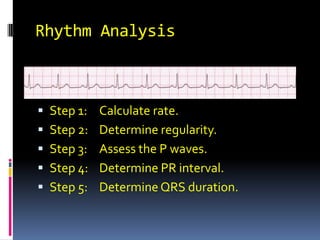
  • 1. Rhythm Analysis Step 1: Calculate rate. Step 2: Determine regularity. Step 3: Assess the P waves. Step 4: Determine PR interval. Step 5: Determine QRS duration.
  • 2. Step 1: Calculate Rate Option 1 Count the # of R waves in a 6 second rhythm strip, then multiply by 10. Interpretation? 3 sec 3 sec 9 x 10 = 90 bpm
  • 3. Step 1: Calculate Rate Option 2 Find a R wave that lands on a bold line. Count the # of large boxes to the next R wave. If the second R wave is 1 large box away the rate is 300, 2 boxes - 150, 3 boxes - 100, 4 boxes - 75, etc. (cont) R wave
  • 4. Step 1: Calculate Rate Option 2 Interpretation? 300 150 100 75 60 50 Approx. 1 box less than 100 = 95 bpm
  • 5.
  • 6. What is the heart rate?
  • 7. Step 2 : Determine Regularity Regular: If the difference between the longest R-R interval in the ECG and the shortest R-R interval is less than 0.12 second Irregular: If the difference between the longest R-R interval in the ECG and the shortest R-R interval is greater than 0.12 second
  • 8. Step 2: Determine regularity Look at the R-R distances (using a caliper or markings on a pen or paper). Interpretation? R R Regular
  • 9. Step 3: Assess the P waves Are there P waves? Do the P waves all look alike? Do the P waves occur at a regular rate? Is there one P wave before each QRS? Interpretation? Normal P waves with 1 P wave for every QRS
  • 10. Step 4: Determine PR interval Normal: 0.12 - 0.20 seconds. (3 - 5 boxes) Interpretation? 0.12 seconds
  • 11. Step 5: QRS duration Normal: 0.04 - 0.12 seconds. (1 - 3 boxes) Interpretation? 0.08 seconds
  • 12. Rhythm Summary Rate 90-95 bpm Regularity regular P waves normal PR interval 0.12 s QRS duration 0.08 s Interpretation? Normal Sinus Rhythm
  • 13. Normal Sinus Rhythm (NSR) The electrical impulse is formed in the SA node and conducted normally. This is the normal rhythm of the heart; other rhythms that do not conduct via the typical pathway are called arrhythmias.
  • 14. NSR Parameters Rate 60 - 100 bpm Regularity regular P waves normal PR interval 0.12 - 0.20 s QRS duration 0.04 - 0.12 s
  • 15. Cardiac Arrhythmias and Their Electrocardiographic Interpretation Abnormal rhythmicity of the pacemaker Shift of the pacemaker from the sinus node to another place in the heart Blocks at different points in the spread of the impulse through the heart Abnormal pathways of impulse transmission through the heart Spontaneous generation of spurious impulses in almost any part of the heart
  • 16. Tachycardia: The term "tachycardia" means fast heart rate, usually defined in an adult person as faster than 100 beats per minute This electrocardiogram is normal except that the heart rate The general causes of tachycardia include increased body temperature, stimulation of the heart by the sympathetic nerves, or toxic conditions of the heart Abnormal Sinus Rhythms
  • 17.
  • 18.
  • 19. Sinus Tachycardia Deviation from NSR - Rate > 100 bpm
  • 20. Sinus Tachycardia SA node is depolarizing faster than normal, impulse is conducted normally.
  • 21. Bradycardia The term "Bradycardia" means a slow heart rate, usually defined as fewer than 60 beats per minute The cause of Bradycardia is Vagal Stimulation
  • 22.
  • 23.
  • 24. Sinus Bradycardia Deviation from NSR - Rate < 60 bpm
  • 25. Sinus Bradycardia SA node is depolarizing slower than normal, impulse is conducted normally (i.e. normal PR and QRS interval).
  • 26. Atrioventricular Block Delay or blocks conduction from the atria to the ventricles. Causes are ischemia, extreme stimulation of the heart by vagus nerves, inflammation of the AV node or A-V bundle
  • 27. 1st Degree AV Block Prolonged conduction delay in the AV node or Bundle of His.
  • 28.
  • 29.
  • 30. 1st Degree AV Block Deviation from NSR PR Interval > 0.20 s
  • 31. 2nd Degree Block When conduction through the A-V bundle is slowed enough to increase the P-R interval to 0.45 second. There will be P wave but no QRS-T wave There is drop beat of the ventricles
  • 32.
  • 33.
  • 34.
  • 35. 2nd Degree AV Block Deviation from NSR PR interval progressively lengthens, then the impulse is completely blocked (P wave not followed by QRS).
  • 36. 3rd Degree AV Block Deviation from NSR The P waves are completely blocked in the AV junction; QRS complexes originate independently from below the junction.
  • 37. 3rd Degree AV Block There is complete block of conduction in the AV junction, so the atria and ventricles form impulses independently of each other. Without impulses from the atria, the ventricles own intrinsic pacemaker kicks in at around 30 - 45 beats/minute.
  • 38.
  • 39.
  • 40. Premature Contractions A premature contraction is a contraction of the heart before the time that normal contraction would have been expected. This condition is also called extrasystole, premature beat, or ectopic beat The causes are local areas of ischemia, toxic irritation of A-V node, purkinje system caused by drugs or caffeine
  • 41. Premature Atrial Contractions Deviation from NSR These ectopic beats originate in the atria (but not in the SA node), therefore the contour of the P wave, the PR interval, and the timing are different than a normally generated pulse from the SA node.
  • 42.
  • 43.
  • 44. Premature Ventricular Contraction Deviation from NSR Ectopic beats originate in the ventricles resulting in wide and bizarre QRS complexes. The QRS complex is usually prolonged. The reason is that the impulse is conducted mainly through slowly conducting muscle of the ventricles
  • 45.
  • 46.
  • 47. Atrial Fibrillation Deviation from NSR No organized atrial depolarization, so no normal P waves The cause of atrial fibrillation is atrial enlargement resulting from heart valve lesions that prevent the atria from emptying adequately into the ventricles
  • 48. Atrial Fibrillation Recent theories suggest that it is due to multiple re-entrant wavelets conducted between the R & L atria. Either way, impulses are formed in a totally unpredictable fashion. The AV node allows some of the impulses to pass through at variable intervals (so rhythm is irregularly irregular).
  • 49.
  • 50.
  • 51. Ventricular Fibrillation The ventricular cells are excitable and depolarizing randomly. Rapid drop in cardiac output and death occurs if not quickly reversed
  • 52. Ventricular Fibrillation Deviation from NSR Completely abnormal.
  • 53.
  • 54.