Finaale pulmonary stenosis
•Télécharger en tant que PPT, PDF•
32 j'aime•13,958 vues

This document discusses pulmonary stenosis (PS), a congenital heart defect where the pulmonary valve is narrowed. It accounts for 20-30% of congenital heart disease. PS can be valvular, subvalvular, or supravalvular. Symptoms range from none to right heart failure. Diagnosis involves listening for a systolic murmur and click, and imaging like echocardiogram and catheterization to measure pressures and gradients. Treatment is usually balloon valvuloplasty or surgery if severe.

Recommandé
Contenu connexe
Tendances
Tendances (20)
Similaire à Finaale pulmonary stenosis
Similaire à Finaale pulmonary stenosis (20)
Plus de Fuad Farooq
Plus de Fuad Farooq (20)
Dernier
Dernier (20)
Finaale pulmonary stenosis
- 2. Pulmonary Stenosis (PS) has beenPulmonary Stenosis (PS) has been described in 1761 by Morgagnidescribed in 1761 by Morgagni It accounts for 20-30% of all CHDIt accounts for 20-30% of all CHD Found in 8% to 10% of patients as isolatedFound in 8% to 10% of patients as isolated PSPS In 50% of cases the septum is intactIn 50% of cases the septum is intact
- 3. Three anatomical varientsThree anatomical varients Valvar (90%)Valvar (90%) Subvalvar (Subvalvar (Infundibular stenosisInfundibular stenosis from hypertrophy of the Cristafrom hypertrophy of the Crista Supraventricularis)Supraventricularis) Supravalvar (pulmonary artery trunkSupravalvar (pulmonary artery trunk or branches )or branches )
- 4. Pulmonary valve stenosis is usuallyPulmonary valve stenosis is usually isolated or part of the complex congenitalisolated or part of the complex congenital heart diseaseheart disease
- 5. Exact embryologic process is not wellExact embryologic process is not well understoodunderstood Mal-development of the distal part of the bulbusMal-development of the distal part of the bulbus cordiscordis Genetic factors may play a roleGenetic factors may play a role Known association between pulmonary valve andKnown association between pulmonary valve and supravalvular stenosis with multiple somaticsupravalvular stenosis with multiple somatic abnormalitiesabnormalities
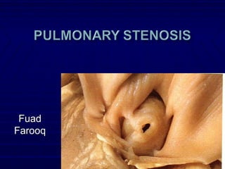
- 6. Valve commisures are partially fused and the 3Valve commisures are partially fused and the 3 leaflets are thin and pliant, resulting in a conicalleaflets are thin and pliant, resulting in a conical or dome-shaped structure with a narrowedor dome-shaped structure with a narrowed central orificecentral orifice It may be diffusely thickened with one, two, orIt may be diffusely thickened with one, two, or three leaflets and commissural fusionthree leaflets and commissural fusion
- 7. Bicuspid valve is found in 90% of patientsBicuspid valve is found in 90% of patients with Tetralogy of Fallotwith Tetralogy of Fallot Rare in individuals with isolated valvular PSRare in individuals with isolated valvular PS
- 8. 10-15% of individuals with valvular PS have10-15% of individuals with valvular PS have dysplastic pulmonary valvesdysplastic pulmonary valves Valves have irregularly shaped, thickenedValves have irregularly shaped, thickened leaflets, with little commissural fusion and exhibitleaflets, with little commissural fusion and exhibit reduced mobilityreduced mobility The leaflets are composed of myxomatous tissue,The leaflets are composed of myxomatous tissue, which may extend to the vessel wallwhich may extend to the vessel wall The valve annulus is usually small and theThe valve annulus is usually small and the supravalvular area of the pulmonary trunk issupravalvular area of the pulmonary trunk is usually hypoplasticusually hypoplastic Poststenotic dilatation of the pulmonary artery isPoststenotic dilatation of the pulmonary artery is uncommonuncommon
- 9. Secondary changes in the right ventricle andSecondary changes in the right ventricle and pulmonary arteriespulmonary arteries Right ventricle espacially infundibular regionRight ventricle espacially infundibular region becomes diffusely hypertrophiedbecomes diffusely hypertrophied Produce dynamic subvalvular obstructionProduce dynamic subvalvular obstruction Right atrium may be thick and dilate as aRight atrium may be thick and dilate as a result of the increased pressure necessaryresult of the increased pressure necessary to fill the hypertrophic right ventricleto fill the hypertrophic right ventricle
- 10. Rise in RV pressure proportional to theRise in RV pressure proportional to the severity of obstructionseverity of obstruction Increase in muscle massIncrease in muscle mass Hyperplasia of the muscle cells with aHyperplasia of the muscle cells with a concomitant increase in the number of capillariesconcomitant increase in the number of capillaries Increased muscle mass may enable theIncreased muscle mass may enable the hypertensive right ventricle to maintain ahypertensive right ventricle to maintain a normal stroke volumenormal stroke volume
- 11. Development of poststenotic dilation ofDevelopment of poststenotic dilation of the pulmonary artery trunk sometimesthe pulmonary artery trunk sometimes extending to the proximal left pulmonaryextending to the proximal left pulmonary arteryartery Result from the high velocity jet of flow ejectedResult from the high velocity jet of flow ejected through the small valve orifice - “jet effect”through the small valve orifice - “jet effect” The degree of dilation is not necessarilyThe degree of dilation is not necessarily proportional to the severity of obstructionproportional to the severity of obstruction
- 12. RV eventually dilate and failRV eventually dilate and fail Exacerbated by:Exacerbated by: Development of tricuspid insufficiencyDevelopment of tricuspid insufficiency Subendocardial (esp. at infundibular region) andSubendocardial (esp. at infundibular region) and papillary muscle ischemia/infarctionpapillary muscle ischemia/infarction
- 13. Mostly diagnosis is made when murmurMostly diagnosis is made when murmur detected in asymptomatic patients on routinedetected in asymptomatic patients on routine examinationexamination Exertional dyspnea, fatigue and cynosis dueExertional dyspnea, fatigue and cynosis due to inability of the right ventricle to increase itsto inability of the right ventricle to increase its output in response to exertionoutput in response to exertion If the stenosis is not relieved then features ofIf the stenosis is not relieved then features of right heart failureright heart failure
- 14. Chest pain, syncope and sudden deathChest pain, syncope and sudden death with strenuous exercisewith strenuous exercise Decreased myocardial perfusion caused byDecreased myocardial perfusion caused by inadequate cardiac output during exercise,inadequate cardiac output during exercise, leading to ischemia and ventricular arrhythmiasleading to ischemia and ventricular arrhythmias
- 15. ‘‘a’ wave becomes progressively largera’ wave becomes progressively larger with increasing severity of obstructionwith increasing severity of obstruction
- 16. Left parasternal heaveLeft parasternal heave Systolic thrill in severe pulmonarySystolic thrill in severe pulmonary stenosisstenosis Located at the 2Located at the 2ndnd to 3to 3rdrd intercostal space butintercostal space but it may also be felt at the suprasternal notchit may also be felt at the suprasternal notch Absent in young infants with severe stenosisAbsent in young infants with severe stenosis and in patients with congestive heart failureand in patients with congestive heart failure and low cardiac outputand low cardiac output
- 17. S1 normal followed by a pulmonaryS1 normal followed by a pulmonary ejection click in patients with mild orejection click in patients with mild or moderate stenosismoderate stenosis Corresponds to the time when the domingCorresponds to the time when the doming pulmonary valve reaches its open positionpulmonary valve reaches its open position Become earlier as severity increased until itBecome earlier as severity increased until it merges with the first heart sound andmerges with the first heart sound and becomes inaudiblebecomes inaudible
- 18. Click is followed by ESM maximal at theClick is followed by ESM maximal at the upper left sternal borderupper left sternal border Intensity – increases with the severity of obstructionIntensity – increases with the severity of obstruction Radiation – entire precordium, neck & backRadiation – entire precordium, neck & back (characteristically)(characteristically) Become soft when severe stenosis with right heartBecome soft when severe stenosis with right heart failure due to low cardiac outputfailure due to low cardiac output
- 19. P2 intensity typically decreases withP2 intensity typically decreases with increase severity of obstructionincrease severity of obstruction
- 20. S4 – lower left sternal border in patients withS4 – lower left sternal border in patients with severe stenosissevere stenosis Pansystolic murmur of tricuspid insufficiencyPansystolic murmur of tricuspid insufficiency may be present lower along the left sternalmay be present lower along the left sternal borderborder S3 – when present think of ASDS3 – when present think of ASD
- 23. Mild stenosis usually have a normalMild stenosis usually have a normal electrocardiogramelectrocardiogram Rightward axis deviation is often the only abnormalityRightward axis deviation is often the only abnormality
- 24. Electrocardiogram is almost alwaysElectrocardiogram is almost always abnormalabnormal Right axis deviationRight axis deviation The R:S ratio in V1 is usually >4:1The R:S ratio in V1 is usually >4:1 R wave is typically <20 mmR wave is typically <20 mm The T waves in the right precordial leads areThe T waves in the right precordial leads are upright in approximately 50% of patientsupright in approximately 50% of patients
- 25. QRS axis >110 degrees and not uncommonly extremeQRS axis >110 degrees and not uncommonly extreme RADRAD A pure R, Rs, or QR is the usual pattern in the rightA pure R, Rs, or QR is the usual pattern in the right precordial leads, and the R wave is usually >20 mmprecordial leads, and the R wave is usually >20 mm The R:S ratio in VThe R:S ratio in V66 may be <1.0may be <1.0 The T waves usually inverted in the right precordial leadsThe T waves usually inverted in the right precordial leads P-pulmonale indicating right atrial enlargementP-pulmonale indicating right atrial enlargement
- 26. Estimation of RV pressure is possibe if a pure REstimation of RV pressure is possibe if a pure R wave is present in lead Vwave is present in lead V4R4R or Vor V11 The height of the R wave in millimeters,The height of the R wave in millimeters, multiplied by 5, approximates the rightmultiplied by 5, approximates the right ventricular pressure in mm of Hgventricular pressure in mm of Hg
- 28. Enlarged main pulmonary artery secondaryEnlarged main pulmonary artery secondary to post-stenotic dilatationto post-stenotic dilatation Enlarged left pulmonary artery (jet streamEnlarged left pulmonary artery (jet stream effect)effect) Normal to decreased peripheral pulmonaryNormal to decreased peripheral pulmonary vasculaturevasculature CardiomegalyCardiomegaly Results from RA and RV enlargementResults from RA and RV enlargement
- 29. Standard and high parasternal short and longStandard and high parasternal short and long axis views and the subcostal sagittal viewsaxis views and the subcostal sagittal views Valve leaflets usually appear prominent becauseValve leaflets usually appear prominent because of thickeningof thickening Systolic motion is restricted, with inward curvingSystolic motion is restricted, with inward curving of the tips of the leaflets, known as domingof the tips of the leaflets, known as doming Poststenotic dilation of the main and branchPoststenotic dilation of the main and branch pulmonary arteries are also easily recognizedpulmonary arteries are also easily recognized
- 33. Assessment of RV anatomy and function andAssessment of RV anatomy and function and assessment of tricuspid valveassessment of tricuspid valve Evidence of dynamic subpulmonary stenosisEvidence of dynamic subpulmonary stenosis should be soughtshould be sought Dysplastic pulmonary valve usually can beDysplastic pulmonary valve usually can be ascertained by echocardiographyascertained by echocardiography The leaflets appear thickened and immobile,The leaflets appear thickened and immobile, without the characteristic doming seen in typicalwithout the characteristic doming seen in typical casescases The pulmonary valve annulus is hypoplastic, andThe pulmonary valve annulus is hypoplastic, and supra-annular narrowing of the proximal mainsupra-annular narrowing of the proximal main
- 34. Allows quantitative assessment of severityAllows quantitative assessment of severity of pulmonary valve stenosisof pulmonary valve stenosis The echo beam must be aligned parallelThe echo beam must be aligned parallel with the main pulmonary artery trunk orwith the main pulmonary artery trunk or the direction of the flow jet as seen onthe direction of the flow jet as seen on color Dopplercolor Doppler Excellent correlation between the Doppler-Excellent correlation between the Doppler- derived gradient and that obtained byderived gradient and that obtained by
- 35. Role of catheterization has becomeRole of catheterization has become largely limited in therapeuticslargely limited in therapeutics
- 36. Measurement of RV pressure andMeasurement of RV pressure and compared with systemic arterial pressurecompared with systemic arterial pressure and the pressure gradient across theand the pressure gradient across the pulmonary valvepulmonary valve Resting RV pressure >30 to 35 mm HgResting RV pressure >30 to 35 mm Hg and pressure gradient across theand pressure gradient across the pulmonary valve of >10 are consideredpulmonary valve of >10 are considered abnormalabnormal
- 37. RV pressures 105 mmHg and PA pressures are 60, so the gradient is 45 mmHg
- 38. If associated infundibular obstruction, pressureIf associated infundibular obstruction, pressure gradients are encountered across the pulmonarygradients are encountered across the pulmonary valve and also across the infundibulumvalve and also across the infundibulum RVEDP may be normal but usually elevated withRVEDP may be normal but usually elevated with severe obstruction or right ventricular failuresevere obstruction or right ventricular failure Right atrial pressure is normal in mild toRight atrial pressure is normal in mild to moderate obstruction, but tall right atrial ‘A’moderate obstruction, but tall right atrial ‘A’ waves usually are seen with severe obstructionwaves usually are seen with severe obstruction
- 39. Provides information about the locationProvides information about the location and severity of pulmonary stenosis that isand severity of pulmonary stenosis that is invaluable for diagnostic and therapeuticinvaluable for diagnostic and therapeutic purposespurposes Anatomy of the pulmonary valve andAnatomy of the pulmonary valve and associated features can be shown best byassociated features can be shown best by right ventricular angiography in theright ventricular angiography in the anteroposterior cranial and lateral viewanteroposterior cranial and lateral view
- 40. Normal pulmonary valve area is 2.0 cmNormal pulmonary valve area is 2.0 cm22 /m/m22 of BSAof BSA Mild stenosis - Valve area larger than 1 cm2 - Right ventricular pressure less than half the left ventricular pressure - Peak gradient 10-35 mm Hg - Peak velocity <3m/sec Moderate stenosis - Valve area 0.5- 1.0 cm2 - Right ventricular pressure is greater than half but <75% of the left ventricular pressure - Peak gradient is 36-64 mm Hg - Peak velocity 3- 4m/sec Severe stenosis - Valve area <0.5cm2 -Right ventricular pressure 75% of the left ventricular pressure - Peak gradient >64 mm Hg - Peak velocity >4/sec
- 41. Idiopathic dilation of the main pulmonary arteryIdiopathic dilation of the main pulmonary artery ASDASD Peripheral pulmonary arterial stenosisPeripheral pulmonary arterial stenosis Mitral valve prolapseMitral valve prolapse Straight back syndromeStraight back syndrome Aortic stenosisAortic stenosis Innocent murmursInnocent murmurs VSD with or without associated pulmonaryVSD with or without associated pulmonary stenosisstenosis Tetralogy of FallotTetralogy of Fallot Pulmonary atresia with intact ventricular septumPulmonary atresia with intact ventricular septum Ebstein anomaly of the tricuspid valveEbstein anomaly of the tricuspid valve
- 42. Noonan syndrome in 50% of patientsNoonan syndrome in 50% of patients Most common lesion is pulmonary stenosis owing to pulmonaryMost common lesion is pulmonary stenosis owing to pulmonary valve dysplasiavalve dysplasia 25% of Hypertrophic CMP of the left ventricle with PS25% of Hypertrophic CMP of the left ventricle with PS Rarely - cardiac tumors can grow on or into the RVRarely - cardiac tumors can grow on or into the RV outflow tract and cause flow obstructionoutflow tract and cause flow obstruction Extrinsic lesions e.g., Sinus of Valsalva aneurysms andExtrinsic lesions e.g., Sinus of Valsalva aneurysms and aortic graft aneurysms compressing cardiac structuresaortic graft aneurysms compressing cardiac structures and can cause PSand can cause PS Multiple lentigines syndrome, or leopard syndrome, hasMultiple lentigines syndrome, or leopard syndrome, has been associated with pulmonary valve and pulmonarybeen associated with pulmonary valve and pulmonary artery stenosisartery stenosis
- 43. Neurofibromatosis, deposits from glycogen storageNeurofibromatosis, deposits from glycogen storage diseases and goutdiseases and gout Carcinoid results in development of myxomatousCarcinoid results in development of myxomatous (fibroelastic) plaques in the RV outflow tract(fibroelastic) plaques in the RV outflow tract Distortion and constriction of the pulmonic ring, as well as fusionDistortion and constriction of the pulmonic ring, as well as fusion or destruction of pulmonary valve leaflets, resulting in bothor destruction of pulmonary valve leaflets, resulting in both stenosis and regurgitationstenosis and regurgitation Rare manifestation of rheumatic heart disease - followsRare manifestation of rheumatic heart disease - follows involvement of the mitral and aortic valvesinvolvement of the mitral and aortic valves
- 44. Valvuloplasty should be performed in anyValvuloplasty should be performed in any symptomatic patient as soon as thesymptomatic patient as soon as the diagnosis is madediagnosis is made Immediate valvoplasty in infants withImmediate valvoplasty in infants with critical pulmonary valve stenosiscritical pulmonary valve stenosis Asymptomatic patients with severeAsymptomatic patients with severe obstruction should be treatedobstruction should be treated semielectively with valvuloplasty shortlysemielectively with valvuloplasty shortly
- 45. Moderate obstruction should undergoModerate obstruction should undergo elective valvuloplasty if the rightelective valvuloplasty if the right ventricular pressure is 50% systemic orventricular pressure is 50% systemic or higherhigher No intervention is necessary for patientsNo intervention is necessary for patients with mild obstructionwith mild obstruction They should not be restricted in their physicalThey should not be restricted in their physical activity and should be treated as normalactivity and should be treated as normal childrenchildren
- 46. Described initially by Kan and associates inDescribed initially by Kan and associates in 19821982 After obtaining appropriate hemodynamic andAfter obtaining appropriate hemodynamic and angiographic information about severity andangiographic information about severity and location of obstruction, an exchange guidewire islocation of obstruction, an exchange guidewire is introduced through an end-hole catheter andintroduced through an end-hole catheter and positioned in the distal left pulmonary arterypositioned in the distal left pulmonary artery A balloon is chosen that is 20% to 40% largerA balloon is chosen that is 20% to 40% larger than the angiographically measured pulmonarythan the angiographically measured pulmonary valve annulus, and it is positioned over avalve annulus, and it is positioned over a guidewire with the valve at its midpointguidewire with the valve at its midpoint In larger patients with an annulus diameter ofIn larger patients with an annulus diameter of >20 mm, the double-balloon technique may be>20 mm, the double-balloon technique may be
- 47. Independent predictors of suboptimalIndependent predictors of suboptimal intermediate-term outcomeintermediate-term outcome Small annular size (characteristic of patientsSmall annular size (characteristic of patients with dysplastic valves)with dysplastic valves) Higher immediate residual gradientHigher immediate residual gradient Earlier year of the initial valvuloplastyEarlier year of the initial valvuloplasty Smaller balloon to annulus ratioSmaller balloon to annulus ratio
- 48. Death (0.2%)Death (0.2%) Laceration of the inferior vena cava, iliac veinLaceration of the inferior vena cava, iliac vein junction during balloon withdrawaljunction during balloon withdrawal Tearing of the pulmonary valve annulus duringTearing of the pulmonary valve annulus during balloon inflationballoon inflation Perforation of the right ventricular outflowPerforation of the right ventricular outflow tract resulting in tamponade and tricuspidtract resulting in tamponade and tricuspid regurgitation requiring surgical interventionregurgitation requiring surgical intervention Vein thrombosis and vein tearsVein thrombosis and vein tears Arrhythmias (1.3%)Arrhythmias (1.3%)
- 49. Stroke, seizures, necrotizing enterocolitis,Stroke, seizures, necrotizing enterocolitis, endocarditis and septic shockendocarditis and septic shock Abrupt closure of the ductus despiteAbrupt closure of the ductus despite prostaglandin infusion requiring urgentprostaglandin infusion requiring urgent aortopulmonary shunt placementaortopulmonary shunt placement Pulmonary insufficiencyPulmonary insufficiency Risk factors: smaller body surface area at theRisk factors: smaller body surface area at the time of intervention, larger balloon to annulustime of intervention, larger balloon to annulus ratio and a higher degree of obstructionratio and a higher degree of obstruction before dilationbefore dilation
- 50. Reserved for dysplastic pulmonary valvesReserved for dysplastic pulmonary valves resistant to dilation or with multiple levels ofresistant to dilation or with multiple levels of fixed obstructionfixed obstruction Often a persistent pressure gradient immediatelyOften a persistent pressure gradient immediately after surgery in patients with isolated valvarafter surgery in patients with isolated valvar pulmonary stenosis attributable to dynamicpulmonary stenosis attributable to dynamic narrowing of the hypertrophied infundibulum (asnarrowing of the hypertrophied infundibulum (as also observed following balloon valvuloplasty)also observed following balloon valvuloplasty) Reduction in gradient occur in the first 24 hoursReduction in gradient occur in the first 24 hours after surgery and continues at a slower rate asafter surgery and continues at a slower rate as the hypertrophy resolves over the subsequentthe hypertrophy resolves over the subsequent
- 51. Presence of symptoms such asPresence of symptoms such as progressive decrease in exerciseprogressive decrease in exercise tolerance or functional class or evidencetolerance or functional class or evidence of right heart failureof right heart failure Progressive RV enlargement oftenProgressive RV enlargement often accompanied by increasing TR owing toaccompanied by increasing TR owing to annular dilationannular dilation Development or progression of atrialDevelopment or progression of atrial
- 52. Homograft valves, xenograft valves,Homograft valves, xenograft valves, pericardial valves, and mechanical valvespericardial valves, and mechanical valves Percutaneous pulmonary valvePercutaneous pulmonary valve replacement with a bovine jugular venousreplacement with a bovine jugular venous valve sutured inside a stent was firstvalve sutured inside a stent was first described in a human in 2000described in a human in 2000
- 53. Symptoms are unreliable in reflectingSymptoms are unreliable in reflecting hemodynamic severity because theyhemodynamic severity because they usually are seen only in patients withusually are seen only in patients with severe or critical obstructionsevere or critical obstruction The course of mild stenosis is benignThe course of mild stenosis is benign No intervention is requiredNo intervention is required The hemodynamic response to exercise inThe hemodynamic response to exercise in these patients is normalthese patients is normal
- 54. Patients with moderate stenosis may developPatients with moderate stenosis may develop progressively greater outflow tract obstruction,progressively greater outflow tract obstruction, especially during periods of rapid growthespecially during periods of rapid growth In most asymptomatic patients with moderateIn most asymptomatic patients with moderate pulmonary valve stenosis formal exercise testingpulmonary valve stenosis formal exercise testing demonstrated subnormal cardiac outputdemonstrated subnormal cardiac output response and abnormal increase in rightresponse and abnormal increase in right ventricular end-diastolic pressure suggestingventricular end-diastolic pressure suggesting that both systolic and diastolic dysfunction maythat both systolic and diastolic dysfunction may be caused by long-standing moderatebe caused by long-standing moderate
- 55. Children with severe stenosis commonlyChildren with severe stenosis commonly develop increasingly severe obstructiondevelop increasingly severe obstruction Result from disproportionate growth of theResult from disproportionate growth of the child relative to the pulmonary valvechild relative to the pulmonary valve Exercise hemodynamics in children andExercise hemodynamics in children and adults with severe obstruction before andadults with severe obstruction before and after valvotomy suggest that irreversibleafter valvotomy suggest that irreversible changes in cardiac function can develop ifchanges in cardiac function can develop if treatment is delayed beyond childhoodtreatment is delayed beyond childhood
- 56. Patients with severe stenosis have a lowerPatients with severe stenosis have a lower cardiac stroke index at rest and during exercisecardiac stroke index at rest and during exercise and also higher RVEDP at rest and abnormaland also higher RVEDP at rest and abnormal increase with exercise than patients with milderincrease with exercise than patients with milder diseasedisease Following valvotomy there is an improvement inFollowing valvotomy there is an improvement in stroke index and reduction in RVEDP at rest andstroke index and reduction in RVEDP at rest and during exercise within 1 year of operationduring exercise within 1 year of operation In contrast this improvement is not observed inIn contrast this improvement is not observed in older patientsolder patients Implying permanent changes e.g., myocardial fibrosisImplying permanent changes e.g., myocardial fibrosis have occurredhave occurred
- 58. 5% of all cases of right ventricular outflow5% of all cases of right ventricular outflow tract obstructiontract obstruction Two types:Two types: Obstructive fibrous band at the junction of theObstructive fibrous band at the junction of the main right ventricular cavity and the proximalmain right ventricular cavity and the proximal infundibulum that closely resembles double-infundibulum that closely resembles double- chambered right ventriclechambered right ventricle Narrowing of the infundibulum is due toNarrowing of the infundibulum is due to fibromuscular thickening of its wall, which mayfibromuscular thickening of its wall, which may extend from immediately below the pulmonaryextend from immediately below the pulmonary valve to the proximal infundibulumvalve to the proximal infundibulum
- 59. ““Double-chambered right ventricle“Double-chambered right ventricle“ Aberrant hypertrophied muscular bands - divideAberrant hypertrophied muscular bands - divide the RV cavity into a proximal high-pressurethe RV cavity into a proximal high-pressure chamber and a distal low-pressure chamber tochamber and a distal low-pressure chamber to the hypertrophied muscle bandsthe hypertrophied muscle bands Orientation differs from that of the moderatorOrientation differs from that of the moderator bandband Septal attachment of the moderator band is usually inSeptal attachment of the moderator band is usually in the apical third of the ventricular septumthe apical third of the ventricular septum Septal attachment of anomalous muscle bundles areSeptal attachment of anomalous muscle bundles are
- 60. Visualized best from – subcostal andVisualized best from – subcostal and parasternal viewsparasternal views Presence or absence of a ventricularPresence or absence of a ventricular septal defect can be documentedseptal defect can be documented Abnormal fluttering of the pulmonary valveAbnormal fluttering of the pulmonary valve is often a concomitant findingis often a concomitant finding Treatment is surgical correctionTreatment is surgical correction