💚Call Girls In Amritsar 💯Anvi 📲🔝8725944379🔝Amritsar Call Girl No💰Advance Cash...
Cardiac Inotropes : Current Agent and Future Directions
1. REVIEW
Clinical update
Cardiac inotropes: current agents and
future directions
Gerd Hasenfuss1* and John R. Teerlink2,3
1
Department of Cardiology and Pneumology, University Medical Center, Heart Research Center Go¨ttingen, Robert-Koch-Strasse 40, 37075 Go¨ttingen, Germany; 2
School of
Medicine, University of California, San Francisco, San Francisco, CA, USA; and 3
Heart Failure and Clinical Echocardiography, Veterans Affairs Medical Center, San Francisco, CA
94121, USA
Received 16 November 2010; revised 6 January 2011; accepted 25 January 2011; online publish-ahead-of-print 8 March 2011
Intrinsic inotropic stimulation of the heart is central to the regulation of cardiovascular function, and exogenous inotropic therapies have
been used clinically for decades. Unfortunately, current inotropic drugs have consistently failed to show beneficial effects beyond short-
term haemodynamic improvement in patients with heart failure. To address these limitations, new agents targeting novel mechanisms are
being developed: (i) istaroxime has been developed as a non-glycoside inhibitor of the sodium-potassium-ATPase with additional stimulatory
effects on the sarcoplasmic reticulum calcium pump (SERCA) and has shown lusitropic and inotropic properties in experimental and early
clinical studies; (ii) from a mechanistic point of view, the cardiac myosin activators, directly activating the acto-myosin cross-bridges, are most
appealing with improved cardiac performance in both animal and early clinical studies; (iii) gene therapy approaches have been successfully
employed to increase myocardial SERCA2a; (iv) nitroxyl donors have been developed and have shown evidence of positive lusitropic and
inotropic, as well as potent vasodilatory effects in early animal studies; (v) the ryanodine receptor stabilizers reduce pathological leak of
calcium from the sarcoplasmic reticulum with initial promising pre-clinical results; and finally, (vi) metabolic energy modulation may represent
a promising means to improve contractile performance of the heart. There is an urgent clinical need for agents that improve cardiac per-
formance with a favourable safety profile. These current novel approaches to improving cardiac function provide the hope that such agents
may soon be available.
-----------------------------------------------------------------------------------------------------------------------------------------------------------
Keywords Inotropes † Drugs † Therapies
Introduction
In 1986, two review articles on new positive inotropic agents for
the treatment of congestive heart failure were published,1,2
pre-
senting a number of promising developments. However, over 24
years later, the only inotropic agent recommended, and weakly
at that, in the European Society of Cardiology (ESC) Guidelines
for the treatment of chronic heart failure is digitalis, a drug intro-
duced in the eighteenth century.3
In the setting of acute heart
failure, inotropic agents are only recommended in patients with
systolic blood pressure ,90 mmHg and evidence of inadequate
organ perfusion despite other therapeutic interventions.
Why have so many promising inotropic drugs failed to demon-
strate beneficial clinical outcomes in patients with heart failure?
The therapeutic hypothesis is compelling; central to the pathogen-
esis of systolic heart failure is decreased left ventricular (LV)
contractile function. The initial insults (myocardial infarction, other
cardiomyopathies, hypertension, etc.) that cause decreased LV func-
tion set into motion an inexorable series of maladaptive haemo-
dynamic, remodelling, and neurohormonal responses that result in
heart failure, clinical deterioration, and ultimately death. It would
seem intuitively obvious that improving LV function should halt
the progression of disease and improve clinical outcomes. A
recent analysis supports this concept, where clinical device or
drug trials that demonstrate long-term improvement in LV function
are associated with improved survival.4
Furthermore, the success of
cardiac resynchronization therapy in reducing heart failure events
and improving survival clearly supports the hypothesis.5
However,
there is no similar supportive evidence for inotropic agents. While
there may be many reasons for the failure of currently available ino-
tropes to improve clinical outcomes, the adverse effect of these
agents on myocardial energetics and intracellular calcium may play
* Corresponding author. Tel: +49 551 39 6351, Fax: +49 551 39 6389, Email: hasenfus@med.uni-goettingen.de
Published on behalf of the European Society of Cardiology. All rights reserved. & The Author 2011. For permissions please email: journals.permissions@oup.com.
European Heart Journal (2011) 32, 1838–1845
doi:10.1093/eurheartj/ehr026
atEuropeanSocietyofCardiologyonNovember6,2013http://eurheartj.oxfordjournals.org/DownloadedfromatEuropeanSocietyofCardiologyonNovember6,2013http://eurheartj.oxfordjournals.org/DownloadedfromatEuropeanSocietyofCardiologyonNovember6,2013http://eurheartj.oxfordjournals.org/DownloadedfromatEuropeanSocietyofCardiologyonNovember6,2013http://eurheartj.oxfordjournals.org/DownloadedfromatEuropeanSocietyofCardiologyonNovember6,2013http://eurheartj.oxfordjournals.org/DownloadedfromatEuropeanSocietyofCardiologyonNovember6,2013http://eurheartj.oxfordjournals.org/DownloadedfromatEuropeanSocietyofCardiologyonNovember6,2013http://eurheartj.oxfordjournals.org/DownloadedfromatEuropeanSocietyofCardiologyonNovember6,2013http://eurheartj.oxfordjournals.org/DownloadedfromatEuropeanSocietyofCardiologyonNovember6,2013http://eurheartj.oxfordjournals.org/Downloadedfrom
2. an important role. While most inotropic agents increase energy
consumption and intracellular calcium, inotropic stimulation
through cardiac resynchronization therapy does not. Thus, the
success story of cardiac resynchronization by biventricular pacing
therapy may indicate that the future search for inotropic interven-
tion in heart failure is not hopeless, and that inotropic mechanisms
that avoid these liabilities may be clinically beneficial.
What defines an inotropic
intervention?
Inotropic interventions comprise all means that increase muscular
contractile force, and in particular, the contractile force of the
myocardium. The mechanisms underlying the regulation of myo-
cardial force production can best be explained at the level of the
smallest force-producing unit, the acto-myosin cross-bridge using
a simplified two-stage cross-bridge model (Figure 1). During a
cross-bridge cycle, the myosin head attaches to actin, rotates to
produce force, which is maintained during the so-called on-time
(Figure 2). Thereafter, the cross-bridge detaches again to enter
its non-force-producing state for the duration of the off-time.
The on-time and the unitary force production of the cross-bridge
define the force–time integral of the individual cross-bridge cycle.
Accordingly, contractile force depends on the number of cross-
bridges attached per unit of time. The cross-bridges are activated
by calcium binding to troponin C with the subsequent confor-
mational changes of tropomyosin and troponin I to facilitate acto-
myosin interaction. The muscle relaxes when calcium is pumped
back into the sarcoplasmic reticulum (SR) by the sarcoplasmic reti-
culum calcium pump (SERCA) and eliminated outside the cell by
the sodium–calcium exchanger (NCX).6
Inotropy, i.e. the
number of cross-bridges activated, depends upon: (i) the amount
of calcium available to bind to troponin C, (ii) the calcium affinity
of troponin C, and (iii) alterations at the level of the cross-bridge
cycle including promotion of the force-producing cross-bridge
state, cross-bridge unitary force production, and prolongation of
the on-time, and thus the duration of the force-producing state.
Furthermore, the ability of attached cross-bridges to increase
calcium affinity of troponin C and activate neighbouring cross-
bridges (i.e. co-operativity) may also increase contractile force.
The mechanisms underlying the increase in inotropy may also
influence the velocity of contraction and relaxation as well as
energy consumption of the myocardium. It is assumed that one
molecule of ATP is hydrolysed during one individual cross-bridge
cycle. Accordingly, the most efficient way to increase contractile
force would be to prolong cross-bridge attachment time, but
this mechanism might reduce the velocity of force development
and relaxation. However, it is also recognized that there is a
basal rate of non-force-generating ATP hydrolysis by myosin,7
such that mechanisms that increase the likelihood of force-
generating hydrolysis with cross-bridge formation would poten-
tially increase performance with no adverse effect on energetics.
Thus, efficiency (economy) of contraction and contractile perform-
ance may diverge depending on the inotropic mechanism.
How does the heart regulate its
inotropic state?
Endogenous inotropic mechanisms include (i) length-dependent
activation of cross-bridges, (ii) contraction frequency-dependent
activation of contractile force, and (iii) catecholamine-mediated
inotropy.6
The most important mechanism to regulate the basal
contractile force of the heart is length-dependent activation of
cross-bridges by increasing the length of the individual sarcomere
(Frank-Starling mechanism). This length-dependent activation
occurs without an increase in calcium release, partially by spatial
changes and increased co-operativity.8
Frequency-dependent
up-regulation of contractile force is calcium dependent. With
increasing heart rate, more calcium enters the cardiomyocyte, is
accumulated into the SR, and is available for release during the
next contraction, resulting in more recruited cross-bridges and
increased contractile force.9
Catecholamines increase contractile
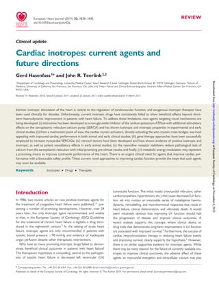
Figure 1 Acto-myosin interaction. The myosin head carrying
the ATPase site combines with actin to produce force. Calcium
binding to troponin C (TnC) results in a conformational change
of tropomyosin, troponin I (TnI), and troponin T (TnT), allowing
the myosin head to attach to actin, facilitating the acto-myosin
cross-bridge to cycle (see also Figures 2 and 6).
Figure 2 Cartoon of a simple two-state on–off cross-bridge
model. Assuming that one molecule of ATP is hydrolysed
during each cycle, the duration of the on-state determines cross-
bridge economy: prolonged attachment increases and shortened
decreases cross-bridge economy. Force development of the
muscle depends on the number of cross-bridges attached per
unit of time. fti, cross-bridge force-time integral.
Cardiac inotropes 1839
3. force by the b-adrenoceptor-adenylyl cyclase system or by stimu-
lation of a-receptors. Through protein kinase A, the
b-adrenoceptor system phosphorylates L-type calcium channels
to increase calcium influx and ryanodine receptors (RyRs) to
increase SR calcium release resulting in activation of cross-bridges.
In addition, phosphorylation of phospholamban accelerates SR
accumulation of calcium and relaxation which is supported by
phosphorylation of troponin I due to reduced calcium sensitivity
of troponin C (positive lusitropy). At the cross-bridge level,
cyclic AMP (cAMP)-mediated increase in contractility has been
reported to reduce the attachment time of the individual cross-
bridge. Consequently, cAMP-mediated inotropy increases the
rate of force development and rate of relaxation at the expense
of a reduced economy of contraction.9
Alterations of the inotropic state
in heart failure
In heart failure, excitation–contraction coupling is significantly
altered, largely by abnormal calcium accumulation of the SR
(Figure 3). Calcium enters the cell following activation of the L-type
calcium current during the upstroke of the action potential. This
calcium triggers the release of a larger amount of calcium by activat-
ing the RyR, which is the calcium release channel of the SR. Released
calcium binds to troponin C to activate acto-myosin cross-bridges,
inducing myocyte contraction. For relaxation, calcium is transported
back to the SR by SERCA and eliminated outside the cell through the
NCX. In heart failure, SR calcium uptake is abnormal due to SR
calcium leak through RyR, decreased re-uptake of calcium secondary
to decreased SERCA protein levels, and increased calcium elimin-
ation outside the cell due to increased levels of the NCX. Disturbed
SR calcium accumulation is also the main mechanism underlying
inversion of the force-frequency relation (see above, ‘How does
the heart regulate its inotropic state?’). In the failing myocardium,
frequency-dependent up-regulation of SR calcium load is absent,
which is associated with a decline of contractile force at higher
heart rates.9
Current inotropes
Current inotropic drugs include cardiac glycosides,
b-adrenoceptor agonists, phosphodiesterase (PDE) inhibitors,
and calcium sensitizers (Figure 4, Table 1).1,2
Cardiac glycosides
inhibit the sodium-potassium-ATPase (Na+
/K+
-ATPase) resulting
in sodium accumulation which in turn promotes cellular calcium
accumulation by influencing driving forces of the NCX. Providing
intact SR function, calcium accumulates in the SR, ready for
release during the next twitch. As explained above,
b-adrenoceptor stimulation increases intracellular cAMP that acti-
vates protein kinase A to phosphorylate key calcium-cycling pro-
teins. Phosphodiesterase inhibitors prevent cAMP degradation,
thus increasing cAMP activation of protein kinase A. Because
b-adrenoceptor density is reduced in heart failure, PDE inhibitors
have been assumed to be more effective in heart failure patients.
The beneficial effects of both catecholamines and PDE inhibitors
are directly a result of their ability to increase intracellular
calcium, which is also the direct mechanism of the adverse
effects of these agents, including myocardial ischaemia and arrhyth-
mias. Calcium sensitizers increase contractile force without
increasing intracellular calcium release. The molecular mechanism
of current calcium sensitizers is at the level of troponin C
through increased calcium affinity or more downstream through
alterations of cross-bridge kinetics (1, 2).
Current clinical use of inotropes
According to current ESC guidelines, cardiac glycosides (digoxin)
are indicated in patients with heart failure and atrial fibrillation to
Figure 3 Excitation–contraction coupling. Calcium enters the cell through the L-type calcium channel during the action potential and triggers
the release of a larger amount of calcium from the sarcoplasmic reticulum. This calcium binds to troponin C resulting in activation of contractile
proteins. The muscle relaxes when calcium is removed into the sarcoplasmic reticulum by sarcoplasmic reticulum calcium pump or outside the
cell by the sodium-calcium exchanger. RyR2, ryanodine receptor 2; TNT, troponin T; TNC, troponin C; TNI, troponin I; PL, phospholamban.
G. Hasenfuss and J.R. Teerlink1840
4. control the ventricular rate.3
In patients with sinus rhythm, digoxin
may be given to symptomatic patients with chronic systolic heart
failure to improve ventricular function and patient well-being,
and to reduce hospitalization, but without improving survival. Non-
glycoside inotropic agents should only be used in patients with
acute heart failure with low blood pressure or cardiac output in
the presence of signs of hypoperfusion or congestion. Available
agents include cAMP-elevating drugs such as dobutamine, dopa-
mine, milrinone, and enoximone.10
However, cAMP-mediated ino-
tropic stimulation may impair survival, in particular, in patients with
coronary artery disease. This adverse effect has been demon-
strated by several clinical trials, including the OPTIME-CHF trial
that randomized patients with acute heart failure to milrinone or
placebo infusion. Milrinone was not superior to placebo regarding
the primary endpoint of number of days hospitalized for
cardiovascular causes within 60 days after randomization, and mil-
rinone was associated with more adverse events, with significant
increases in atrial fibrillation/flutter, ventricular tachycardia/fibrilla-
tion, and sustained hypotension.11
In addition, milrinone patients
suffering from coronary artery disease had worse outcomes,
including increased mortality.12
Moreover, in the ESCAPE trial,
use of inotropes was associated with a significantly increased risk
of all-cause mortality.13
Use of the calcium sensitizer levosimendan may be more favour-
able. Levosimendan has a unique mechanism of action. It binds to
troponin C depending on the actual calcium concentration. This
may result in drug binding only during high systolic calcium levels
but not during diastole when calcium is low. By this on–off mech-
anism, levosimendan may increase calcium sensitivity only during
systole without impairing diastolic relaxation.14
In addition, levosi-
mendan activates ATP-dependent potassium channels in smooth
muscle cells resulting in vasodilation.15
At higher concentrations,
levosimendan also acts as a PDE inhibitor.14
A number of clinical
trials have been performed demonstrating inotropic and vasodilat-
ing properties of levosimendan in patients with heart failure includ-
ing cardiogenic shock: an increase in stroke volume and cardiac
output and a decrease in pulmonary capillary wedge pressure
(PCWP).10
Levosimendan possesses unusual pharmacokinetics
with a biological half-life of its active metabolite extending over
several days, explaining the recommendation to administer the
drug for only 24 h using continuous infusion. However, some
caution in the use of levosimendan, particularly with bolus
loading doses, may be warranted given the results of the only
placebo-controlled acute heart failure trial, REVIVE II, which
demonstrated an early increase in mortality, as well as increased
atrial fibrillation/flutter, ventricular ectopy, and sustained hypoten-
sion.16
A recent meta-analysis of studies in patients with acute
severe heart failure showed that levosimendan has favourable
haemodynamic effects superior to placebo or dobutamine and
Figure 4 Inotropic mechanisms and current inotropic interventions. Activation of the b-adrenoceptor stimulates adenylyl cyclase to produce
cAMP, which activates protein kinase A (PKA) to phosphorylate intracellular calcium-cycling proteins. Phosphodiesterases (PDEs) degrade
cAMP. Phosphodiesterases are inhibited by Phosphodiesterase inhibitors. Digitalis inhibits transport of three sodium ions for two potassium
ions through Na/K-ATPase. Calcium sensitizers increase the affinity of troponin C for calcium.
................................................................................
Table 1 Inotropic mechanisms and drugs
Inotropic mechanism Drugs
Sodium-potassium-ATPase inhibition Digoxin
b-Adrenoceptor stimulation Dobutamine, dopamine
Phosphodiesterase inhibition Enoximone, milrinone
Calcium sensitization Levosimendan
Sodium-potassium-ATPase inhibition
plus SERCA activation
Istaroxime
Acto-myosin cross-bridge activation Omecamtiv mecarbil
SERCA activation Gene transfer
SERCA activation plus vasodilation Nitroxyl donor;
CXL-1020
Ryanodine receptor stabilization Ryanodine receptor
stabilizer; S44121
Energetic modulation Etomoxir, pyruvate
Cardiac inotropes 1841
5. suggested that levosimendan was associated with reduced mor-
tality compared with dobutamine.17
Future directions
Promising new inotropic agents have been developed during the
last decade (Table 1). They are based on pathophysiological
defects identified in heart failure and include (i) istaroxime,
which is an inhibitor of Na+
/K+
-ATPase and an activator of
SERCA, (ii) cardiac myosin activators, (iii) increasing myocardial
SERCA through gene therapy, (iv) nitroxyl donors, (v) stabilizers
of the RyR, and (vi) energetic modulation (Figure 5).
Istaroxime—a new luso-inotropic
agent?
Istaroxime [(E,Z)-3-((2-aminoethoxy)imino)androstane-6,17-dione]
has been identified in the search for new inotropic agents acting at
the Na+
/K+
-ATPase. Istaroxime does not have a glycoside-like struc-
ture and in addition to its inhibitory effects on Na+
/K+
-ATPase, it has
been suggested to stimulate SERCA.18
Inhibition of Na+
/K+
-ATPase
increases intracellular sodium, which reducesthe driving force for the
NCX, decreasing calcium elimination outside the cell. Moreover,
increased sodium may stimulate the NCX to function in the
reverse mode of transporting calcium intracellularly. Calcium influx
into the cytosol may, however, be harmful in the failing heart with
reduced SERCA activity and elevated diastolic calcium levels. Under
those circumstances, Na+
/K+
-ATPase inhibition by elevating cytoso-
lic calcium may not only impair diastolic function, but it may also
induce delayed after-depolarization and cardiac arrhythmias. There-
fore, additional effects on top of Na+
/K+
-ATPase inhibition, which
promote calcium uptake of the SR, may be crucial to the potential
success of this type of inotropic agent. In a study of guinea pig myo-
cytes, Micheletti et al.19
demonstrated that istaroxime increased
twitch amplitude and accelerated relaxation without the after-
contractions seen with digoxin. In various dog studies, istaroxime
increased the maximum rates of rise and fall in LV pressure and
decreased end-diastolic pressure and volume without a change in
the heart rate and the blood pressure. Most importantly, these ino-
tropic and lusitropic effects were different from those of digoxin
and have not been associated with an increase in myocardial
oxygen consumption.20,21
Thus, from animal experiments istaroxime
shows a favourable profile with increased inotropy and accelerated
relaxation without associated increased energy consumption.
The HORIZON trial evaluated the haemodynamic, echocardio-
graphic, and neurohormonal effects of intravenous istaroxime in
120 patients hospitalized with heart failure and reduced ejection
fraction.22
In this randomized, double-blind, placebo-controlled,
dose-escalating study, three doses of istaroxime or a placebo
were given as intravenous infusions over 6 h to patients with a
history of heart failure and a PCWP .20 mmHg. A reduction in
PCWP was the primary endpoint, which was attained in all three
dose groups during the entire observation period of 6 h. There
was an increase in systolic blood pressure and a transient increase
in cardiac index with the highest dose and a decrease in heart rate
Figure 5 Future inotropic compounds: the ryanodine receptor (RyR) stabilizers reduce sarcoplasmic reticulum leak through the ryanodine
receptor and reconstitute ryanodine receptor channel function. Istaroxime inhibits sodium-potassium-ATPase and stimulates SERCA2a. Cardiac
myosin activators promote transition of cross-bridges from the weakly to the strongly bound force-producing state. Energetic modulators
improve myocardial energetics through switching from fatty acid to glucose oxidation or by other mechanisms including means to increase
the cellular phosphorylation potential. Virus-mediated sarcoplasmic reticulum calcium pump gene transfer (AV-SERCA) increases sarcoplasmic
reticulum calcium uptake. Nitroxyl (HNO) may increase sarcoplasmic reticulum calcium uptake by modification of sarcoplasmic reticulum
calcium pump and/or phospholamban (PL).
G. Hasenfuss and J.R. Teerlink1842
6. and diastolic and systolic volume, without a change in ejection frac-
tion. As indicators for improved diastolic function, E-wave decel-
eration time and Ea velocity increased, and the E/Ea ratio
decreased. Istaroxime shortened QTc and was well tolerated
with a short half-life of 1 h. Thus, the haemodynamic profile of
istaroxime reflects inotropic and lusitropic effects without any indi-
cation of vasodilatory properties. The limitation of this study is
related to the fact that patients included presented with milder
forms of acute heart failure, not requiring inotropic interventions
according to current guidelines.
Cardiac myosin activators—
increasing myocardial
performance
Cardiac myosin activators represent a new class of compounds,
which directly influence the cross-bridge cycle. These molecules
accelerate the rate of actin-dependent phosphate release of the
weakly bound acto-myosin cross-bridge (the rate-limiting step of
the cross-bridge cycle). This promotes transition to the force-
producing on-state of the cross-bridge. (Figure 6)23
As a conse-
quence, more cross-bridges enter the force-producing state,
more cross-bridges are activated per unit of time, and contractile
force increases (Figure 2). Several compounds have been investi-
gated so far.24
These agents stimulate myosin-ATPase and increase
fractional shortening of myocytes without increasing intracellular
calcium transients. The increase in myocyte shortening is associ-
ated with an increase in time-to-peak contraction with unaltered
velocity of contraction.
The molecule omecamtiv mecarbil (formerly CK-1827452) has
recently been studied in two different dog models of heart
failure.25
Both included tachycardia-pacing-induced failure on top
of myocardial infarction in one model and pressure overload by
constriction of the ascending aorta in the other. In both models,
omecamtiv mecarbil increased stroke volume and cardiac
output, and decreased LV end-diastolic pressure and heart rate.
In addition, omecamtiv mecarbil increased LV systolic ejection
time (SET) by 26%. Importantly, these improvements in cardiac
function were not associated with increased myocardial oxygen
consumption.
Omecamtiv mecarbil has advanced into clinical studies. The
first-in-human study assessed the effect of omecamtiv mecarbil in
ascending dose cohorts of healthy volunteers (n ¼ 34) and demon-
strated dose- and concentration-dependent increases in the SET,
stroke volume, fractional shortening, and ejection fraction.26
A sub-
sequent study in patients with heart failure presented similar find-
ings.27
Due to concerns that prolongation of SET might adversely
impact diastolic-filling, particularly during exercise, another study,
enrolling 94 patients with documented ischaemic cardiomyopathy,
exercise-induced angina, reduced ejection fraction, and sympto-
matic heart failure, evaluated the effect of intravenous omecamtiv
mecarbil on symptom-limited exercise tolerance.28
In these
patients, there was no deleterious effect of omecamtiv mecarbil
on exercise tolerance. No doubt, the cardiac myosin activators
are a very interesting new development in inotropic heart failure
therapy. While the characteristic increase of SET may be a
matter of concern, as long as relaxation is not prolonged and
heart rate not too high, the increased ejection period should be
well tolerated, as suggested by the exercise study.
Figure 6 Mode of action of cardiac myosin activators. The agents promote actin-dependent phosphate release (Pi–release) moving the cross-
bridge into its strongly bound force-producing state (see text). A, actin; M, myosin.
Cardiac inotropes 1843
7. Gene therapy approaches to
increase sarcoplasmic reticulum
calcium pump activity—
stimulating the calcium pumps
Various gene therapy strategies have been proposed to correct
abnormal excitation–contraction coupling in heart failure and
increase inotropy. Most approaches are related to reduced SR
calcium uptake, however, abnormal SR leak has also been con-
sidered. It has been shown in isolated myocytes that overexpres-
sion of the RyR-regulatory protein FKBP12.6 increases SR
calcium content and fractional shortening.29
A number of studies have been performed to evaluate the possi-
bility of SERCA gene transfer. Up-regulating SERCA2a, the cardiac
isoformofthesarco-endoplasmic reticulum calciumATPase,indiffer-
ent animal models of heart failure results in improvement in systolic
and diastolic function30,31
and may reduce arrhythmias.32
A recent
paper on a sheep model of myocardial infarction and mitral regurgita-
tion showed that SERCA up-regulation improves function and
reduces the remodelling processes.33
The Calcium Up-regulation
by Percutaneous Administration of Gene Therapy in Cardiac
Disease (CUPID) study, enrolled with a total of 39 patients with
severe heart failure randomized to adeno-associated virus-mediated
transfer of SERCA2a or placebo, has been recently presented.34
As a
phase 2 study, there was no single primary endpoint, but when a
broad range of efficacy and safety endpoints were evaluated, there
were very encouraging signals in improvement in symptoms and ven-
tricular remodelling.35
Problems related to gene transfer approaches
to increase inotropy include (i) immunological reactions, (ii) duration
of gene expression, (iii) control of gene expression, and (iv) unknown
toxic effects, which are related to the vector used to transfer a gene.
Currently, new-generation adeno-associated viruses have brought
considerable progress in the field.
Nitroxyl—nitric oxide’s soon-to-be
famous sibling?
Nitric oxide (NO) is well known as an important signalling molecule
central to the regulation of vascular tone and other cardiovascular
processes, but NO’s one-electron-reduced and protonated sibling,
nitroxyl (HNO), is currently less well known. While HNO and
NO are both gaseous signalling molecules and can be potent vasodi-
lators, HNO appears to have additional unique signalling pathways
and mechanisms independent of NO.36,37
In vitro experiments have
demonstrated HNO-induced vasorelaxation in isolated large
conduit and small resistance arteries, as well as intact coronary and
pulmonary vascular beds, and HNO is a potent arterio- and venodi-
latorin intact animalstudies.Whilesomeof thesevasorelaxant prop-
erties may be mediated by soluble guanylate cyclase (sGC), other
mechanisms are also important in HNO-induced vasodilation,
including increased circulating neuropeptide calcitonin gene-
related peptide levels and activation of vascular smooth muscle pot-
assium channels (perhaps both Kv and KATP-channels). While the
importance of these non-sGC mechanisms in patients is unclear,
there is one characteristic that clearly differentiates HNO from
traditional nitrovasodilators: the potential absence of tolerance or
tachyphylaxis.38
If confirmed in human studies, the absence of vascu-
lar tolerance could provide an important clinical advantage for HNO
in the setting of heart failure. Additional effects of interest include
inhibiting platelet aggregation and limiting vascular smooth muscle
proliferation.39
While these vascular properties are intriguing, the positive ino-
tropic effect of HNO is potentially of greater clinical interest. Early
in vitro experiments suggested positive inotropic and lusitropic
properties of HNO, while subsequent studies in healthy and
heart failure dog models with the HNO donor Angeli’s salt
(Na2N2O3) demonstrated significant improvements in
load-independent LV contractility, associated with reductions in
pre-load volume and diastolic pressure.40,41
The mechanisms of
these beneficial inotropic and lusitropic effects continue to be elu-
cidated, but they appear to be independent of cAMP/protein
kinase A (PKA) and cGMP/PKG signalling,42
with no modification
of L-type calcium channel activity,43
and related to modification
of specific cysteine residues on either phospholamban44
and/or
SERCA2a,45
resulting in augmented SR calcium transients. These
and other studies39
were encouraging, but the clinical utility of
HNO was limited by the poor pharmacological properties of the
available HNO donors, such as Na2N2O3. Recently, a clinical
development programme has used the new HNO donor
CXL-1020 in animal studies, which have confirmed its positive ino-
tropic and lusitropic effects.46
Clinical studies are currently being
conducted (clinicaltrials.gov NCT01092325, clinicaltrials.gov
NCT01096043). Nitroxyl donors, such as CXL-1020, may be a
new generation of inodilators that avoid the safety issues of
current inodilators, such as catecholamines, PDE inhibitors, and
levosimendan, and offer significant promise, however, the extent
of the vasodilating properties may determine their clinical utility.
Ryanodine receptor stabilizers—
stopping the leak
As discussed above, calcium leak through RyRs significantly con-
tributes to abnormal calcium cycling in human heart failure.
Accordingly, restoration of RyR function seems to be an interesting
target for its treatment. A leak of calcium from the SR may not
only decrease SR calcium load and availability for systolic contrac-
tion, but it may also promote diastolic dysfunction due to diastolic
activation of contractile proteins. In addition, SR calcium leak may
be a trigger for arrhythmias and contribute to altered gene
expression in heart failure. Finally, a leak has unfavourable ener-
getic consequences because ATP consumption for SR calcium
accumulation increases with recycling calcium. Several compounds
that reduce calcium leak through the RyR have been developed.
JTV519, a 1,4-benzothiazepine, was one of the first compounds
that restored abnormal RyR function and preserved contractile
performance in heart failure models.47,48
In addition, JTV519
improved diastolic and systolic function in isolated myocardium
from failing human hearts.49
In addition to RyR stabilization,
JTV519 has inhibitor properties on L-type calcium channels, potass-
ium channels, and possibly other transporters. Subsequently,
molecules that may specifically act on cardiac RyRs have been
G. Hasenfuss and J.R. Teerlink1844
8. developed, including S44121. The study drug S44121 is currently
being evaluated in a phase 2 multicentre clinical study (ISRCTN
Registration number 14227980).
Energetic modulators—fuelling
the engine
Disturbed energetic metabolism is considered to play a major role
in human heart failure.50
This may result from inadequate vessel
formation during cardiac hypertrophy, altered substrate uptake
of the myocyte or disturbed mitochondrial oxidative phosphoryl-
ation, and ATP availability for contractile processes. Several
drugs that switch energy metabolism from fatty acids to glucose
oxidation have been investigated. Etomoxir, an inhibitor of mito-
chondrial carnitine palmitoyltransferase 1, had shown promising
results in animal experiments. It was also shown that substrate
switching is associated with changes in gene expression such as
myosin heavy chain gene or SERCA, but in a recent study in rats
with aortic banding, an improvement of cardiac function has not
been observed.51
A clinical trial had to be stopped prematurely
because of liver toxicity of the substance in some patients.52
Administration of the glycolytic substrate pyruvate may result in
profound inotropic effects under experimental conditions as well
as in patients with heart failure. Pyruvate has numerous molecular
effects that may contribute to its inotropic action. These include:
(i) an increase in phosphorylation potential, (ii) a reduction of inor-
ganic phosphate, (iii) a decrease in hydrogen ion concentration, and
(iv) a modulation of the cytosolic redox state. The most important
mechanism for its inotropic action may be an increase in the phos-
phorylation potential and an increase in free energy of ATP hydroly-
sis. In isolated muscle strip preparations from patients with end-stage
heart failure, pyruvate resulted in a concentration-dependent
increase in developed force and a decrease in diastolic force.53
There was a dose-dependent prolongation of time-to-peak tension
and relaxation time. This was associated with increased intracellular
calcium transients and increased SR calcium content. The data
support the hypothesis that pyruvate increases energy availability
to the SERCA resulting in improved SR calcium handling.53
When
pyruvate was injected into the coronary circulation of patients
with dilated cardiomyopathy, it exhibited a profile of an ideal inotro-
pic agent with an increase in cardiac index and stroke volume index, a
decrease in PCWP and heart rate. Mean aortic pressure and system
vascular resistance did not change.54,55
These pyruvate data suggest
that energetic modulation has a significant potential for the treat-
ment of heart failure. A major difficulty for using pyruvate to treat
acute heart failure in patients results from the fact that high arterial
concentrations are needed which can only be achieved by
intra-arterial application of pyruvate. Nevertheless, the favourable
pronounced inotropic effects of pyruvate suggest that efforts
should be invested to search for energetic targets in the treatment
of heart failure.
Conclusion
Despite the compelling therapeutic hypothesis that increasing ven-
tricular performance should improve clinical outcomes, the
development of positive inotropes in the past has generated a
litany of failures. These failures have been largely due to increases
in cardiovascular complications directly related to the mechanism
of action of these agents. Newer agents targeting different mech-
anisms of action hold new promise for the future, as long as com-
plications of myocardial ischaemia, arrhythmias, and hypotension
can be avoided.
Funding
Supported by German Research Foundation (G.H.).
Conflict of interest: J.R.T. received research grants/consultant to
the following companies: Abbott Laboratories, Amgen, Cardioxyl,
Cytokinetics, Celladon, Orion Pharmaceuticals. G.H. received research
grants/consultant to the following companies: Servier, Orion
Pharmaceuticals.
References
1. Colucci WS, Wright RF, Braunwald E. New positive inotropic agents in the treat-
ment of congestive heart failure. Mechanisms of action and recent clinical devel-
opments. 2. N Engl J Med 1986;314:349–358.
2. Colucci WS, Wright RF, Braunwald E. New positive inotropic agents in the treat-
ment of congestive heart failure. Mechanisms of action and recent clinical devel-
opments. 1. N Engl J Med 1986;314:290–299.
3. Dickstein K, Cohen-Solal A, Filippatos G, McMurray JJ, Ponikowski P,
Poole-Wilson PA, Stromberg A, van Veldhuisen DJ, Atar D, Hoes AW,
Keren A, Mebazaa A, Nieminen M, Priori SG, Swedberg K, Vahanian A,
Camm J, De Caterina R, Dean V, Funck-Brentano C, Hellemans I,
Kristensen SD, McGregor K, Sechtem U, Silber S, Tendera M, Widimsky P,
Zamorano JL. ESC Guidelines for the diagnosis and treatment of acute and
chronic heart failure 2008: the Task Force for the Diagnosis and Treatment of
Acute and Chronic Heart Failure 2008 of the European Society of Cardiology.
Developed in collaboration with the Heart Failure Association of the ESC
(HFA) and endorsed by the European Society of Intensive Care Medicine
(ESICM). Eur Heart J 2008;29:2388–2442.
4. Kramer DG, Trikalinos TA, Kent DM, Antonopoulos GV, Konstam MA,
Udelson JE. Quantitative evaluation of drug or device effects on ventricular remo-
deling as predictors of therapeutic effects on mortality in patients with heart
failure and reduced ejection fraction: a meta-analytic approach. J Am Coll Cardiol
2010;56:392–406.
5. Cleland JG, Daubert JC, Erdmann E, Freemantle N, Gras D, Kappenberger L,
Tavazzi L. The effect of cardiac resynchronization on morbidity and mortality in
heart failure. N Engl J Med 2005;352:1539–1549.
6. Bers DM. Calcium cycling and signaling in cardiac myocytes. Annu Rev Physiol 2008;
70:23–49.
7. Stewart MA, Franks-Skiba K, Chen S, Cooke R. Myosin ATP turnover rate is a
mechanism involved in thermogenesis in resting skeletal muscle fibers. Proc Natl
Acad Sci USA 2010;107:430–435.
8. Holubarsch C, Ruf T, Goldstein DJ, Ashton RC, Nickl W, Pieske B, Pioch K,
Ludemann J, Wiesner S, Hasenfuss G, Posival H, Just H, Burkhoff D. Existence
of the Frank-Starling mechanism in the failing human heart. Investigations on
the organ, tissue, and sarcomere levels. Circulation 1996;94:683–689.
9. Hasenfuss G, Pieske B. Calcium cycling in congestive heart failure. J Mol Cell Cardiol
2002;34:951–969.
10. Teerlink JR, Metra M, Zaca V, Sabbah HN, Cotter G, Gheorghiade M, Cas LD.
Agents with inotropic properties for the management of acute heart failure syn-
dromes. Traditional agents and beyond. Heart Fail Rev 2009;14:243–253.
11. Cuffe MS, Califf RM, Adams KF Jr, Benza R, Bourge R, Colucci WS, Massie BM,
O’Connor CM, Pina I, Quigg R, Silver MA, Gheorghiade M. Short-term intrave-
nous milrinone for acute exacerbation of chronic heart failure: a randomized con-
trolled trial. JAMA 2002;287:1541–1547.
12. Felker GM, Benza RL, Chandler AB, Leimberger JD, Cuffe MS, Califf RM,
Gheorghiade M, O’Connor CM. Heart failure etiology and response to milrinone
in decompensated heart failure: results from the OPTIME-CHF study. J Am Coll
Cardiol 2003;41:997–1003.
13. Elkayam U, Tasissa G, Binanay C, Stevenson LW, Gheorghiade M, Warnica JW,
Young JB, Rayburn BK, Rogers JG, DeMarco T, Leier CV. Use and impact of ino-
tropes and vasodilator therapy in hospitalized patients with severe heart failure.
Am Heart J 2007;153:98–104.
Cardiac inotropes 1845
9. 14. Hasenfuss G, Pieske B, Castell M, Kretschmann B, Maier LS, Just H. Influence of
the novel inotropic agent levosimendan on isometric tension and calcium
cycling in failing human myocardium. Circulation 1998;98:2141–2147.
15. Yokoshiki H, Sperelakis N. Vasodilating mechanisms of levosimendan. Cardiovasc
Drugs Ther 2003;17:111–113.
16. Cleland JG, Freemantle N, Coletta AP, Clark AL. Clinical trials update from the
American Heart Association: REPAIR-AMI, ASTAMI, JELIS, MEGA, REVIVE-II,
SURVIVE, and PROACTIVE. Eur J Heart Fail 2006;8:105–110.
17. Delaney A, Bradford C, McCaffrey J, Bagshaw SM, Lee R. Levosimendan for the
treatment of acute severe heart failure: a meta-analysis of randomised controlled
trials. Int J Cardiol 2010;138:281–289.
18. Rocchetti M, Besana A, Mostacciuolo G, Micheletti R, Ferrari P, Sarkozi S, Szegedi C,
Jona I, Zaza A. Modulation of sarcoplasmic reticulum function by Na+/K+ pump
inhibitors with different toxicity: digoxin and PST2744 [(E,Z)-3-((2-aminoethoxy)imi-
no)androstane-6,17-dione hydrochloride]. J Pharmacol Exp Ther 2005;313:207–215.
19. Micheletti R, Mattera GG, Rocchetti M, Schiavone A, Loi MF, Zaza A, Gagnol RJ, De
Munari S, Melloni P, Carminati P, Bianchi G, Ferrari P. Pharmacological profile of the
novel inotropic agent (E,Z)-3-((2-aminoethoxy)imino)androstane-6,17-dione
hydrochloride (PST2744). J Pharmacol Exp Ther 2002;303:592–600.
20. Mattera GG, Lo Giudice P, Loi FM, Vanoli E, Gagnol JP, Borsini F, Carminati P.
Istaroxime: a new luso-inotropic agent for heart failure. Am J Cardiol 2007;99:
33A–40A.
21. Sabbah HN, Imai M, Cowart D, Amato A, Carminati P, Gheorghiade M. Hemody-
namic properties of a new-generation positive luso-inotropic agent for the acute
treatment of advanced heart failure. Am J Cardiol 2007;99:41A–46A.
22. Gheorghiade M, Blair JE, Filippatos GS, Macarie C, Ruzyllo W, Korewicki J,
Bubenek-Turconi SI, Ceracchi M, Bianchetti M, Carminati P, Kremastinos D,
Valentini G, Sabbah HN. Hemodynamic, echocardiographic, and neurohormonal
effects of istaroxime, a novel intravenous inotropic and lusitropic agent: a ran-
domized controlled trial in patients hospitalized with heart failure. J Am Coll
Cardiol 2008;51:2276–2285.
23. Vale RD, Milligan RA. The way things move: looking under the hood of molecular
motor proteins. Science 2000;288:88–95.
24. Teerlink JR. A novel approach to improve cardiac performance: cardiac myosin
activators. Heart Fail Rev 2009;14:289–298.
25. Shen YT, Malik FI, Zhao X, Depre C, Dhar SK, Abarzua P, Morgans DJ, Vatner SF.
Improvement of cardiac function by a cardiac Myosin activator in conscious dogs
with systolic heart failure. Circ Heart Fail 2010;3:522–527.
26. Teerlink JR, Malik FI, Clarke CP, Saikali KG, Escandon RD, Lee JH, Wolff AA. The
selective cardiac myosin activator, CK-1827452, increases left ventricular systolic
function by increasing ejection time: results of a first-in-human study of a unique
and novel mechanism. J Card Fail 2006;12:763.
27. Cleland JGF, Malik FI. The selective cardiac myosin activator, CK-1827452,
increases systolic function in heart failure. J Card Fail 2008;14:67.
28. Greenberg BH, Chou W, Escandon R, Lee JH, Saikali KG, Malik F, Wolff AA,
Shaburishvili T. Phase II safety study evaluating the novel cardiac myosin activator,
CK-1827452, in patients with ischemic cardiomyopathy and angina. J Card Fail
2009;15:S67–S67.
29. Prestle J, Janssen PM, Janssen AP, Zeitz O, Lehnart SE, Bruce L, Smith GL,
Hasenfuss G. Overexpression of FK506-binding protein FKBP12.6 in cardiomyo-
cytes reduces ryanodine receptor-mediated Ca(2+) leak from the sarcoplasmic
reticulum and increases contractility. Circ Res 2001;88:188–194.
30. Sakata S, Lebeche D, Sakata N, Sakata Y, Chemaly ER, Liang LF, Takewa Y,
Jeong D, Park WJ, Kawase Y, Hajjar RJ. Targeted gene transfer increases contrac-
tility and decreases oxygen cost of contractility in normal rat hearts. Am J Physiol
Heart Circ Physiol 2007;292:H2356–H2363.
31. Schmidt U, del Monte F, Miyamoto MI, Matsui T, Gwathmey JK, Rosenzweig A,
Hajjar RJ. Restoration of diastolic function in senescent rat hearts through adeno-
viral gene transfer of sarcoplasmic reticulum Ca(2+)-ATPase. Circulation 2000;
101:790–796.
32. del Monte F, Lebeche D, Guerrero JL, Tsuji T, Doye AA, Gwathmey JK, Hajjar RJ.
Abrogation of ventricular arrhythmias in a model of ischemia and reperfusion by
targeting myocardial calcium cycling. Proc Natl Acad Sci USA 2004;101:5622–5627.
33. Beeri R, Chaput M, Guerrero JL, Kawase Y, Yosefy C, Abedat S, Karakikes I,
Morel C, Tisosky A, Sullivan S, Handschumacher MD, Gilon D, Vlahakes GJ,
Hajjar RJ, Levine RA. Gene delivery of sarcoplasmic reticulum calcium ATPase
inhibits ventricular remodeling in ischemic mitral regurgitation. Circ Heart Fail
2010;3:627–634.
34. Jaski BE, Jessup ML, Mancini DM, Cappola TP, Pauly DF, Greenberg B, Borow K,
Dittrich H, Zsebo KM, Hajjar RJ. Calcium upregulation by percutaneous adminis-
tration of gene therapy in cardiac disease (CUPID Trial), a first-in-human phase 1/
2 clinical trial. J Card Fail 2009;15:171–181.
35. Stiles S. CUPID: first-in-human gene therapy for advanced heart failure promising
in small study. http://www.theheart.org/article/1083549.do.
36. Irvine JC, Ritchie RH, Favaloro JL, Andrews KL, Widdop RE, Kemp-Harper BK.
Nitroxyl (HNO): the Cinderella of the nitric oxide story. Trends Pharmacol Sci
2008;29:601–608.
37. Paolocci N, Jackson MI, Lopez BE, Miranda K, Tocchetti CG, Wink DA, Hobbs AJ,
Fukuto JM. The pharmacology of nitroxyl (HNO) and its therapeutic potential:
not just the Janus face of NO. Pharmacol Ther 2007;113:442–458.
38. Irvine J, Kemp-Harper BK, Widdop RE. Chronic administration of the HNO
donor, Angeli’s salt does not lead to tolerance, cross-tolerance or endothelial
dysfunction: comparison with GTN and DEA/NO. Antioxid Redox Signal 2010;
doi:10.1089/ars.2010.3269. Published online ahead of print 19 September 2010.
39. Bullen ML, Miller AA, Andrews KL, Irvine J, Ritchie RH, Sobey C, Kemp-Harper B.
Nitroxyl (HNO) as a vasoprotective signaling molecule. Antioxid Redox Signal
2010; doi:10.1089/ars.2010.3327. Published online ahead of print 2 November
2010.
40. Paolocci N, Katori T, Champion HC, St John ME, Miranda KM, Fukuto JM,
Wink DA, Kass DA. Positive inotropic and lusitropic effects of HNO/NO- in
failing hearts: independence from beta-adrenergic signaling. Proc Natl Acad Sci
USA 2003;100:5537–5542.
41. Paolocci N, Saavedra WF, Miranda KM, Martignani C, Isoda T, Hare JM, Espey MG,
Fukuto JM, Feelisch M, Wink DA, Kass DA. Nitroxyl anion exerts redox-sensitive
positive cardiac inotropy in vivo by calcitonin gene-related peptide signaling. Proc
Natl Acad Sci USA 2001;98:10463–10468.
42. Tocchetti CG, Wang W, Froehlich JP, Huke S, Aon MA, Wilson GM, Di
Benedetto G, O’Rourke B, Gao WD, Wink DA, Toscano JP, Zaccolo M,
Bers DM, Valdivia HH, Cheng H, Kass DA, Paolocci N. Nitroxyl improves cellular
heart function by directly enhancing cardiac sarcoplasmic reticulum Ca2+ cycling.
Circ Res 2007;100:96–104.
43. Kohr MJ, Kaludercic N, Tocchetti CG, Dong Gao W, Kass DA, Janssen PM,
Paolocci N, Ziolo MT. Nitroxyl enhances myocyte Ca2+ transients by exclusively
targeting SR Ca2+-cycling. Front Biosci (Elite Ed) 2010;2:614–626.
44. Froehlich JP, Mahaney JE, Keceli G, Pavlos CM, Goldstein R, Redwood AJ,
Sumbilla C, Lee DI, Tocchetti CG, Kass DA, Paolocci N, Toscano JP. Phospholam-
ban thiols play a central role in activation of the cardiac muscle sarcoplasmic reti-
culum calcium pump by nitroxyl. Biochemistry 2008;47:13150–13152.
45. Lancel S, Zhang J, Evangelista A, Trucillo MP, Tong X, Siwik DA, Cohen RA,
Colucci WS. Nitroxyl activates SERCA in cardiac myocytes via glutathiolation
of cysteine 674. Circ Res 2009;104:720–723.
46. Wang MJ, Mazhari R, Ilsar I, Wang A, Sabbah MS, Sabbah HN. Intravenous infusion
of CXL-1020, a novel nitroxyl (HNO) donor, improves left ventricular systolic
and diastolic function in dogs with advanced heart failure. J Card Fail 2009;15:
S73–S74.
47. Yano M, Kobayashi S, Kohno M, Doi M, Tokuhisa T, Okuda S, Suetsugu M,
Hisaoka T, Obayashi M, Ohkusa T, Kohno M, Matsuzaki M. FKBP12.6-mediated
stabilization of calcium-release channel (ryanodine receptor) as a novel thera-
peutic strategy against heart failure. Circulation 2003;107:477–484.
48. Lehnart SE, Wehrens XH, Reiken S, Warrier S, Belevych AE, Harvey RD,
Richter W, Jin SL, Conti M, Marks AR. Phosphodiesterase 4D deficiency in the
ryanodine-receptor complex promotes heart failure and arrhythmias. Cell 2005;
123:535–536.
49. Toischer K, Lahnart SE, Tenderich G, Milting H, Ko¨rfer R, Schmitto JD,
Scho¨ndube FA, Kaneko N, Laughrey CM, Smith GL, Hasenfuss G, Seidler T.
K201 improves aspects of the contractile performance of human failing myocar-
dium via reduction in CA2+
leak from the sarcoplasmic reticulum. Basic Res Cardiol
2010;105:279–287.
50. Ingwall JS. Energy metabolism in heart failure and remodelling. Cardiovasc Res
2009;81:412–419.
51. Schwarzer M, Faerber G, Rueckauer T, Blum D, Pytel G, Mohr FW, Doenst T.
The metabolic modulators, etomoxir and NVP-LAB121, fail to reverse pressure
overload induced heart failure in vivo. Basic Res Cardiol 2009;104:547–557.
52. Holubarsch CJ, Rohrbach M, Karrasch M, Boehm E, Polonski L, Ponikowski P,
Rhein S. A double-blind randomized multicentre clinical trial to evaluate the effi-
cacy and safety of two doses of etomoxir in comparison with placebo in patients
with moderate congestive heart failure: the ERGO (etomoxir for the recovery of
glucose oxidation) study. Clin Sci (Lond) 2007;113:205–212.
53. Hasenfuss G, Maier LS, Hermann HP, Luers C, Hunlich M, Zeitz O, Janssen PM,
Pieske B. Influence of pyruvate on contractile performance and Ca(2+) cycling in
isolated failing human myocardium. Circulation 2002;105:194–199.
54. Hermann HP, Pieske B, Schwarzmuller E, Keul J, Just H, Hasenfuss G. Haemo-
dynamic effects of intracoronary pyruvate in patients with congestive heart
failure: an open study. Lancet 1999;353:1321–1323.
55. Hermann HP, Arp J, Pieske B, Kogler H, Baron S, Janssen PM, Hasenfuss G.
Improved systolic and diastolic myocardial function with intracoronary pyruvate
in patients with congestive heart failure. Eur J Heart Fail 2004;6:213–218.
G. Hasenfuss and J.R. Teerlink1845a