SlideShare une entreprise Scribd logo
1  sur  12
Chapter 77Protozoa: Structure, Classification, Growth, and Development
Robert G. Yaeger.
General Concepts
Protozoa
Protozoa are one-celled animals found worldwide in most habitats. Most species are free living,
but all higher animals are infected with one or more species of protozoa. Infections range from
asymptomatic to life threatening, depending on the species and strain of the parasite and the
resistance of the host.
Structure
Protozoa are microscopic unicellular eukaryotes that have a relatively complex internal
structure and carry out complex metabolic activities. Some protozoa have structures for
propulsion or other types of movement.
Classification
On the basis of light and electron microscopic morphology, the protozoa are currently classified
into six phyla. Most species causing human disease are members of the phyla
Sacromastigophora and Apicomplexa.
Life Cycle Stages
The stages of parasitic protozoa that actively feed and multiply are frequently called
trophozoites; in some protozoa, other terms are used for these stages. Cysts are stages with a
protective membrane or thickened wall. Protozoan cysts that must survive outside the host
usually have more resistant walls than cysts that form in tissues.
Reproduction
Binary fission, the most common form of reproduction, is asexual; multiple asexual division
occurs in some forms. Both sexual and asexual reproduction occur in the Apicomplexa.
Nutrition
All parasitic protozoa require preformed organic substances—that is, nutrition is holozoic as in
higher animals.
Introduction
The Protozoa are considered to be a subkingdom of the kingdom Protista, although in the
classical system they were placed in the kingdom Animalia. More than 50,000 species have
been described, most of which are free-living organisms; protozoa are found in almost every
possible habitat. The fossil record in the form of shells in sedimentary rocks shows that
protozoa were present in the Pre-cambrian era. Anton van Leeuwenhoek was the first person
to see protozoa, using microscopes he constructed with simple lenses. Between 1674 and 1716,
he described, in addition to free-living protozoa, several parasitic species from animals,
and Giardia lamblia from his own stools. Virtually all humans have protozoa living in or on their
body at some time, and many persons are infected with one or more species throughout their
life. Some species are considered commensals, i.e., normally not harmful, whereas others are
pathogens and usually produce disease. Protozoan diseases range from very mild to life-
threatening. Individuals whose defenses are able to control but not eliminate a parasitic
infection become carriers and constitute a source of infection for others. In geographic areas of
high prevalence, well-tolerated infections are often not treated to eradicate the parasite
because eradication would lower the individual's immunity to the parasite and result in a high
likelihood of reinfection.
Many protozoan infections that are inapparent or mild in normal individuals can be life-
threatening in immunosuppressed patients, particularly patients with acquired immune
deficiency syndrome (AIDS). Evidence suggests that many healthy persons harbor low numbers
of Pneumocystis carinii in their lungs. However, this parasite produces a frequently fatal
pneumonia in immunosuppressed patients such as those with AIDS. Toxoplasma gondii, a very
common protozoan parasite, usually causes a rather mild initial illness followed by a long-
lasting latent infection. AIDS patients, however, can develop fatal toxoplasmic
encephalitis. Cryptosporidium was described in the 19th century, but widespread human
infection has only recently been recognized. Cryptosporidium is another protozoan that can
produce serious complications in patients with AIDS. Microsporidiosis in humans was reported
in only a few instances prior to the appearance of AIDS. It has now become a more common
infection in AIDS patients. As more thorough studies of patients with AIDS are made, it is likely
that other rare or unusual protozoan infections will be diagnosed.
Acanthamoeba species are free-living amebas that inhabit soil and water. Cyst stages can be
airborne. Serious eye-threatening corneal ulcers due to Acanthamoeba species are being
reported in individuals who use contact lenses. The parasites presumably are transmitted in
contaminated lens-cleaning solution. Amebas of the genus Naegleria, which inhabit bodies of
fresh water, are responsible for almost all cases of the usually fatal disease primary amebic
meningoencephalitis. The amebas are thought to enter the body from water that is splashed
onto the upper nasal tract during swimming or diving. Human infections of this type were
predicted before they were recognized and reported, based on laboratory studies of
Acanthamoeba infections in cell cultures and in animals.
The lack of effective vaccines, the paucity of reliable drugs, and other problems, including
difficulties of vector control, prompted the World Health Organization to target six diseases for
increased research and training. Three of these were protozoan infections—malaria,
trypanosomiasis, and leishmaniasis. Although new information on these diseases has been
gained, most of the problems with control persist.
Structure
Most parasitic protozoa in humans are less than 50 μm in size. The smallest (mainly intracellular
forms) are 1 to 10 μm long, but Balantidium coli may measure 150 μm. Protozoa are unicellular
eukaryotes. As in all eukaryotes, the nucleus is enclosed in a membrane. In protozoa other than
ciliates, the nucleus is vesicular, with scattered chromatin giving a diffuse appearance to the
nucleus, all nuclei in the individual organism appear alike. One type of vesicular nucleus
contains a more or less central body, called an endosome or karyosome. The endosome lacks
DNA in the parasitic amebas and trypanosomes. In the phylum Apicomplexa, on the other hand,
the vesicular nucleus has one or more nucleoli that contain DNA. The ciliates have both a
micronucleus and macronucleus, which appear quite homogeneous in composition.
The organelles of protozoa have functions similar to the organs of higher animals. The plasma
membrane enclosing the cytoplasm also covers the projecting locomotory structures such as
pseudopodia, cilia, and flagella. The outer surface layer of some protozoa, termed a pellicle, is
sufficiently rigid to maintain a distinctive shape, as in the trypanosomes andGiardia. However,
these organisms can readily twist and bend when moving through their environment. In most
protozoa the cytoplasm is differentiated into ectoplasm (the outer, transparent layer) and
endoplasm (the inner layer containing organelles); the structure of the cytoplasm is most easily
seen in species with projecting pseudopodia, such as the amebas. Some protozoa have a
cytosome or cell “mouth” for ingesting fluids or solid particles. Contractile vacuoles for
osmoregulation occur in some, such as Naegleria and Balantidium. Many protozoa have
subpellicular microtubules; in the Apicomplexa, which have no external organelles for
locomotion, these provide a means for slow movement. The trichomonads and trypanosomes
have a distinctive undulating membrane between the body wall and a flagellum. Many other
structures occur in parasitic protozoa, including the Golgi apparatus, mitochondria, lysosomes,
food vacuoles, conoids in the Apicomplexa, and other specialized structures. Electron
microscopy is essential to visualize the details of protozoal structure. From the point of view of
functional and physiologic complexity, a protozoan is more like an animal than like a single cell.
Figure 77-1 shows the structure of the bloodstream form of a trypanosome, as determined by
electron microscopy.
Figure 77-1
Fine structure of a protozoan parasite, Typanosoma evansi, as revealed by transmission
electron microcopy of thin sections. (Adapted from Vickerman K: Protozoology. Vol. 3 London
School of Hygiene and Tropical Medicine, London, 1977, with permission.)
Classification
In 1985 the Society of Protozoologists published a taxonomic scheme that distributed the
Protozoa into six phyla. Two of these phyla—the Sarcomastigophora and the Apicomplexa--
contain the most important species causing human disease. This scheme is based on
morphology as revealed by light, electron, and scanning microscopy. Dientamoeba fragilis, for
example, had been thought to be an ameba and placed in the family Entamoebidae. However,
internal structures seen by electron microscopy showed that it is properly placed in the order
Trichomonadida of flagellate protozoa. In some instances, organisms that appear identical
under the microscope have been assigned different species names on the basis of such criteria
as geographic distribution and clinical manifestations; a good example is the genusLeishmania,
for which subspecies names are often used. Biochemical methods have been employed on
strains and species to determine isoenzyme patterns or to identify relevant nucleotide
sequences in RNA, DNA, or both. Extensive studies have been made on the kinetoplast, a
unique mitochondrion found in the hemoflagellates and other members of the order
Kinetoplastida. The DNA associated with this organelle is of great interest. Cloning is widely
used in taxonomic studies, for example to study differences in virulence or disease
manifestations in isolates of a single species obtained from different hosts or geographic
regions. Antibodies (particularly monoclonal antibodies) to known species or to specific
antigens from a species are being employed to identify unknown isolates. Eventually, molecular
taxonomy may prove to be a more reliable basis than morphology for protozoan taxonomy, but
the microscope is still the most practical tool for identifying a protozoan parasite.
Table 77-1 lists the medically important protozoa.
Table 77-1
Classification of Parasitic Protozoa and Associated Diseases.
Life Cycle Stages
During its life cycle, a protozoan generally passes through several stages that differ in structure
and activity. Trophozoite (Greek for “animal that feeds”) is a general term for the active,
feeding, multiplying stage of most protozoa. In parasitic species this is the stage usually
associated with pathogenesis. In the hemoflagellates the terms amastigote, promastigote,
epimastigote, and trypomastigote designate trophozoite stages that differ in the absence or
presence of a flagellum and in the position of the kinetoplast associated with the flagellum. A
variety of terms are employed for stages in the Apicomplexa, such as tachyzoite and bradyzoite
for Toxoplasma gondii. Other stages in the complex asexual and sexual life cycles seen in this
phylum are the merozoite (the form resulting from fission of a multinucleate schizont) and
sexual stages such as gametocytes and gametes. Some protozoa form cysts that contain one or
more infective forms. Multiplication occurs in the cysts of some species so that excystation
releases more than one organism. For example, when the trophozoite of Entamoeba
histolytica first forms a cyst, it has a single nucleus. As the cyst matures nuclear division
produces four nuclei and during excystation four uninucleate metacystic amebas appear.
Similarly, a freshly encysted Giardia lamblia has the same number of internal structures
(organelles) as the trophozoite. However, as the cyst matures the organelles double and two
trophozoites are formed. Cysts passed in stools have a protective wall, enabling the parasite to
survive in the outside environment for a period ranging from days to a year, depending on the
species and environmental conditions. Cysts formed in tissues do not usually have a heavy
protective wall and rely upon carnivorism for transmission. Oocysts are stages resulting from
sexual reproduction in the Apicomplexa. Some apicomplexan oocysts are passed in the feces of
the host, but the oocysts of Plasmodium, the agent of malaria, develop in the body cavity of the
mosquito vector.
Reproduction
Reproduction in the Protozoa may be asexual, as in the amebas and flagellates that infect
humans, or both asexual and sexual, as in the Apicomplexa of medical importance. The most
common type of asexual multiplication is binary fission, in which the organelles are duplicated
and the protozoan then divides into two complete organisms. Division is longitudinal in the
flagellates and transverse in the ciliates; amebas have no apparent anterior-posterior axis.
Endodyogeny is a form of asexual division seen in Toxoplasma and some related organisms.
Two daughter cells form within the parent cell, which then ruptures, releasing the smaller
progeny which grow to full size before repeating the process. In schizogony, a common form of
asexual division in the Apicomplexa, the nucleus divides a number of times, and then the
cytoplasm divides into smaller uninucleate merozoites. In Plasmodium, Toxoplasma, and other
apicomplexans, the sexual cycle involves the production of gametes (gamogony), fertilization to
form the zygote, encystation of the zygote to form an oocyst, and the formation of infective
sporozoites (sporogony) within the oocyst.
Some protozoa have complex life cycles requiring two different host species; others require
only a single host to complete the life cycle. A single infective protozoan entering a susceptible
host has the potential to produce an immense population. However, reproduction is limited by
events such as death of the host or by the host's defense mechanisms, which may either
eliminate the parasite or balance parasite reproduction to yield a chronic infection. For
example, malaria can result when only a few sporozoites of Plasmodium falciparum—perhaps
ten or fewer in rare instances—are introduced by a feeding Anopheles mosquito into a person
with no immunity. Repeated cycles of schizogony in the bloodstream can result in the infection
of 10 percent or more of the erythrocytes—about 400 million parasites per milliliter of blood.
Nutrition
The nutrition of all protozoa is holozoic; that is, they require organic materials, which may be
particulate or in solution. Amebas engulf particulate food or droplets through a sort of
temporary mouth, perform digestion and absorption in a food vacuole, and eject the waste
substances. Many protozoa have a permanent mouth, the cytosome or micropore, through
which ingested food passes to become enclosed in food vacuoles. Pinocytosis is a method of
ingesting nutrient materials whereby fluid is drawn through small, temporary openings in the
body wall. The ingested material becomes enclosed within a membrane to form a food vacuole.
Protozoa have metabolic pathways similar to those of higher animals and require the same
types of organic and inorganic compounds. In recent years, significant advances have been
made in devising chemically defined media for the in vitro cultivation of parasitic protozoa. The
resulting organisms are free of various substances that are present in organisms grown in
complex media or isolated from a host and which can interfere with immunologic or
biochemical studies. Research on the metabolism of parasites is of immediate interest because
pathways that are essential for the parasite but not the host are potential targets for
antiprotozoal compounds that would block that pathway but be safe for humans. Many
antiprotozoal drugs were used empirically long before their mechanism of action was known.
The sulfa drugs, which block folate synthesis in malaria parasites, are one example.
The rapid multiplication rate of many parasites increases the chances for mutation; hence,
changes in virulence, drug susceptibility, and other characteristics may take place. Chloroquine
resistance in Plasmodium falciparum and arsenic resistance in Trypanosoma rhodesiense are
two examples.
Competition for nutrients is not usually an important factor in pathogenesis because the
amounts utilized by parasitic protozoa are relatively small. Some parasites that inhabit the small
intestine can significantly interfere with digestion and absorption and affect the nutritional
status of the host; Giardia and Cryptosporidium are examples. The destruction of the host's
cells and tissues as a result of the parasites' metabolic activities increases the host's nutritional
needs. This may be a major factor in the outcome of an infection in a malnourished individual.
Finally, extracellular or intracellular parasites that destroy cells while feeding can lead to organ
dysfunction and serious or life-threatening consequences.
References
1. Englund PT, Sher A (eds): The Biology of Parasitism. A Molecular and Immunological
Approach. Alan R. Liss, New York, 1988 .
2. Goldsmith R, Heyneman D (eds): Tropical Medicine and Parasitology. Appleton and
Lange, East Norwalk, CT, 1989 .
3. Lee JJ, Hutner SH, Bovee EC (eds): An Illustrated Guide to the Protozoa. Society of
Protozoologists, Lawrence, KS, 1985 .
4. Kotler DP, Orenstein JM. Prevalence of Intestinal Microsporidiosis in HIV-infected
individuals referred for gastrointestinal evaluation. J
Gastroenterol. 1994;89:1998. [PubMed]
5. Neva FA, Brown H: Basic Clinical Parasitology, 6th edition, Appleton & Lange, Norwalk,
CT, 1994 .
Acanthamoeba – Free Living Organism
FREE-LIVING AMOEBAAmphizoic amoebae - They have also beencalled amphizoic
amoebaebecause these amoebaehave the ability to exist asfree-living organisms innature and
only occasionallyinvade a host and live asparasites within host tissue.
ACANTHAMOEBA - A microscopic, free-living amoeba that can cause rare, but severe infections
of the eye, skin, and central nervous system.Several species of Acanthamoeba, including A.
culbertsoni, A. polyphaga, A. castellanii, A. astronyxis, A. hatchetti, A. rhysodes, A. divionensis,
A. lugdunensis, and A. lenticulata are implicated in human disease. The important species is
A.culbertsoni
ACANTHAMOEBA - Acanthamoeba spp. have been found in: • soil • heating, ventilating, and •
fresh, brackish, and sea air conditioning systems water • mammalian cell cultures • Sewage •
Vegetables • swimming pools • human nostrils and • contact lens equipment; throats •
medicinal pools • human and animal brain, • dental treatment units skin, and lung tissues. •
dialysis machines
ACANTHAMOEBA - has two stages, cysts and trophozoites, in its life cycle. No flagellated stage
exists as part of the life cycle. The trophozoites replicate by mitosis. When Acanthamoeba spp.
enters the eye it can cause severe keratitis in otherwise healthy individuals, particularly contact
lens users . When it enters the respiratory system or through the skin, it can invade the central
nervous system by hematogenous dissemination causing granulomatous amebic encephalitis
(GAE) or disseminated disease, or skin lesions in individuals with compromised immune systems
ACANTHAMOEBALIFE CYCLE STAGES Free-living trophozoites and cysts occur in both the soil
and freshwater.
ACANTHAMOEBALife cycle:
ACANTHAMOEBAThere are two morphological forms:  Trophozoite - A trophozoite is 20-
50µm in size - Rough exterior with several spine like projections(acanthopoda).  Cyst -
Spherical and 15µm in diameter. Both forms can be the source of infection
ACANTHAMOEBATrophozoite Cyst Feeding & dividing  Response to adversity Asexual 
Dormant, resistant Cyst forming  Double-walled with pores
ACANTHAMOEBAPathogenicity and Clinical Features:  Granulomatous Amebic Encephalitis
(GAE) and disseminated infection primarily affect people with compromised immune systems.
 Commonly seen in immunocompromised patients, including those with neoplasia, systemic
lupus erythematosus, human immunodeficiency virus and tuberculosis Incubation period: 
Unknown but estimated at weeks to months. The route of infection is aerosol or direct
inoculation with hematogenous spread to the CNS.
ACANTHAMOEBARisk Factors: Symptoms:• Alcoholism • Headache• Drug abuse •
Confusion• Chemotherapy • fever,• Corticosteroids • Lethargy• Organ transplantation •
Nausea and vomiting • SeizuresSigns: • Photophobia• Neck stiffness • Neck stiffness.• Focal
neurological deficits • Patients may become• Patients may also develop frankly psychotic.
raised intracranial pressure
Acanthamoeba Keratitis A progressive disease of the cornea, which is sight-threatening
Commonly seen in: - immunocompetent patients. - However, infection does not confer
immunity and reinfection is common. Risk factors:• poor contact lens hygiene• corneal
abrasion• exposure of the eye to contaminated water
Acanthamoeba Keratitis Affected individual  Signs: may complain of: • Conjunctival
hyperemia• Eye pain • Episcleritis• Eye redness • Scleritis• Blurred vision • Loosening of the
corneal• Sensitivity to light epithelium. (photophobia) • Rarely, trophozoites can• Sensation of
something infiltrate the corneal in the eye nerve and retina, leading• Excessive tearing to
chorioretinitis
Acanthamoeba KeratitisDiagnosis CSF wet mount -usually lymphocyte predominance and
low glucose (motile trophozoites) Culture-Agar plates seeded with E.coli
Immunofluorescence or polymerase chain reaction (PCR) Corneal scrape or biopsy
Acanthamoeba KeratitisPrevention and ControlThese guidelines should be followed by all
contact lens users to helpreduce the risk of eye infections: Visit your eye care provider for
regular eye examinations. Wear and replace contact lenses according to the schedule
prescribed by your eye care provider. Remove contact lenses before any activity involving
contact with water, including showering, using a hot tub, or swimming. Wash hands with soap
and water and dry before handling contact lenses.
Acanthamoeba KeratitisPrevention and Control Clean contact lenses according to
instructions from your eye care provider and the manufacturers guidelines. 1. Never reuse or
top off old solution. Use fresh cleaning or disinfecting solution each time lenses are cleaned and
stored. 2. Never use saline solution or rewetting drops to disinfect lenses. Neither solution is an
effective or approved disinfectant. 3. Be sure to clean, rub, and rinse your lenses each time you
remove your lenses. Rubbing and rinsing your contact lenses will aid in removing harmful
microbes and residues.
Acanthamoeba KeratitisPrevention and Control Store reusable lenses in the proper storage
case. 1. Storage cases should be rubbed and rinsed with sterile contact lens solution (never use
tap water), emptied, and left open to dry after each use. 2. Replace storage cases at least once
every three months. Contact lens users with questions regarding which solutions are best for
them should consult their eye care providers. They should also consult their eye care providers
if they have any of the following symptoms: eye pain or redness, blurred vision, sensitivity to
light, sensation of something in the eye, or excessive tearing.
Granulomatous Amebic Encephalitis A serious infection of the brain and spinal cord that
typically occurs in persons with a compromised immune system.
Granulomatous Amebic EncephalitisSymptoms include:• Mental status changes body• Loss of
coordination • Double vision• Fever • Sensitivity to light• Muscular weakness or • Other
neurologic partial paralysis problems affecting one side of the
ACANTHAMOEBATreatment: Granulomatous Amebic Encephalitis (GAE) is treated with
pentamidine, usually in combination with one or more of the following: • Ketoconazole •
Hydroxystilbamidine • Paromomycin • 5-fluorocytosine polymyxin • Sulfadiazine •
Trimethoprim-sulfamethoxazole • Azithromycin
ACANTHAMOEBATreatment: Acanthamoeba keratitis - Therapy should include the cationic
antiseptic agents, of which chlorhexidine or polyhexamethylene biguanide (PHMB) is the most
effective. Ocular lesions - Enucleation of ulcer and corneal transplant
REFERENCES• Contact Lens News and Information. (2012, February 20). Contact Lens Solutions
Ineffective Against Acanthamoeba, Study Finds. Retrieved August 10, 2012, from
http://t3.gstatic.com/images?q=tbn:ANd9GcTB4chV7tr0UeG44UKnU9qSBv4IOy_0_vA
JrnYHIvhM75iGwMF• Centers for Disease Control and Prevention (CDC). Acanthamoeba -
Granulomatous Amebic Encephalitis (GAE); Keratitis. Retrieved August 11, 2012, from
http://www.cdc.gov/parasites/acanthamoeba/disease.html• Centers for Disease Control and
Prevention (CDC). Laboratory Identification of Parasites of Public Health Concern; Free-Living
Amebic Infections. Retrieved August 11, 2012, from
http://www.dpd.cdc.gov/dpdx/HTML/FreeLivingAmebic.htm• Simon Kilvington, PhD. (2008).
Physiological Response of Acanthamoeba to Contact Lens Disinfectants [Powerpoint Format].
Retrieved August 11, 2012, from
http://www.google.com.ph/url?sa=t&rct=j&q=physiological%20response%20of%20ac• Animal
Planet. Monsters inside me. Acanthamoeba picture. Retrieved August 15, 2012, from
http://animal.discovery.com/invertebrates/monsters-inside-me/acanthamoeba- keratitis/
REFERENCES• The University of Edinburgh. (2003, May 28). Acanthamoeba. Retrieved August
15, 2012, from http://www.bms.ed.ac.uk/research/others/smaciver/Comp1.jpg• Baylor College
of Medicine. (2008, January, 30). Human Genome Sequencing Center; About Acanthamoeba
castellani Neff. Retrieved August 15, 2012, from http://www.hgsc.bcm.tmc.edu/microbial-
detail.xsp?project_id=163• Naveed, Khan. (2009). Acanthamoeba Biology and Pathogenesis.
Retrieved August 15, 2012, from
http://www.caister.com/supplementary/acanthamoeba/c10.html
THANK YOU!• Edna Mae C. Genzola, RMT• Katherine Royce L. Panizales, RMT• Mary Jean D.
Somcio, RMT
Brain eating amoeba (Naegleria Fowleri)
1. 1. Naegleria Fowleri: The Brain-Eating Amoeba Neurobiology’s Worse Nightmare
2. 2. So what is Naegleria Fowleri?
3. 3. Naegleria Fowleri is:  A parasite  Typically found in fresh water sources  A rare
infection  Cause of Primary Amebic Meningoencephalitis  But if infected, almost
always results in death!
4. 4. What parts of the nervous system does it involve? • Central nervous system • The
processing center that sends, receives, and interprets information from all parts of the
body • Olfactory nerve (CN1) • Relays sensory information about smell to the brain •
Olfactory bulbs • Structure that receives and processes the neural input about odours
that is detected by nasal cavity cells • Brain cells • Aka nerve cells • Receive and relay
information to other nerve cells
5. 5. What happens during an infection of N. Fowleri? • It attacks the central nervous
system (via nose) • Attaches to olfactory nerve and heads to olfactory bulbs to feed on
nerve tissue  necrosis & hemorrhaging • Travels further via nerve fibres to cranium
floor and into brain • Consumes brain cells
6. 6. Brain = Life  This example of N. Fowleri demonstrates how necessary the brain is to
life  Without the building blocks of the nervous system (i.e. neurons), the nervous
system cannot function  Without the nervous system, we cannot live.
7. 7. Concluding Remarks on the Course  Through this course, I am now able to better
analyze neurobiological events  It has first and foremost, provided me with the
knowledge necessary to understand the parts of the nervous system, their functions and
underlying mechanisms and the impacts of their malfunction. In the case of N. Fowleri,
knowledge of how the nerve cells operate, allows me to understand that if they are
impaired, there is a loss of communication between core body organs and this can lead
to a poor functioning body or even death  By providing everyday examples throughout
the lectures, I have seen that the brain really is at the core of every activity I engage in.
Without a functioning brain, breathing patterns would not be accurate, initiation of
movement would not occur, and body temperature would not be regulated. All of these
things that I take for granted would not be functioning if it weren’t for the brain. 
Lastly, application of the knowledge gained through the weekly quizzes have provided
me with a firm understanding of the brain’s functions. Without having discussed N.
Fowleri or olfaction in the course, I am able to understand the basic mechanism through
which this microorganism operates, as it pertains to the nervous system. Given the
knowledge that neurons do not regenerate, it is clear to me that if an individual were
infected with N. Fowleri, that he or she would likely die since this microorganism kills
brain cells which are necessary for life.
8. 8. Acknowledgements  Thanks to Dr. Peggy Mason and the staff at Coursera! 
References:  http://en.wikipedia.org/wiki/Naegleria_fowleri 
http://fox4kc.com/2014/07/11/brain-eating-amoeba-kills-johnson-county-resident/ 
http://tribune.com.pk/story/733683/brain-eating-amoeba-kills-two-in-karachi/ 
https://www.coursera.org/course/neurobio  Images/Photos from Google Images
Chapter 77 protozoa

Contenu connexe

Tendances

7 - Protozoal Pathogens
7 - Protozoal Pathogens7 - Protozoal Pathogens
7 - Protozoal Pathogens
Rachel Belton
 
Characteristics of protozoa
Characteristics of protozoaCharacteristics of protozoa
Characteristics of protozoa
Noe Mendez
 
Free Living Protozoans
Free Living ProtozoansFree Living Protozoans
Free Living Protozoans
Jessa Carino
 
Taxonomy of protozoa
Taxonomy of protozoaTaxonomy of protozoa
Taxonomy of protozoa
Noe Mendez
 

Tendances (20)

medical protozology introduction
medical protozology introductionmedical protozology introduction
medical protozology introduction
 
Morphology of protozoa
Morphology of protozoaMorphology of protozoa
Morphology of protozoa
 
1 nutrition in protozoa
1 nutrition in protozoa1 nutrition in protozoa
1 nutrition in protozoa
 
7 - Protozoal Pathogens
7 - Protozoal Pathogens7 - Protozoal Pathogens
7 - Protozoal Pathogens
 
Characteristics of protozoa
Characteristics of protozoaCharacteristics of protozoa
Characteristics of protozoa
 
Phylum Protozoa
Phylum ProtozoaPhylum Protozoa
Phylum Protozoa
 
Free Living Protozoans
Free Living ProtozoansFree Living Protozoans
Free Living Protozoans
 
General characteristics of protozoa
General characteristics of protozoaGeneral characteristics of protozoa
General characteristics of protozoa
 
FURTHER PHYLOGENETIC CONSIDERATION OF PROTOZOANS
FURTHER PHYLOGENETIC CONSIDERATION OF PROTOZOANSFURTHER PHYLOGENETIC CONSIDERATION OF PROTOZOANS
FURTHER PHYLOGENETIC CONSIDERATION OF PROTOZOANS
 
Protoza
ProtozaProtoza
Protoza
 
Taxonomy of protozoa
Taxonomy of protozoaTaxonomy of protozoa
Taxonomy of protozoa
 
Protozoa over all view.
Protozoa over all view.Protozoa over all view.
Protozoa over all view.
 
Protozoa
ProtozoaProtozoa
Protozoa
 
Introduction to Protozoan parasites
Introduction to Protozoan parasitesIntroduction to Protozoan parasites
Introduction to Protozoan parasites
 
Invertebrate diversity Phylums
Invertebrate diversity Phylums Invertebrate diversity Phylums
Invertebrate diversity Phylums
 
Full chapter 8 miller and harley, bsc
Full chapter 8 miller and harley, bscFull chapter 8 miller and harley, bsc
Full chapter 8 miller and harley, bsc
 
Protozoa and disease
Protozoa and diseaseProtozoa and disease
Protozoa and disease
 
Overview of protozoa
Overview of protozoaOverview of protozoa
Overview of protozoa
 
Animal-like protozoan
Animal-like protozoanAnimal-like protozoan
Animal-like protozoan
 
Protozoology theory 2
Protozoology theory 2Protozoology theory 2
Protozoology theory 2
 

En vedette (8)

Trichomona tenax trichomona vaginalis y entamoeba gingivalis
Trichomona tenax trichomona vaginalis y entamoeba gingivalis Trichomona tenax trichomona vaginalis y entamoeba gingivalis
Trichomona tenax trichomona vaginalis y entamoeba gingivalis
 
Trichomonas tenax
Trichomonas tenaxTrichomonas tenax
Trichomonas tenax
 
Clinical parasitology samples signs and symptoms
Clinical parasitology samples signs and symptomsClinical parasitology samples signs and symptoms
Clinical parasitology samples signs and symptoms
 
Trichomonas Tenax
Trichomonas TenaxTrichomonas Tenax
Trichomonas Tenax
 
Trypanosomiasis
TrypanosomiasisTrypanosomiasis
Trypanosomiasis
 
Parasitology
ParasitologyParasitology
Parasitology
 
Parasitic infection
Parasitic infectionParasitic infection
Parasitic infection
 
Trichomoniasis
TrichomoniasisTrichomoniasis
Trichomoniasis
 

Similaire à Chapter 77 protozoa

Diagnosing medical parasites
Diagnosing medical parasitesDiagnosing medical parasites
Diagnosing medical parasites
nizhgma.ru
 
HCM 124 lecture 5 .ppt
HCM 124 lecture 5 .pptHCM 124 lecture 5 .ppt
HCM 124 lecture 5 .ppt
elphaswalela
 
Food Microorganisms Food microbiology encompasses the stu.docx
Food Microorganisms  Food microbiology encompasses the stu.docxFood Microorganisms  Food microbiology encompasses the stu.docx
Food Microorganisms Food microbiology encompasses the stu.docx
AKHIL969626
 
lecture3jdjdjdjdjdjdjdjjdjd Gt.pdfuojsisososoksksssk
lecture3jdjdjdjdjdjdjdjjdjd Gt.pdfuojsisososoksksssklecture3jdjdjdjdjdjdjdjjdjd Gt.pdfuojsisososoksksssk
lecture3jdjdjdjdjdjdjdjjdjd Gt.pdfuojsisososoksksssk
taniaothman32
 

Similaire à Chapter 77 protozoa (20)

Protozoa. Plasmodia, Toxoplasma Gondii. Diagnosis of Malaria & Toxoplasmosis....
Protozoa. Plasmodia, Toxoplasma Gondii. Diagnosis of Malaria & Toxoplasmosis....Protozoa. Plasmodia, Toxoplasma Gondii. Diagnosis of Malaria & Toxoplasmosis....
Protozoa. Plasmodia, Toxoplasma Gondii. Diagnosis of Malaria & Toxoplasmosis....
 
PROTOZOANS features.pptx
PROTOZOANS features.pptxPROTOZOANS features.pptx
PROTOZOANS features.pptx
 
pptx.pptx
pptx.pptxpptx.pptx
pptx.pptx
 
Introprotozoa
IntroprotozoaIntroprotozoa
Introprotozoa
 
MICRO-ORGANISMS.pptx
MICRO-ORGANISMS.pptxMICRO-ORGANISMS.pptx
MICRO-ORGANISMS.pptx
 
Protozoa
ProtozoaProtozoa
Protozoa
 
module 9 biology.pdf
module 9 biology.pdfmodule 9 biology.pdf
module 9 biology.pdf
 
morphology & structure of spirochete, fungi & protozoa
morphology & structure of spirochete, fungi & protozoamorphology & structure of spirochete, fungi & protozoa
morphology & structure of spirochete, fungi & protozoa
 
viruses algae fungi bacteria.pdf
viruses algae fungi bacteria.pdfviruses algae fungi bacteria.pdf
viruses algae fungi bacteria.pdf
 
Classification of microrganisms
Classification of microrganismsClassification of microrganisms
Classification of microrganisms
 
Entamoeba histolytica
Entamoeba histolyticaEntamoeba histolytica
Entamoeba histolytica
 
Diagnosing medical parasites
Diagnosing medical parasitesDiagnosing medical parasites
Diagnosing medical parasites
 
HCM 124 lecture 5 .ppt
HCM 124 lecture 5 .pptHCM 124 lecture 5 .ppt
HCM 124 lecture 5 .ppt
 
Cryptosporidiosis
CryptosporidiosisCryptosporidiosis
Cryptosporidiosis
 
Microbiology
Microbiology Microbiology
Microbiology
 
Protozoa(unit 1)
Protozoa(unit 1)Protozoa(unit 1)
Protozoa(unit 1)
 
E. histolytica
E. histolyticaE. histolytica
E. histolytica
 
Microbes, diversity, discovery & development
Microbes, diversity, discovery & developmentMicrobes, diversity, discovery & development
Microbes, diversity, discovery & development
 
Food Microorganisms Food microbiology encompasses the stu.docx
Food Microorganisms  Food microbiology encompasses the stu.docxFood Microorganisms  Food microbiology encompasses the stu.docx
Food Microorganisms Food microbiology encompasses the stu.docx
 
lecture3jdjdjdjdjdjdjdjjdjd Gt.pdfuojsisososoksksssk
lecture3jdjdjdjdjdjdjdjjdjd Gt.pdfuojsisososoksksssklecture3jdjdjdjdjdjdjdjjdjd Gt.pdfuojsisososoksksssk
lecture3jdjdjdjdjdjdjdjjdjd Gt.pdfuojsisososoksksssk
 

Dernier

Making and Justifying Mathematical Decisions.pdf
Making and Justifying Mathematical Decisions.pdfMaking and Justifying Mathematical Decisions.pdf
Making and Justifying Mathematical Decisions.pdf
Chris Hunter
 
Seal of Good Local Governance (SGLG) 2024Final.pptx
Seal of Good Local Governance (SGLG) 2024Final.pptxSeal of Good Local Governance (SGLG) 2024Final.pptx
Seal of Good Local Governance (SGLG) 2024Final.pptx
negromaestrong
 
Russian Escort Service in Delhi 11k Hotel Foreigner Russian Call Girls in Delhi
Russian Escort Service in Delhi 11k Hotel Foreigner Russian Call Girls in DelhiRussian Escort Service in Delhi 11k Hotel Foreigner Russian Call Girls in Delhi
Russian Escort Service in Delhi 11k Hotel Foreigner Russian Call Girls in Delhi
kauryashika82
 

Dernier (20)

PROCESS RECORDING FORMAT.docx
PROCESS      RECORDING        FORMAT.docxPROCESS      RECORDING        FORMAT.docx
PROCESS RECORDING FORMAT.docx
 
Making and Justifying Mathematical Decisions.pdf
Making and Justifying Mathematical Decisions.pdfMaking and Justifying Mathematical Decisions.pdf
Making and Justifying Mathematical Decisions.pdf
 
Unit-IV; Professional Sales Representative (PSR).pptx
Unit-IV; Professional Sales Representative (PSR).pptxUnit-IV; Professional Sales Representative (PSR).pptx
Unit-IV; Professional Sales Representative (PSR).pptx
 
Ecological Succession. ( ECOSYSTEM, B. Pharmacy, 1st Year, Sem-II, Environmen...
Ecological Succession. ( ECOSYSTEM, B. Pharmacy, 1st Year, Sem-II, Environmen...Ecological Succession. ( ECOSYSTEM, B. Pharmacy, 1st Year, Sem-II, Environmen...
Ecological Succession. ( ECOSYSTEM, B. Pharmacy, 1st Year, Sem-II, Environmen...
 
Unit-IV- Pharma. Marketing Channels.pptx
Unit-IV- Pharma. Marketing Channels.pptxUnit-IV- Pharma. Marketing Channels.pptx
Unit-IV- Pharma. Marketing Channels.pptx
 
APM Welcome, APM North West Network Conference, Synergies Across Sectors
APM Welcome, APM North West Network Conference, Synergies Across SectorsAPM Welcome, APM North West Network Conference, Synergies Across Sectors
APM Welcome, APM North West Network Conference, Synergies Across Sectors
 
Key note speaker Neum_Admir Softic_ENG.pdf
Key note speaker Neum_Admir Softic_ENG.pdfKey note speaker Neum_Admir Softic_ENG.pdf
Key note speaker Neum_Admir Softic_ENG.pdf
 
Nutritional Needs Presentation - HLTH 104
Nutritional Needs Presentation - HLTH 104Nutritional Needs Presentation - HLTH 104
Nutritional Needs Presentation - HLTH 104
 
This PowerPoint helps students to consider the concept of infinity.
This PowerPoint helps students to consider the concept of infinity.This PowerPoint helps students to consider the concept of infinity.
This PowerPoint helps students to consider the concept of infinity.
 
Holdier Curriculum Vitae (April 2024).pdf
Holdier Curriculum Vitae (April 2024).pdfHoldier Curriculum Vitae (April 2024).pdf
Holdier Curriculum Vitae (April 2024).pdf
 
Basic Civil Engineering first year Notes- Chapter 4 Building.pptx
Basic Civil Engineering first year Notes- Chapter 4 Building.pptxBasic Civil Engineering first year Notes- Chapter 4 Building.pptx
Basic Civil Engineering first year Notes- Chapter 4 Building.pptx
 
Advance Mobile Application Development class 07
Advance Mobile Application Development class 07Advance Mobile Application Development class 07
Advance Mobile Application Development class 07
 
Seal of Good Local Governance (SGLG) 2024Final.pptx
Seal of Good Local Governance (SGLG) 2024Final.pptxSeal of Good Local Governance (SGLG) 2024Final.pptx
Seal of Good Local Governance (SGLG) 2024Final.pptx
 
microwave assisted reaction. General introduction
microwave assisted reaction. General introductionmicrowave assisted reaction. General introduction
microwave assisted reaction. General introduction
 
Mehran University Newsletter Vol-X, Issue-I, 2024
Mehran University Newsletter Vol-X, Issue-I, 2024Mehran University Newsletter Vol-X, Issue-I, 2024
Mehran University Newsletter Vol-X, Issue-I, 2024
 
Unit-V; Pricing (Pharma Marketing Management).pptx
Unit-V; Pricing (Pharma Marketing Management).pptxUnit-V; Pricing (Pharma Marketing Management).pptx
Unit-V; Pricing (Pharma Marketing Management).pptx
 
Sports & Fitness Value Added Course FY..
Sports & Fitness Value Added Course FY..Sports & Fitness Value Added Course FY..
Sports & Fitness Value Added Course FY..
 
Application orientated numerical on hev.ppt
Application orientated numerical on hev.pptApplication orientated numerical on hev.ppt
Application orientated numerical on hev.ppt
 
fourth grading exam for kindergarten in writing
fourth grading exam for kindergarten in writingfourth grading exam for kindergarten in writing
fourth grading exam for kindergarten in writing
 
Russian Escort Service in Delhi 11k Hotel Foreigner Russian Call Girls in Delhi
Russian Escort Service in Delhi 11k Hotel Foreigner Russian Call Girls in DelhiRussian Escort Service in Delhi 11k Hotel Foreigner Russian Call Girls in Delhi
Russian Escort Service in Delhi 11k Hotel Foreigner Russian Call Girls in Delhi
 

Chapter 77 protozoa

  • 1. Chapter 77Protozoa: Structure, Classification, Growth, and Development Robert G. Yaeger. General Concepts Protozoa Protozoa are one-celled animals found worldwide in most habitats. Most species are free living, but all higher animals are infected with one or more species of protozoa. Infections range from asymptomatic to life threatening, depending on the species and strain of the parasite and the resistance of the host. Structure Protozoa are microscopic unicellular eukaryotes that have a relatively complex internal structure and carry out complex metabolic activities. Some protozoa have structures for propulsion or other types of movement. Classification On the basis of light and electron microscopic morphology, the protozoa are currently classified into six phyla. Most species causing human disease are members of the phyla Sacromastigophora and Apicomplexa. Life Cycle Stages The stages of parasitic protozoa that actively feed and multiply are frequently called trophozoites; in some protozoa, other terms are used for these stages. Cysts are stages with a protective membrane or thickened wall. Protozoan cysts that must survive outside the host usually have more resistant walls than cysts that form in tissues. Reproduction Binary fission, the most common form of reproduction, is asexual; multiple asexual division occurs in some forms. Both sexual and asexual reproduction occur in the Apicomplexa. Nutrition All parasitic protozoa require preformed organic substances—that is, nutrition is holozoic as in higher animals.
  • 2. Introduction The Protozoa are considered to be a subkingdom of the kingdom Protista, although in the classical system they were placed in the kingdom Animalia. More than 50,000 species have been described, most of which are free-living organisms; protozoa are found in almost every possible habitat. The fossil record in the form of shells in sedimentary rocks shows that protozoa were present in the Pre-cambrian era. Anton van Leeuwenhoek was the first person to see protozoa, using microscopes he constructed with simple lenses. Between 1674 and 1716, he described, in addition to free-living protozoa, several parasitic species from animals, and Giardia lamblia from his own stools. Virtually all humans have protozoa living in or on their body at some time, and many persons are infected with one or more species throughout their life. Some species are considered commensals, i.e., normally not harmful, whereas others are pathogens and usually produce disease. Protozoan diseases range from very mild to life- threatening. Individuals whose defenses are able to control but not eliminate a parasitic infection become carriers and constitute a source of infection for others. In geographic areas of high prevalence, well-tolerated infections are often not treated to eradicate the parasite because eradication would lower the individual's immunity to the parasite and result in a high likelihood of reinfection. Many protozoan infections that are inapparent or mild in normal individuals can be life- threatening in immunosuppressed patients, particularly patients with acquired immune deficiency syndrome (AIDS). Evidence suggests that many healthy persons harbor low numbers of Pneumocystis carinii in their lungs. However, this parasite produces a frequently fatal pneumonia in immunosuppressed patients such as those with AIDS. Toxoplasma gondii, a very common protozoan parasite, usually causes a rather mild initial illness followed by a long- lasting latent infection. AIDS patients, however, can develop fatal toxoplasmic encephalitis. Cryptosporidium was described in the 19th century, but widespread human infection has only recently been recognized. Cryptosporidium is another protozoan that can produce serious complications in patients with AIDS. Microsporidiosis in humans was reported in only a few instances prior to the appearance of AIDS. It has now become a more common infection in AIDS patients. As more thorough studies of patients with AIDS are made, it is likely that other rare or unusual protozoan infections will be diagnosed. Acanthamoeba species are free-living amebas that inhabit soil and water. Cyst stages can be airborne. Serious eye-threatening corneal ulcers due to Acanthamoeba species are being reported in individuals who use contact lenses. The parasites presumably are transmitted in contaminated lens-cleaning solution. Amebas of the genus Naegleria, which inhabit bodies of fresh water, are responsible for almost all cases of the usually fatal disease primary amebic meningoencephalitis. The amebas are thought to enter the body from water that is splashed onto the upper nasal tract during swimming or diving. Human infections of this type were
  • 3. predicted before they were recognized and reported, based on laboratory studies of Acanthamoeba infections in cell cultures and in animals. The lack of effective vaccines, the paucity of reliable drugs, and other problems, including difficulties of vector control, prompted the World Health Organization to target six diseases for increased research and training. Three of these were protozoan infections—malaria, trypanosomiasis, and leishmaniasis. Although new information on these diseases has been gained, most of the problems with control persist. Structure Most parasitic protozoa in humans are less than 50 μm in size. The smallest (mainly intracellular forms) are 1 to 10 μm long, but Balantidium coli may measure 150 μm. Protozoa are unicellular eukaryotes. As in all eukaryotes, the nucleus is enclosed in a membrane. In protozoa other than ciliates, the nucleus is vesicular, with scattered chromatin giving a diffuse appearance to the nucleus, all nuclei in the individual organism appear alike. One type of vesicular nucleus contains a more or less central body, called an endosome or karyosome. The endosome lacks DNA in the parasitic amebas and trypanosomes. In the phylum Apicomplexa, on the other hand, the vesicular nucleus has one or more nucleoli that contain DNA. The ciliates have both a micronucleus and macronucleus, which appear quite homogeneous in composition. The organelles of protozoa have functions similar to the organs of higher animals. The plasma membrane enclosing the cytoplasm also covers the projecting locomotory structures such as pseudopodia, cilia, and flagella. The outer surface layer of some protozoa, termed a pellicle, is sufficiently rigid to maintain a distinctive shape, as in the trypanosomes andGiardia. However, these organisms can readily twist and bend when moving through their environment. In most protozoa the cytoplasm is differentiated into ectoplasm (the outer, transparent layer) and endoplasm (the inner layer containing organelles); the structure of the cytoplasm is most easily seen in species with projecting pseudopodia, such as the amebas. Some protozoa have a cytosome or cell “mouth” for ingesting fluids or solid particles. Contractile vacuoles for osmoregulation occur in some, such as Naegleria and Balantidium. Many protozoa have subpellicular microtubules; in the Apicomplexa, which have no external organelles for locomotion, these provide a means for slow movement. The trichomonads and trypanosomes have a distinctive undulating membrane between the body wall and a flagellum. Many other structures occur in parasitic protozoa, including the Golgi apparatus, mitochondria, lysosomes, food vacuoles, conoids in the Apicomplexa, and other specialized structures. Electron microscopy is essential to visualize the details of protozoal structure. From the point of view of functional and physiologic complexity, a protozoan is more like an animal than like a single cell.
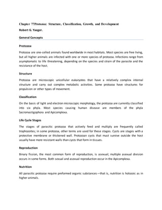
  • 4. Figure 77-1 shows the structure of the bloodstream form of a trypanosome, as determined by electron microscopy. Figure 77-1 Fine structure of a protozoan parasite, Typanosoma evansi, as revealed by transmission electron microcopy of thin sections. (Adapted from Vickerman K: Protozoology. Vol. 3 London School of Hygiene and Tropical Medicine, London, 1977, with permission.) Classification In 1985 the Society of Protozoologists published a taxonomic scheme that distributed the Protozoa into six phyla. Two of these phyla—the Sarcomastigophora and the Apicomplexa-- contain the most important species causing human disease. This scheme is based on morphology as revealed by light, electron, and scanning microscopy. Dientamoeba fragilis, for example, had been thought to be an ameba and placed in the family Entamoebidae. However, internal structures seen by electron microscopy showed that it is properly placed in the order Trichomonadida of flagellate protozoa. In some instances, organisms that appear identical under the microscope have been assigned different species names on the basis of such criteria as geographic distribution and clinical manifestations; a good example is the genusLeishmania, for which subspecies names are often used. Biochemical methods have been employed on strains and species to determine isoenzyme patterns or to identify relevant nucleotide sequences in RNA, DNA, or both. Extensive studies have been made on the kinetoplast, a unique mitochondrion found in the hemoflagellates and other members of the order Kinetoplastida. The DNA associated with this organelle is of great interest. Cloning is widely used in taxonomic studies, for example to study differences in virulence or disease manifestations in isolates of a single species obtained from different hosts or geographic regions. Antibodies (particularly monoclonal antibodies) to known species or to specific antigens from a species are being employed to identify unknown isolates. Eventually, molecular taxonomy may prove to be a more reliable basis than morphology for protozoan taxonomy, but the microscope is still the most practical tool for identifying a protozoan parasite.
  • 5. Table 77-1 lists the medically important protozoa. Table 77-1 Classification of Parasitic Protozoa and Associated Diseases. Life Cycle Stages During its life cycle, a protozoan generally passes through several stages that differ in structure and activity. Trophozoite (Greek for “animal that feeds”) is a general term for the active, feeding, multiplying stage of most protozoa. In parasitic species this is the stage usually associated with pathogenesis. In the hemoflagellates the terms amastigote, promastigote, epimastigote, and trypomastigote designate trophozoite stages that differ in the absence or presence of a flagellum and in the position of the kinetoplast associated with the flagellum. A variety of terms are employed for stages in the Apicomplexa, such as tachyzoite and bradyzoite for Toxoplasma gondii. Other stages in the complex asexual and sexual life cycles seen in this phylum are the merozoite (the form resulting from fission of a multinucleate schizont) and sexual stages such as gametocytes and gametes. Some protozoa form cysts that contain one or more infective forms. Multiplication occurs in the cysts of some species so that excystation releases more than one organism. For example, when the trophozoite of Entamoeba histolytica first forms a cyst, it has a single nucleus. As the cyst matures nuclear division produces four nuclei and during excystation four uninucleate metacystic amebas appear. Similarly, a freshly encysted Giardia lamblia has the same number of internal structures (organelles) as the trophozoite. However, as the cyst matures the organelles double and two trophozoites are formed. Cysts passed in stools have a protective wall, enabling the parasite to survive in the outside environment for a period ranging from days to a year, depending on the species and environmental conditions. Cysts formed in tissues do not usually have a heavy protective wall and rely upon carnivorism for transmission. Oocysts are stages resulting from sexual reproduction in the Apicomplexa. Some apicomplexan oocysts are passed in the feces of the host, but the oocysts of Plasmodium, the agent of malaria, develop in the body cavity of the mosquito vector.
  • 6. Reproduction Reproduction in the Protozoa may be asexual, as in the amebas and flagellates that infect humans, or both asexual and sexual, as in the Apicomplexa of medical importance. The most common type of asexual multiplication is binary fission, in which the organelles are duplicated and the protozoan then divides into two complete organisms. Division is longitudinal in the flagellates and transverse in the ciliates; amebas have no apparent anterior-posterior axis. Endodyogeny is a form of asexual division seen in Toxoplasma and some related organisms. Two daughter cells form within the parent cell, which then ruptures, releasing the smaller progeny which grow to full size before repeating the process. In schizogony, a common form of asexual division in the Apicomplexa, the nucleus divides a number of times, and then the cytoplasm divides into smaller uninucleate merozoites. In Plasmodium, Toxoplasma, and other apicomplexans, the sexual cycle involves the production of gametes (gamogony), fertilization to form the zygote, encystation of the zygote to form an oocyst, and the formation of infective sporozoites (sporogony) within the oocyst. Some protozoa have complex life cycles requiring two different host species; others require only a single host to complete the life cycle. A single infective protozoan entering a susceptible host has the potential to produce an immense population. However, reproduction is limited by events such as death of the host or by the host's defense mechanisms, which may either eliminate the parasite or balance parasite reproduction to yield a chronic infection. For example, malaria can result when only a few sporozoites of Plasmodium falciparum—perhaps ten or fewer in rare instances—are introduced by a feeding Anopheles mosquito into a person with no immunity. Repeated cycles of schizogony in the bloodstream can result in the infection of 10 percent or more of the erythrocytes—about 400 million parasites per milliliter of blood. Nutrition The nutrition of all protozoa is holozoic; that is, they require organic materials, which may be particulate or in solution. Amebas engulf particulate food or droplets through a sort of temporary mouth, perform digestion and absorption in a food vacuole, and eject the waste substances. Many protozoa have a permanent mouth, the cytosome or micropore, through which ingested food passes to become enclosed in food vacuoles. Pinocytosis is a method of ingesting nutrient materials whereby fluid is drawn through small, temporary openings in the body wall. The ingested material becomes enclosed within a membrane to form a food vacuole. Protozoa have metabolic pathways similar to those of higher animals and require the same types of organic and inorganic compounds. In recent years, significant advances have been made in devising chemically defined media for the in vitro cultivation of parasitic protozoa. The resulting organisms are free of various substances that are present in organisms grown in complex media or isolated from a host and which can interfere with immunologic or
  • 7. biochemical studies. Research on the metabolism of parasites is of immediate interest because pathways that are essential for the parasite but not the host are potential targets for antiprotozoal compounds that would block that pathway but be safe for humans. Many antiprotozoal drugs were used empirically long before their mechanism of action was known. The sulfa drugs, which block folate synthesis in malaria parasites, are one example. The rapid multiplication rate of many parasites increases the chances for mutation; hence, changes in virulence, drug susceptibility, and other characteristics may take place. Chloroquine resistance in Plasmodium falciparum and arsenic resistance in Trypanosoma rhodesiense are two examples. Competition for nutrients is not usually an important factor in pathogenesis because the amounts utilized by parasitic protozoa are relatively small. Some parasites that inhabit the small intestine can significantly interfere with digestion and absorption and affect the nutritional status of the host; Giardia and Cryptosporidium are examples. The destruction of the host's cells and tissues as a result of the parasites' metabolic activities increases the host's nutritional needs. This may be a major factor in the outcome of an infection in a malnourished individual. Finally, extracellular or intracellular parasites that destroy cells while feeding can lead to organ dysfunction and serious or life-threatening consequences. References 1. Englund PT, Sher A (eds): The Biology of Parasitism. A Molecular and Immunological Approach. Alan R. Liss, New York, 1988 . 2. Goldsmith R, Heyneman D (eds): Tropical Medicine and Parasitology. Appleton and Lange, East Norwalk, CT, 1989 . 3. Lee JJ, Hutner SH, Bovee EC (eds): An Illustrated Guide to the Protozoa. Society of Protozoologists, Lawrence, KS, 1985 . 4. Kotler DP, Orenstein JM. Prevalence of Intestinal Microsporidiosis in HIV-infected individuals referred for gastrointestinal evaluation. J Gastroenterol. 1994;89:1998. [PubMed] 5. Neva FA, Brown H: Basic Clinical Parasitology, 6th edition, Appleton & Lange, Norwalk, CT, 1994 .
  • 8. Acanthamoeba – Free Living Organism FREE-LIVING AMOEBAAmphizoic amoebae - They have also beencalled amphizoic amoebaebecause these amoebaehave the ability to exist asfree-living organisms innature and only occasionallyinvade a host and live asparasites within host tissue. ACANTHAMOEBA - A microscopic, free-living amoeba that can cause rare, but severe infections of the eye, skin, and central nervous system.Several species of Acanthamoeba, including A. culbertsoni, A. polyphaga, A. castellanii, A. astronyxis, A. hatchetti, A. rhysodes, A. divionensis, A. lugdunensis, and A. lenticulata are implicated in human disease. The important species is A.culbertsoni ACANTHAMOEBA - Acanthamoeba spp. have been found in: • soil • heating, ventilating, and • fresh, brackish, and sea air conditioning systems water • mammalian cell cultures • Sewage • Vegetables • swimming pools • human nostrils and • contact lens equipment; throats • medicinal pools • human and animal brain, • dental treatment units skin, and lung tissues. • dialysis machines ACANTHAMOEBA - has two stages, cysts and trophozoites, in its life cycle. No flagellated stage exists as part of the life cycle. The trophozoites replicate by mitosis. When Acanthamoeba spp. enters the eye it can cause severe keratitis in otherwise healthy individuals, particularly contact lens users . When it enters the respiratory system or through the skin, it can invade the central nervous system by hematogenous dissemination causing granulomatous amebic encephalitis (GAE) or disseminated disease, or skin lesions in individuals with compromised immune systems ACANTHAMOEBALIFE CYCLE STAGES Free-living trophozoites and cysts occur in both the soil and freshwater. ACANTHAMOEBALife cycle: ACANTHAMOEBAThere are two morphological forms:  Trophozoite - A trophozoite is 20- 50µm in size - Rough exterior with several spine like projections(acanthopoda).  Cyst - Spherical and 15µm in diameter. Both forms can be the source of infection ACANTHAMOEBATrophozoite Cyst Feeding & dividing  Response to adversity Asexual  Dormant, resistant Cyst forming  Double-walled with pores ACANTHAMOEBAPathogenicity and Clinical Features:  Granulomatous Amebic Encephalitis (GAE) and disseminated infection primarily affect people with compromised immune systems.  Commonly seen in immunocompromised patients, including those with neoplasia, systemic lupus erythematosus, human immunodeficiency virus and tuberculosis Incubation period:  Unknown but estimated at weeks to months. The route of infection is aerosol or direct inoculation with hematogenous spread to the CNS.
  • 9. ACANTHAMOEBARisk Factors: Symptoms:• Alcoholism • Headache• Drug abuse • Confusion• Chemotherapy • fever,• Corticosteroids • Lethargy• Organ transplantation • Nausea and vomiting • SeizuresSigns: • Photophobia• Neck stiffness • Neck stiffness.• Focal neurological deficits • Patients may become• Patients may also develop frankly psychotic. raised intracranial pressure Acanthamoeba Keratitis A progressive disease of the cornea, which is sight-threatening Commonly seen in: - immunocompetent patients. - However, infection does not confer immunity and reinfection is common. Risk factors:• poor contact lens hygiene• corneal abrasion• exposure of the eye to contaminated water Acanthamoeba Keratitis Affected individual  Signs: may complain of: • Conjunctival hyperemia• Eye pain • Episcleritis• Eye redness • Scleritis• Blurred vision • Loosening of the corneal• Sensitivity to light epithelium. (photophobia) • Rarely, trophozoites can• Sensation of something infiltrate the corneal in the eye nerve and retina, leading• Excessive tearing to chorioretinitis Acanthamoeba KeratitisDiagnosis CSF wet mount -usually lymphocyte predominance and low glucose (motile trophozoites) Culture-Agar plates seeded with E.coli Immunofluorescence or polymerase chain reaction (PCR) Corneal scrape or biopsy Acanthamoeba KeratitisPrevention and ControlThese guidelines should be followed by all contact lens users to helpreduce the risk of eye infections: Visit your eye care provider for regular eye examinations. Wear and replace contact lenses according to the schedule prescribed by your eye care provider. Remove contact lenses before any activity involving contact with water, including showering, using a hot tub, or swimming. Wash hands with soap and water and dry before handling contact lenses. Acanthamoeba KeratitisPrevention and Control Clean contact lenses according to instructions from your eye care provider and the manufacturers guidelines. 1. Never reuse or top off old solution. Use fresh cleaning or disinfecting solution each time lenses are cleaned and stored. 2. Never use saline solution or rewetting drops to disinfect lenses. Neither solution is an effective or approved disinfectant. 3. Be sure to clean, rub, and rinse your lenses each time you remove your lenses. Rubbing and rinsing your contact lenses will aid in removing harmful microbes and residues. Acanthamoeba KeratitisPrevention and Control Store reusable lenses in the proper storage case. 1. Storage cases should be rubbed and rinsed with sterile contact lens solution (never use tap water), emptied, and left open to dry after each use. 2. Replace storage cases at least once every three months. Contact lens users with questions regarding which solutions are best for them should consult their eye care providers. They should also consult their eye care providers if they have any of the following symptoms: eye pain or redness, blurred vision, sensitivity to light, sensation of something in the eye, or excessive tearing.
  • 10. Granulomatous Amebic Encephalitis A serious infection of the brain and spinal cord that typically occurs in persons with a compromised immune system. Granulomatous Amebic EncephalitisSymptoms include:• Mental status changes body• Loss of coordination • Double vision• Fever • Sensitivity to light• Muscular weakness or • Other neurologic partial paralysis problems affecting one side of the ACANTHAMOEBATreatment: Granulomatous Amebic Encephalitis (GAE) is treated with pentamidine, usually in combination with one or more of the following: • Ketoconazole • Hydroxystilbamidine • Paromomycin • 5-fluorocytosine polymyxin • Sulfadiazine • Trimethoprim-sulfamethoxazole • Azithromycin ACANTHAMOEBATreatment: Acanthamoeba keratitis - Therapy should include the cationic antiseptic agents, of which chlorhexidine or polyhexamethylene biguanide (PHMB) is the most effective. Ocular lesions - Enucleation of ulcer and corneal transplant REFERENCES• Contact Lens News and Information. (2012, February 20). Contact Lens Solutions Ineffective Against Acanthamoeba, Study Finds. Retrieved August 10, 2012, from http://t3.gstatic.com/images?q=tbn:ANd9GcTB4chV7tr0UeG44UKnU9qSBv4IOy_0_vA JrnYHIvhM75iGwMF• Centers for Disease Control and Prevention (CDC). Acanthamoeba - Granulomatous Amebic Encephalitis (GAE); Keratitis. Retrieved August 11, 2012, from http://www.cdc.gov/parasites/acanthamoeba/disease.html• Centers for Disease Control and Prevention (CDC). Laboratory Identification of Parasites of Public Health Concern; Free-Living Amebic Infections. Retrieved August 11, 2012, from http://www.dpd.cdc.gov/dpdx/HTML/FreeLivingAmebic.htm• Simon Kilvington, PhD. (2008). Physiological Response of Acanthamoeba to Contact Lens Disinfectants [Powerpoint Format]. Retrieved August 11, 2012, from http://www.google.com.ph/url?sa=t&rct=j&q=physiological%20response%20of%20ac• Animal Planet. Monsters inside me. Acanthamoeba picture. Retrieved August 15, 2012, from http://animal.discovery.com/invertebrates/monsters-inside-me/acanthamoeba- keratitis/ REFERENCES• The University of Edinburgh. (2003, May 28). Acanthamoeba. Retrieved August 15, 2012, from http://www.bms.ed.ac.uk/research/others/smaciver/Comp1.jpg• Baylor College of Medicine. (2008, January, 30). Human Genome Sequencing Center; About Acanthamoeba castellani Neff. Retrieved August 15, 2012, from http://www.hgsc.bcm.tmc.edu/microbial- detail.xsp?project_id=163• Naveed, Khan. (2009). Acanthamoeba Biology and Pathogenesis. Retrieved August 15, 2012, from http://www.caister.com/supplementary/acanthamoeba/c10.html THANK YOU!• Edna Mae C. Genzola, RMT• Katherine Royce L. Panizales, RMT• Mary Jean D. Somcio, RMT
  • 11. Brain eating amoeba (Naegleria Fowleri) 1. 1. Naegleria Fowleri: The Brain-Eating Amoeba Neurobiology’s Worse Nightmare 2. 2. So what is Naegleria Fowleri? 3. 3. Naegleria Fowleri is:  A parasite  Typically found in fresh water sources  A rare infection  Cause of Primary Amebic Meningoencephalitis  But if infected, almost always results in death! 4. 4. What parts of the nervous system does it involve? • Central nervous system • The processing center that sends, receives, and interprets information from all parts of the body • Olfactory nerve (CN1) • Relays sensory information about smell to the brain • Olfactory bulbs • Structure that receives and processes the neural input about odours that is detected by nasal cavity cells • Brain cells • Aka nerve cells • Receive and relay information to other nerve cells 5. 5. What happens during an infection of N. Fowleri? • It attacks the central nervous system (via nose) • Attaches to olfactory nerve and heads to olfactory bulbs to feed on nerve tissue  necrosis & hemorrhaging • Travels further via nerve fibres to cranium floor and into brain • Consumes brain cells 6. 6. Brain = Life  This example of N. Fowleri demonstrates how necessary the brain is to life  Without the building blocks of the nervous system (i.e. neurons), the nervous system cannot function  Without the nervous system, we cannot live. 7. 7. Concluding Remarks on the Course  Through this course, I am now able to better analyze neurobiological events  It has first and foremost, provided me with the knowledge necessary to understand the parts of the nervous system, their functions and underlying mechanisms and the impacts of their malfunction. In the case of N. Fowleri, knowledge of how the nerve cells operate, allows me to understand that if they are impaired, there is a loss of communication between core body organs and this can lead to a poor functioning body or even death  By providing everyday examples throughout the lectures, I have seen that the brain really is at the core of every activity I engage in. Without a functioning brain, breathing patterns would not be accurate, initiation of movement would not occur, and body temperature would not be regulated. All of these things that I take for granted would not be functioning if it weren’t for the brain.  Lastly, application of the knowledge gained through the weekly quizzes have provided me with a firm understanding of the brain’s functions. Without having discussed N. Fowleri or olfaction in the course, I am able to understand the basic mechanism through which this microorganism operates, as it pertains to the nervous system. Given the knowledge that neurons do not regenerate, it is clear to me that if an individual were infected with N. Fowleri, that he or she would likely die since this microorganism kills brain cells which are necessary for life. 8. 8. Acknowledgements  Thanks to Dr. Peggy Mason and the staff at Coursera!  References:  http://en.wikipedia.org/wiki/Naegleria_fowleri  http://fox4kc.com/2014/07/11/brain-eating-amoeba-kills-johnson-county-resident/  http://tribune.com.pk/story/733683/brain-eating-amoeba-kills-two-in-karachi/  https://www.coursera.org/course/neurobio  Images/Photos from Google Images