1. Scientific letter
Fulminant myocarditis due to COVID-19
Miocarditis fulminante por COVID-19
To the Editor,
Coronaviruses are single-stranded RNA viruses that are widely
distributed in humans and other mammals. Although most
coronavirus infections in humans are mild, they have recently
caused 2 major pandemics: severe acute respiratory syndrome
(SARS) and Middle East respiratory syndrome (MERS), with
mortality rates of 10% and 37%, respectively.1
SARS coronavirus
2 (SARS-CoV-2) is a type of coronavirus first discovered and
isolated in December 2019 in Wuhan, central China, that is the
cause of the current pandemic known as COVID-19.
Common symptoms of the disease are fever, cough, myalgia,
and shortness of breath. The most serious complications include
acute respiratory distress syndrome (ARDS), cardiac injury, and
secondary superinfection.
The pathophysiology of this virus is still unknown. Various
studies indicate that patients infected with COVID-19 have high
concentrations of interleukin (IL)-1 beta, interferon (IFN) gamma,
IFN-inducible protein (IP)-10, and monocyte chemoattractant
protein (MCP)-1. It has been shown that patients with severe
illness have higher concentrations of granulocyte colony-stimu-
lating factor (GCSF), IP-10, MCP-1, macrophage inflammatory
protein (MIP)-1A, and tumor necrosis factor (TNF) alpha, indicating
that the severity of the illness could be determined by cytokine
storm.2
Patients infected by COVID-19 with cardiac injury have
noticeably higher plasma concentrations of IL-6,3,4
N-terminal
fraction of pro–brain natriuretic peptide (NT-proBNP), and cardiac
troponins (cTnI/T). Because cytokine storm is also the main
pathophysiologic mechanism in fulminant myocarditis, it is
reasonable to consider heart damage due to COVID-19.
The etiology of myocarditis is highly varied and includes a wide
range of infectious agents, systemic diseases, medications, and
toxins. The literature on myocarditis due to coronaviruses is scant,
but cardiac injury seems to be more common in patients infected
by COVID-19 than in patients infected by other coronaviruses.5
We describe the case of a 59-year-old woman with a history of
hypertension, cervical degenerative arthropathy, chronic lumbar
radiculopathy, lymph node tuberculosis diagnosed due to erythe-
ma nodosum, and migraine. Most notably, the patient’s regular
therapy included candesartan 32 mg/d.
In March 2020 the patient presented to the Emergency
Department due to a feverish feeling that had lasted 5 days,
accompanied by squeezing anginal chest pain in the absence of
respiratory symptoms. On arrival, O2 saturation was 96% with
nasal cannula at 2 L/min, and blood pressure was 75/53 mmHg.
Physical examination revealed signs of peripheral hypoperfusion
with normal pulmonary auscultation.
Despite fluid overloading and norepinephrine, the patient
remained hypotensive with signs of hypoperfusion (cool skin and
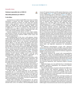
elevated lactic acid at 3.9 mmol/L). Electrocardiography showed
concave ST-segment elevation and PR-segment depression, as well
as low voltages (figure 1A). Chest X-ray indicated mild signs of
vascular redistribution, with no infiltrations (figure 1B). Polymer-
ase chain reaction (PCR) for viruses in the nasopharyngeal swab
specimen was positive for SARS-CoV-2 and negative for adenovirus
and influenza A and B viruses, with a positive epidemiologic
environment (family members with fever and respiratory symp-
toms in previous days). Laboratory work revealed elevated
troponins (TnT, 220-1100 ng/dL) and NT-proBNP (4421 ng/L),
mild leukocytosis (14.17 Â 109
/L), lymphocytes (2.59 Â 109
/L), CRP
10 mg/L, and D-dimer at 24 hours (23 242 ng/mL). Echocardiogra-
phy disclosed moderate concentric hypertrophy, diminished
intraventricular volumes with preserved left ventricular ejection
fraction without segmental abnormalities, and moderate pericar-
dial effusion with no clear signs of hemodynamic deterioration.
Due to a clinical picture indicative of myocarditis (diffuse concave
ST-segment elevation, fever, pericardial effusion, and myocardial
thickening) and preserved left ventricular ejection fraction without
segmental abnormalities, coronary angiography was not per-
formed due to a low clinical suspicion of acute coronary syndrome.
In the Coronary Unit, during implantation of a Swan-Ganz catheter,
the patient experienced rapid hemodynamic deterioration, exhi-
biting electrical activity with no pulse and requiring cardiopul-
monary resuscitation, emergent pericardiocentesis (drainage of
serous fluid), and high-dose vasopressors for hemodynamic
recovery.
An additional echocardiogram (2 hours after admission)
showed severe biventricular failure and diffuse myocardial edema
(figure 2A) and, therefore, the patient underwent balloon counter-
pulsation and venoarterial extracorporeal membrane oxygenation
(ECMO) via the femoral artery.
Myocarditis treatment was started with immunoglobulins
(80 mg/d) for 4 days and methylprednisolone (500 mg/d) at
tapering doses for 14 days and antiviral treatment consisting of IFN
B (0.25 mg/48 h) and ritonavir/lopinavir (400 mg/100 mg/12 h). By
the fifth day of hospitalization, biventricular function was normal,
but the ECMO device was maintained due to dyspnea (figure 2B)
and refractory hypoxemia, with respiratory progress currently
pending.
Myocardial biopsy was not performed due to hemodynamic
instability, significant coagulopathy, and subsequent improve-
ment in cardiac function.
The clinical presentation of patients infected with SARS-CoV-2
is highly variable, and respiratory symptoms are the most
common. In view of the current epidemiologic situation, this
etiologic agent should be considered as a possible cause in other
clinical conditions such as acute myocarditis, even in the absence
of consistent respiratory symptoms.
Fulminant myocarditis is a syndrome with high morbidity and
mortality; hence, early diagnosis and appropriate treatment are
vital. In our patient, the clinical picture was consistent with acute
myocarditis, with no initial respiratory symptoms and with rapid
clinical progression to cardiogenic shock and need for venoarterial
ECMO support. Normal biventricular function was regained within
Rev Esp Cardiol. 2020;73(6):503–515
2. a few days, with severe subsequent dyspnea that required
continued ECMO.
A´ ngela Irabien-Ortiz,a,
* Jose´ Carreras-Mora,b
Alessandro Sionis,b
Julia Pa`mies,b
Jose´ Montiel,a
and Manel Taurona
a
Servicio de Cirugı´a Cardiovascular, Hospital de la Santa Creu i Sant
Pau, Barcelona, Spain
b
Servicio de Cardiologı´a, Hospital de la Santa Creu i Sant Pau,
Barcelona, Spain
*Corresponding author:
E-mail address: angelairabien@hotmail.com (A´ . Irabien-Ortiz).
Available online 15 April 2020
REFERENCES
1. Ksiazek TG, Erdman D, Goldsmith CS, et al. A novel coronavirus associated with
severe acute respiratory syndrome. N Engl J Med. 2003;348:1953–1966.
2. Huang C, Wang Y, Li X, et al. Clinical features of patients infected with 2019 novel
coronavirus in Wuhan, China. Lancet. 2020. http://dx.doi.org/10.1016/S0140-
6736(20)30183-5.
3. Hongde Hu. Fenglian Ma. Xin Wei. Yuan Fangn.. Coronavirus fulminant myocarditis
saved with glucocorticoid and human immunoglobulin. Eur Heart J. 2020. http://
dx.doi.org/10.1093/eurheartj/ehaa190.
4. Fontes JA, Rose NR, Cˇiha´kova´ D. The varying faces of IL-6: from cardiac protection to
cardiac failure. Cytokine. 2015;74:62–68.
5. WHO Statement on the third meeting of the IHR Emergency committee concerning
Middle East respiratory syndrome coronavirus (MERS-CoV). Wkly Epidemiol Rec.
2013;88:435-436.
https://doi.org/10.1016/j.rec.2020.04.005
1885-5857/
C 2020 Sociedad Espan˜ola de Cardiologı´a. Published by Elsevier Espan˜a, S.L.U. All
rights reserved.
Arterial tortuosity syndrome: a late and unexpected
diagnosis and description of a novel likely pathogenic
mutation
Sı´ndrome de tortuosidad arterial: un diagno´stico tardı´o e
inesperado y la descripcio´n de una nueva mutacio´n
probablemente patoge´nica
To the Editor,
We describe a male patient born in June 1965. His parents are
first cousins, he is their second and last child, and he has no
descendants. At the age of 33 years, the patient experienced
palpitations, and the episode was diagnosed as atrial fibrillation
with rapid ventricular response. At that time, he started to be
studied at the cardiology service, where he was also diagnosed
with dyslipidemia, hypertension, hypertensive cardiomyopathy,
and mild aortic regurgitation with a tricuspid aortic valve. Physical
examination revealed the presence of palpebral bilateral ptosis,
hypertelorism, and long face. In addition, the patient has multiple
diseases requiring follow-up by other specialties. These diseases
include high bilateral myopia, producing left-eye blindness due to
retinal detachment, chronic bilateral otitis media requiring
bilateral tympanoplasty, and erectile dysfunction, as well as
Figure 1. A, electrocardiogram on admission showing concave ST-segment
elevation, PR-segment depression, and low voltages. B, chest X-ray on
admission.
Figure 2. A, echocardiogram showing myocardial edema and pericardial
effusion. B, chest X-ray indicative of dyspnea.
Scientific letter / Rev Esp Cardiol. 2020;73(6):503–515504