Breast cancer diagnosis staging screening....koustav
•Télécharger en tant que PPT, PDF•
13 j'aime•2,676 vues

COMMONEST CANCER AMONG FEMALE IN INDIA.....SEMINER PRESENTED IN MEDICAL COLLEGE & HOSPITAL, KOLKATA ..

Recommandé
Contenu connexe
Tendances
Tendances (20)
En vedette
En vedette (20)
Similaire à Breast cancer diagnosis staging screening....koustav
Similaire à Breast cancer diagnosis staging screening....koustav (20)
Dernier
Dernier (20)
Breast cancer diagnosis staging screening....koustav
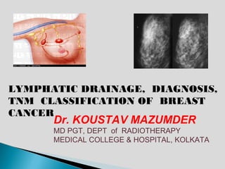
- 1. LYMPHATIC DRAINAGE, DIAGNOSIS, TNM CLASSIFICATION OF BREAST CANCER Dr. KOUSTAV MAZUMDER MD PGT, DEPT of RADIOTHERAPY MEDICAL COLLEGE & HOSPITAL, KOLKATA
- 2. • Breast cancer may be one of the oldest known forms of cancerous tumors in humans. • The oldest description of cancer was discovered in Egypt and dates back to approximately 1600 BC. The Edwin Smith Papyrus describes 8 cases of tumors or ulcers of the breast that were treated by cauterization. • The French surgeon Jean Louis Petit (1674–1750) and later the Scottish surgeon Benjamin Bell (1749–1806) were the first to remove the lymph nodes, breast tissue, and underlying chest muscle . • Their successful work was carried on by William Stewart Halsted who started performing mastectomies in 1882 • The first case-controlled study on breast cancer epidemiology was done by Janet Lane-Claypon, who published a comparative study in 1926 of 500 breast cancer cases and 500 control patients of the same background and lifestyle for the British Ministry of Health
- 3. • LYMPHATIC DRAINAGE • DIAGNOSIS • TNM CLASSIFICATION
- 4. SUPLACLAVICULAR LN INTERNAL MAMMARY LN AXILLARY LN AXILLARY LN
- 5. LEVEL III LEVEL II LEVEL I
- 6. Axillary vein Central Axillary Nodes Apical axillary nodes Lateral Axillary Anterior Nodes axillary nodes Posterior Pectoralis Axillary nodes minor Lateral Thoracic vein Subscapular vein
- 7. Pectoralis minor Pectoralis major Interpectoral node Internal mammary node
- 8. LYMPHTIC DRAINAGE OF BREAST Draining the PARENCHYMA Draining the overlying SKIN except of BREAST AREOLA and NIPPLE Including AREOLA and NIPPLE
- 9. Draining the overlying SKIN except AREOLA and NIPPLE Supraclavicular Infraclavicula LN LN Internal mammary LN Axillary LN Anterior abdominal wall
- 10. Draining the overlying SKIN except AREOLA and NIPPLE Supraclavicular Infraclavicula LN LN Internal mammary LN Axillary LN Subperitoneal lymphatic plexus Sub Diaphragmatic Hepatic Nodes node
- 11. Draining the PARENCHYMA of BREAST Including AREOLA and NIPPLE Chest wall Subareolar plexus of sappay Pectoralis major lobules nipple areola Lymphatic Lake of Haller Lactiferous duct Retromammary fat
- 12. Draining the PARENCHYMA of BREAST Including AREOLA and NIPPLE Supraclavicular Infraclavicula LN LN Internal mammary LN Axillary LN 75%
- 13. Draining the PARENCHYMA of BREAST Including AREOLA and NIPPLE Supraclavicular Infraclavicula LN LN Internal mammary LN Axillary LN
- 17. SCREENING • CLINICAL BREAST EXAMINATION • BREAST AWARENESS • RADIOLOGICAL INVESTIGATION
- 19. MAMMOGRAPHY BI-RADS (Breast Imaging Reporting And Data System)
- 24. PERFORMS= PERsonal perFORmance in Mammographic Screening
- 25. SCREENING GUIDELINE in NCCN 2012 Woman at normal risk Woman at increased risk •Prior Thoracic irradiation •>35 yrs 20-39 yrs >40 yrs •Lifetime risk >20% •CBE every 1-3 yrs •Annual CBE •F/H or genetic predisposition •Breast awareness •Breast awareness •LCIS/ Atypical hyperplasia •Mammography •H/O Breast Cancer
- 28. DIAGNOSIS • HISTORY & CLINICAL EXAMINATION • RADIOLOGICAL EVALUATION • BIOPSY
- 29. RADIOLOGICAL EVALUATION • Diagnostic Mammography Spot compression view or magnifiacation view • Breast ultrasonography woman< 30 yrs of age, woman>30 yrs age (BIRADS 1-3) spontaneous nipple discharge/ skin change BIRADS category 0 • Diagnostic Breast MRI BIRADS 1-3, IBC
- 30. BREAST BIOPSY • Fine needle aspiration(FNA) Biopsy • Core needle Biopsy Non palpable lesion • Excisional Biopsy Atypical hyperplasia, LCIS, mucin producing tumor, Phylloids • Duct excision(with or without ductography) Non sponteneous discharge from duct with BIRADS 1-3
- 31. Guidelines for the basic elements of a pathology report for breast cancer have been established by the College of American Pathologists
- 35. • LYMPHATIC DRAINAGE • DIAGNOSIS • TNM CLASSIFICATION
- 36. • PRIMARY TUMOR (T) • REGIONAL LYMPH NODE(N) • DISTANT METASTASES(M)
- 39. PRIMARY TUMOR (T) REGIONAL LYMPH NODE(N) CLINICAL PATHOLOGICAL
- 41. N1 Metastasis to movable ipsilateral level I, II axillary lymph node(s)
- 42. N2a Metastasis in ipsilateral level I, II axillary lymph nodes fixed to one another (matted) or to other structures
- 43. N2b Metastasis only in clinically detected ipsilateral internal mammary nodes and in the absence of clinically evident level I, II axillary lymph node metastasis
- 44. N3a Metastasis in ipsilateral infraclavicular lymph node(s)
- 45. N3b Metastasis in ipsilateral internal mammary lymph node(s) and axillary lymph node(s)
- 46. N3c Metastasis in ipsilateral supraclavicular lymph node(s)
- 49. pN1a Metastasis in 1 to 3 axillary lymph nodes(at least one >2 mm)
- 50. pN1b Metastasis in internal mammary nodes with micrometastasis or macrometastasis detected by SLNB but not clinically detected
- 51. pN1c Metastasis in 1 to 3 axillary lymph nodes and in internal mammary lymph nodes with micrometastasis or macrometastasis detected in SLNB but not clinically detected
- 52. pN2a Metastasis in 4 to 9 axillary lymph nodes (at least one tumor deposit greater than 2.0 mm)
- 53. pN2b Metastasis in clinically detected internal mammary lymph nodes in the absence of axillary lymph node metastases
- 54. pN3a Metastasis in 10 or more axillary lymph nodes(at least one tumor deposit >2 mm
- 55. pN3a metastasis to the infraclavicular (level III) lymph nodes
- 56. >1 pN3b Metastases in clinically detected ipsilateral internal mammary lymph nodes in the presence of 1 or more positive axillary lymph nodes
- 57. 2 pN3b more than 3 axillary lymph nodes and in internal mammary lymph nodes with micrometastases or macrometastases detected by sentinel lymph node biopsy but not clinically detected
- 58. pN3c Metastasis in ipsilateral supraclavicular lymph nodes
- 59. • PRIMARY TUMOR (T) • REGIONAL LYMPH NODE(N) • DISTANT METASTASES(M)
- 61. Stage 0 Stage IIIA • Tis, N0, M0 T0, N2, M0 T1, N2, M0 Stage IA T2, N2, M0 • T1, N0, M0 T3, N2, M0 T3, N1, M0 Stage IB • • T0, N1mi, M0 T1, N1mi, M0 Stage IIIB T4, N0, M0 Stage IIA T4, N1, M0 T4, N2, M0 • T0, N1, M0 • T1, N1, M0 • T2, N0, M0 Stage IIIC Any T, N3, M0 Stage IIB • T2, N1, M0 Stage IV • T3, N0, M0 Any T, Any N, M1
- 64. HISTOLOGIC GRADE (G) ELSTON- ELLIS modification of SCARFF- BLOOM- RECHARDSON grading system • TUBULE FORMATION • NUCLEAR PLEOMORPHISM • MITOTIC COUNT