Kodo Millet PPT made by Ghanshyam bairwa college of Agriculture kumher bhara...
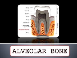
Alveolar bone
1.
2. Introduction
Bone Histology
Cells and Intercellular Matrix
Bone Development
Remodelling
Age Changes
Clinical Considerations
Conclusion
References
3.
4. Bone- used to designate
both an organ and a
tissue
Specialized mineralized
connective tissue
5. mineralised supporting tissue
act as a reservoir for ions
(especially calcium).
provide a framework for bone marrow
gives attachment to muscles
its "plasticity', allows it to remodel according to the
functional demand placed upon it.
6. DEVELOPMENTALLY,
Endochondral bone
Intramembranous bone
HISTOLOGICALLY, according to its density, mature bone can
be divided into;
Compact (cortical) bone
Cancellous (spongy) bone
7. MICROSCOPICALLY:
Lamellar bone
Fibrous bone
LAMELLAR BONE:
Most of the bones, whether compact or cancellous, are composed of
thin plates of bony tissues called lamellae.
These are arranged in piles in a cancellous bone, but in concentric
cylinders (Haversian system or secondary osteon) in a compact bone.
8. FIBROUS BONE (WOVEN BONE):
It is found in young fetal bone.
Collagen fibers - more variable diameter
Irregular orientation giving it matted appearance
9. Alveolar process is dependent on the presence of teeth for its
development and maintenance.
At the late bell stage, bony septa and bony bridge start to form,
and separate the individual tooth germs from another, keeping
individual tooth germs in clearly outlined bony compartment.
(BERKOVITZ)
10. Initially, this bone forms a
thin egg shell of support,
termed as the ‘tooth crypt’,
around each tooth germ.
11. FIG. 9-5 A developing root shown by a
divergent apex around the dental
papilla (arrow), which is enclosed by
an opaque bony crypt.
12. Relationship between
a deciduous tooth & its
accompanying
succedaneous tooth
detailing the formation
of the alveolar bone
- Scoh, Symonds 1974
12/85
AT BIRTH AT 7MONTHS
AT 2½YRS 7YRS
14. Osteocalcin, Osteonectin, Bone morphogenic proteins,
Phosphoproteins and Proteoglycans
Ground substance- Glycosaminoglycans, proteoglycans and
water
Osteopontin, Bone Sialoprotein- cell adhesion proteins
(Mackie et al, 2003)
15. Osteocalcin (bone GLA protein)
Found in bone matrix
Expressed only by fully differentiated cells
Specifically localized to developing bone
Produced by osteoblasts and odontoblasts
Role in bone formation
16. Osteopontin
Glycosylated phosphoprotein
Role in bone formation and resorption
Synthetized by osteoblasts, osteoclasts, osteocytes, smooth
muscles and epithelial cells
Role in cell adhesion
Significant amounts at mineralizing front
19. Inorganic material- calcium, phosphate ,hydroxyl, carbonate,
citrate
Trace amounts of sodium, magnesium and fluorine (Glimcher
1990)
Hydroxyapetite crystals of ultramicroscopic size
Enzymes like alkaline phosphatase, ATP and pyrophosphatase
Parallel to collagen fibres and contribute to lamellar appearance
of bone
20. Portion of maxilla and mandible that forms and supports the
tooth sockets (alveoli)
Forms when tooth erupts to provide osseous attachment to
PDL
Disappears gradually after tooth loss
‘Tooth dependent bony structure’ (Schroeder et al, 1991)
24. Holds the tooth firmly in position during mastication
Aids in movement
Adapts to occlusal loads
Helps to move the teeth for better occlusion.
Functions of alveolar bone
25. Supplies vessels to the PDL.
Houses & protects developing permanent teeth while
supporting primary teeth.
Organizes successive eruptions of primary & secondary teeth.
26.
27. Three parts
1) External plate of cortical bone
2) Inner socket wall
3) Cancellous trabeculae (between two compact layers)-
function of support
28. 1) Circumferential lamellae (encloses entire adult bone and
forms the outer perimeter
29. 2) Concentric lamellae (make up bulk of compact bone and
forms the basic metabolic unit of bone, the osteon)
3) Interstitial lamellae (inter-spread between adjacent concentric
lamellae and fill the spaces between them..actually fragments of
pre-existing concentric lamellae and can be of many shapes)
30. Osteon –cylinder of bone parallel to
long axis of bone (structural and
metabolic units)
Haversian canal –in centre of osteon,
lined by single layer of bone cells
Each canal has a capillary
31. Haversian canals
interconnected by Volkmann
canals
System for dense bones like
cortical plates and alveolar
bone proper, where surface
vessels are unable to supply
blood
32. Dense , lamellated bone – alveolar bone proper (contains
sharpeys fibers and circumferential lamellae)
34. Bone adjacent to PDL that contain sharpeys fibers
Contains higher calcium than other areas
Many features in common with cementum layer on root
surface
Collagen fibers larger in diameter, less numerous , less mature
35. Localized within alveolar bone proper
Sharpeys fibers completely calcified or partially calcified with
uncalcified core
Not unique to jaw -occurs wherever ligaments and muscles
are attached
Thickness of 100-200 microns
High turnover rate
36. FIBER ARRANGEMENT IN ABP
DOUBLE FIBRILLAR ORIENTATION:
Extrinsic fibers- Sharpey’s fibers
run perpendicular to bone surface
produced by PDL fibroblast
At their insertion in bone, they become mineralized, with their periphery
being hypermineralized than cores.
Intrinsic fibers
Laid down by osteoblasts between Sharpey’s fibers
Irregularly arranged & less dense.
37. Presence of trabeculae enclosing irregular marrow spaces
lined with a layer of thin, flattened endosteal cells
Variation in trabeculae pattern depending upon occlusal forces
and genetically
Matrix consists of irregularly arranged lamellae separated by
incremental and resorption lines
38. Found in inter-radicular and inter-dental spaces
Maxilla>mandible
Trabeculae alligned in path of tensile and compressive stresses
to provide maximal resistance to occlusal forces with
minimum bone substance (Glickman et al 1970)
in thickness and number with force
39. Spongy bone (anatomic term)
Trabecular bone (radiographic term)
Cancellous bone (histologic term)
40. Type 1: The interdental and interradicular trabeculae are regular
and horizontal in a ladder like arrangement.
Type 2: Shows irregularly arranged numerous delicate
interdental and interradicular trabeculae
41. CORTICAL BONE SPONGY BONE
About 85% of bone About 15% of bone
Lesser turnover than spongy Higher turnover
Remodel about 3% of its mass
each year
remodel about 25% of its mass
each year
Mechanical/protective role More metabolic function
42. Consists of cancellous bone
bordered by alveolar bone
proper of approximating
teeth and facial and lingual
cortical plates
Narrow septa- only
cribriform plate
Irregular window
43. Study by Heins et al 1986
Area Cribriform
plate+cancell
ous bone
Only
cribriform
plate
Irregular
window
Maxillary
molars
66.6% 20.8% 12.5%
Mandibular
premolar and
molar
85% 15% 0%
44. Mesiodistal angulation of IDS is parallel to line drawn
between CEJ of approximating teeth (Ritchey et al, 1953)
Shape and size of IDS depends on
1) Size and convexity of crowns of approximating teeth
2) Position of teeth
3) Degree of eruption
45. Crest of IDS located 1-2 mm apical to CEJ of adjacent teeth
46. Diagram of relation between CE junction of adjacent teeth shape of
crest of alveolar septa
47. • Embryo and newborn,
• Ribs, sternum, vertebrae, skull, humerus
• Hemopoiesis
Red
hematopoietic
marrow
• Adult
• Red marrow foci found sometimes in
maxillary tuberosity, symphysis and angle
of ramus
• Storage of energy
Yellow fatty
marrow
52. Produce organic matrix of bone
Differentiated from pluripotent
follicle cells
No decrease with age
Uninuclear cells
Secrets collagen as well as non
collagenous proteins
Present on outer bone surface
53. Have high levels of alkaline
phosphatase (this feature
distinguishes it from fibroblasts)
Alkaline phosphatase believed
to cleave organically bound
phosphate and help in bone
growth
Active-plump, cuboidal
Inactive-flattened
54. Secrete type Ӏ and V collagen,
variety of cytokines and several
members of BMP such as BMP-
2, BMP-7, TGF-ß, IGF-1, IGF-2
BMP family helps in bone
formation and repair
Under physiologic condition
which support resorption- release
of IL-6 and hydrolytic enzymes
55. Enclosed within spaces
called lacunae within
calcified matrix
Entrapped Osteoblasts
Reduction in size and loss of
matrix synthesizing ability
after being entrapped
Excess space-lacunae
56. Extend processes into canaliculi
that radiate from lacunae
Anastomosing system
Bring O2 and nutrients to
osteocytes through blood and
remove metabolic waste products
57. More rapid the bone formation-more osteoblasts get
entrapped – more osteocytes (eg- bone formed during repair)
Osteolytic osteolysis- osteocytes capable of resorption
58. Quiescent osteocytes:
paucity of rER, diminished golgi apparatus
An osmiophilic lamina representing mature calcified matrix is seen in close apposition to cell
membrane.
Formative osteocyte:
abundant rER & golgi apparatus
evidence of osteoid in pericellular space within the lacuna.
Resorptive osteocyte:
Numerous ER & well developed golgi apparatus.
The pericellular space is devoid of collagen fibrils & may contain a flocculent material
suggestive of breakdown product.
‘Osteocytic osteolysis’.
59. Originate from hematopoietic tissue
Fusion of mononuclear cells (blood
derived monocytes) to form a
multinucleated cell
Very large, 5-50 nuclei
Active on less than 1% of bone surface
Mobile and capable of migrating
60. Lie in Howships lacunae
Acidophilic cytoplasm
Active osteoclasts- ruffled
border facing bone
(hydrolytic enzymes are
secreted)
Increases surface area
61. Clear zone devoid of organelles
but rich in actin filament,
vinculin, talin (site of adhesion of
osteoclast to bone)
Sealing zone
Ruffled border-enzymes like
tartarate resistant acid
phosphatase, carbonic anhydrase,
proton pump ATP’s
Cathepsin containing
cytoplasmic vesicles near
ruffled border
62.
63.
64. 1. Attachment of the osteoclast to mineralized bone surface
2. Creation of sealed acidic environment through action of proton
pump which demineralizes bone & exposes the organic matrix
3. Degradation of the exposed organic matrix to its constituent
amino acids by the action of released enzymes like acid
phosphatase & cathepsin
4. Sequestering of the mineral ions & amino acids within the
osteoclasts.
Tencate 1994- Described sequence of events of resorptive
process:
65. - When bone is no longer forming…..surface
osteoblasts become inactive ….. Lining cells.
- Thin flat nucleus, few cytoplasmic organelles
- Retain gap junctions with osteocytes….functions
to control mineral homeostasis & endure bone
vitality.
66. Both are layers of differentiated osteogenic connective tissue
Periosteum covers outer surface of bone and endosteum lines
the internal bone cavities
Bundles of collagen fibres from outer layer penetrate bone and
bind periosteum to bone
Endosteum composed of a single layer of osteoblasts with
some connective tissue
67. • Rich in blood vessels, nerves
• Contains collagen fibres and
fibroblasts
• Fibrous periosteum
Outer
layer(fibrous)
• Composed of osteoblasts and
osteoprogenitor cells
• Cellular periosteum
Inner layer
(osteogenic)
68.
69. Medium through which muscles, tendons and
ligaments are attached to bone
Nutritive function to the bone
Osteoprogenitor cells – Important role during
development and repair after fracture
Fibrous layer- acts as limiting membrane
(exostoses in cases of periosteal tear)
70.
71. 1) Endochondral bone formation
2) Intramembranous bone formation
3) Sutural bone formation
72. Cartilage replaced by bone
Shape of cartilage resembles miniature
version of bone to be formed
At end of long bones, vertebrae, ribs,
head of mandible and base of skull
Condensation of mesenchymal cells
73. Perichondrium at the periphery
Rapid growth of cartilage
Cartilage replaced by bone gradually
by osteoblasts at periphery
74. Occurs directly within mesenchyme
Bone develops directly within the soft connective tissue
Vascularity increases and osteoblasts differentiate and lay
down bone
Occurs at multiple sites (primary ossification center)
75. Ossification centers grow radially
Cranial vault, maxilla, body of mandible and mid shafts of long
bones
Proceeds at extremely rapid rate
Woven bone formed first in form of radiating spikules which
ultimately fuse to form plates
Transition of woven bone to lamellar bone
77. Some differentiate into osteoblasts and lay down osteoid
Which then gets calcified
Mineralization always lags behind the production of bone matrix
78. Bone forms along suture
margins
Found in skull
Fibrous joints between bones
Allow only limited movement
Helps skull and face to
accommodate growing organs
like eyes and brain
79. Vascular supply
Derived from inferior and superior alveolar arteries of maxilla and
mandible
Lymphatic drainage
Submandibular lymph nodes
Nerve supply
Branches from anterior, middle and posterior superior alveolar
nerves for maxilla and branches from inferior alveolar nerve for
mandible
80. Bone contour follows root prominence
Intervening vertical depressions that taper
towards margin
Height of facial/lingual plates affected by
1) Allignment of teeth
2) Angulation of root to bone
3) Occlusal force
81. Normally: prominence of the roots
with the intervening vertical
depressions that taper toward the
margin.
On the labial version: the margins
of the labial bone is thinned to a
knife edge & presents an
accentuated arc in the direction of
the apex.
On the lingual version: the
margins of the labial bone is blunt
& rounded & horizontal rather
than arcuate.
82. Buttressing bone- adaptive mechanism against occlusal force
(thickened cervical portion of alveolar plate)
83. Fenestration- Isolated areas in
which root is denuded of bone
and root surface covered only
by periosteum and overlying
gingiva
Dehiscence- Denuded area
extends through marginal bone
84. Facial > lingual
Anteriors > posteriors
Frequently bilateral
20% of all teeth affected
Caused due to malposition, root prominence, labial protrusion
and a thin cortical plate
Can complicate procedure and outcome of periodontal surgery
85.
86. Least stable of periodontal tissues
Structure in a constant state of flux
• Functional requirements
• Age related changes in
bone cells
Local
influences
• Hormones (PTH, vit D,
calcitonin)Systemic
influences
87. Remodeling is the major pathway of bone
changes in
shape,
resistance to forces,
repair of wounds, and
calcium and phosphate homeostasis in the body.
REMODELING
88. Regulation of bone remodelling is a complex process
involving hormones and local factors acting in a autocrine and
paracrine manner on the generation and activity of
differentiated bone cells – Sodek et al 2000
Bone-99% of body calcium ions
Major source of calcium release when blood Ca
Monitored by parathyroid gland
89. Decrease in blood Ca
Detected by receptors on chief cells
of parathyroid gland
Release of PTH
Stimulate osteoblasts
to release IL-1 and IL-6
Stimulates monocytes
to migrate to area
Monocytes coalesces to form
multinucleated osteoclasts in
presence of LIF-
Leukemia inhibiting factor
released by osteoblasts
Bone resorption
Release of Ca ions
from hydroxyapetite
crystals
Normal blood calcium levels
PTH secretion stopped by
feedback mechanism
Organic matrix resorbed
with hydroxyapetite
Collagen breakdown
Release of organic substrate which
are covalently bound to collagen
Stimulates differentiation Bone deposition
90. ‘COUPLING’ refers to interdependency of osteoclasts and
osteoblasts in remodelling
Bone multicellular unit (BMU)
Reversal line
94. Similar to those occurring in remainder of skeletal system
Osteoporosis with ageing
Decreased vascularity
Reduction in metabolic rate and healing capacity(implants,
extraction sockets, bone grafts)
Bone resorption may be increased or decreased
More irregular periodontal surface
95. Thinning of cortical plates
Rarification of bone
Reduction in no of trabeculae
Lacunar resorption more prominent
Susceptibility to fracture
Thickening of collagen fibers
Decrease in water content
96.
97. - Gingival margins …follows the contour of alveolar process.
Abnormalities such as ledges, exostosis & tori…reflect on
gingiva.
- Areas of fenestrations & dehiscence - partial thickness flap.
98. - Process of bone remodeling - in orthodontic treatment.
- Knowledge of the various factors regulating bone formation
has resulted in their use for regeneration of bone.
99. Buccal-lingual/palatal ridge resorption
during first 3 months after extraction
about 30%... Reaching 50% at the end of
1 year (Schropp et al , 2003)
Resorption more pronounced at buccal
than lingual/palatal aspect of ridge
leading to shift of center of ridge towards
lingual/palatal side
101. Classification (Lekhom and Zarb- 1985)
4 bone qualities for the anterior regions of the jaw bone:
Quality1, Quality 2, Quality 3, Quality 4
102. Misch Bone Density Classification
D1-dense cortical
D2-porus cortical and coarse trabecular
D3-porus cortical and fine trabecular
D4-fine trabecular
103. Local response to a noxious stimulus.
A process by which tissue forms faster than the normal regional
regeneration process.
- Frost et al, 1983
By enhancing the various healing stages, this phenomena makes the
healing process occur 2 – 10 times faster than normal physiologic
healing.
RAP begins within a few days of injury, typically peaks at 1 - 2
months, usually lasts 4 months in bone, & may take 6 - >24 months
to subside.
104. Duration & intensity of RAP α type & amount of stimulus & the
site where it was produced.
Noxious stimuli of sufficient magnitude: can evoke RAP.
Fractures
Mechanical abuses
Noninfectious inflammatory injuries: dental implant procedures
Bone grafting surgeries
Internal fixation procedures
Mucoperiosteal surgery
105.
106. Injury to bone: Pathologic process
Arthrofibrosis
Neuropathic soft tissue problems
Rheumatoid phenomena
Secondary osteoporosis
Excessice heat
RAP is delayed / not initiated.
Formation of biologically delayed union / nonunion.
107. RAP does not result in a change in bone volume.
Restricted to bone remodelling.
More evident in cortical bone.
Usually accompanied by a systemic response: Systemic
Acceleratory Phenomena
Biochemical agents also appear to facilitate the RAP.
PG E1
Bisphosphonate
108. Inadequate RAP is associated with:
DM
Peripheral neuropathies
Regional sensory denervation
Severe radiation damage
Severe malnutrition
109. Thus a sound knowledge of bone anatomy,
histology and physiology, will help the clinician in
diagnosing and treatment planning, and lead to a
favorable outcome of surgical procedures
performed
110. Carranza’s Clinical Periodontology- 10th edition
Clinical Periodontology and Implant Dentistry- Jan Lindhe-
5th edition
Contemporary Implant Dentistry- Carl Misch- 3rd edition
Orban’s Oral Histology and Embryology- 11th edition
Structure of Periodontal Tissues in Health and Disease-
Periodontology 2000, vol 40, 2006, 11-28
Compact Bone: It is dense in texture like ivory, but is extremely porous. It is best developed in the cortex of long bones. This is an adaptation to bending & twisting forces.
Cancellous Bone (Trabecular Bone):It is made up of a meshwork of trabeculae (rods & plates) between which are marrow containing spaces. This is an adaptation to compressive forces.
Determined osteogenic precursor cells are present in bone marrow, in the endosteum and in the periosteum that cover the surfaces of the bone. These cells possess an intrinsic capacity to proliferate and differentiate into osteoblasts.
Inducible osteogenic precursor cells represent mesenchymal cells present in the other organs and tissues (eg; muscles) that may become bone forming cells when exposed to specific stimuli.