SlideShare une entreprise Scribd logo
1  sur  60
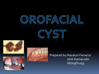
Prepared by:Nazatun Fameiza
             binti Kamarudin
            :NU09DL055
INDEX

 1)Definition of cyst
 2)Classification of cyst
 3)Radicular cyst
 4)Residual cyst
 5)Dentigerous cyst
 6)Odontogenic Keratocyst
 7)Basal Cell Nevus Syndrome
 8)Calcifying cystic Odontogenic tumor
 9)Nasopalatine Duct cyst
 10)Nasolabial cyst.
DEFINITION OF CYST

   Cyst is defined as pathologic cavity,
   having fluid,semifluid, or gaseous contents and
   which is not created by accumulation of pus.
   It is frequently but not always lined by epithelium.
                      cyst


                               Lining epithelium
                    lumen
                                Wall of fibrous
                               connective tissue
CLASSIFICATION OF CYST

                             Cyst of the jaw

          Epithelial (true         Non Epithelial (pseudo cyst)
               cyst)

   Odontogenic                                 Non odontogenic

       Based on etiology               Based on site of origin


                    Inflammatory
 Developmental                                                     unclassified
                             Reduced enamel
                                epitelium                  Cell rest of
                                                            Malassez
                                          Cell rest of Serre
EPITHELIAL/TRUE CYST


ODONTOGENIC               NON ODONTOGENIC


FISSURAL CYST                DEVELOPMENTAL CYST
-median anterior             -palatal cyst of neonate
maxillary cyst               -thyroglossal tract cyst
-nasopalatine duct cyst      -benign cevical
-nasolabial cyst             lymphoepithelial cyst
-globulomaxillary cyst       -epidermoid and dermoid
                             cyst
-median mandibular
cyst                         Heterotopic oral
                             gastrointestinal cyst
ODONTOGENIC

                                                BASED ON SITE OF ORIGIN
    BASED ON ETIOLOGY

                                               1)REDUCED ENAMEL EPITHELIUM
  DEVELOPMENTAL                                       -dentigerous cyst
       CYST                 INFLAMMATORY
                                                        -eruption cyst
-gingival cyst of infants   -periapical cyst
                                                   2)CELL REST OF SERRE
 -gingival cyst of adults    -residual cyst
                                                   -odontogenic keratocyst
-odontogenic keratocyst     -paradental cyst
                                                  -gingival cyst of newborn
   -dentigerous cyst
                                                    -gingival cyst of adults
     -eruption cyst
                                                   -lateral periodontal cyst
-lateral periodantal cyst
                                                 -glandular odontogenic cyst
-botryoid odontogenic
         cyst                                    3)CELL REST OF MALASSEZ
-glandular odontogenic                                 -periapical cyst
         cyst                                           -residual cyst
-calcifying odontogenic                               4)UNCLASSIFIED
           cyst
                                                  -calcified odontogenic cyst
                                                       -paradental cyst
NON EPITHELIAL (PSEUDO
        CYST)
   -solitary bone cyst
  -aneurysm bone cyst
RADICULAR CYST
   synonyms:periapical cyst,apical periodontal cyst,dental cyst

   Definition:
   Cyst that results when cell rest of Malassez in periodontal ligament are
    stimulated to proliferate and undergo cystic degeneration by inflammatory
    products from NON VITAL tooth.

   Clinical Features:
   The most common type of cyst in the jaw
   Age:3rd-6th decade
   Sex:MALE predominance
   Arise from NON VITAL TOOTH ;
   -due to extensive caries,large restorations,trauma
   Asymptomatic
   Larger cyst may cause swelling
   On palpation,the swelling may feel
   A)bony and hard if cortex is intact
   B)crepitant as the bone thins
   C)rubbery and fluactuant if the outer cortex is lost

 Radiographic Features;
 1)location:
 -approximately at the apex of a
  non vital tooth,
 -60% are found in MAXILLA
 -Especially around INCISORS and CANINES
 -(mesial/distal surface of tooth root at the opening of accessory canal or in
  deep periodontal pocket)
 -because of distal inclination of the root,cyst that arise from the maxillary
  lateral incisor may invaginate the antrum
 -may associated with NONVITAL DECIDUOS MOLAR which is situated
  buccal to developing bicuspid.

 Periphery and Shape:
 -well-defined cortical border


 -if the cyst is secondarily infected,the
 inflammatory reaction surrounding the
   bone may result in loss of this cortex or
 alteration lead to sclerotic border.
 -outline:curved or circular
 Internal Structure:
 -radiolucent
 -in long standing cyst,dystrophic calcification may developed,appearing
   as sparsely distributed small particulate radiopacities.


 Effects on surrounding structures:
 -large cyst lead to displacement and resorption of adjacent teeth.
 -resorption patern have curved outline
 -the cyst may invaginate the antrum,but there should be evidence of
   cortical boundaries between the contents of the cyst and the internal
   structure of antrum.
 -cyst may displace mandibular alveolar canal in an inferior direction.
 -the outer cortical plates of maxilla or mandible may expand in curved or
   circular shape
   Differential Diagnosis:
   1)Apical granuloma
   -cyst characterized by round shape,well-defined cortical border,and size
    greater than 2cm in diameter.

   2)early stage of periapical cemental dysplasia,an apical scar or surgical defect
   -patient’s history helps with the differentiation

   3)Odontogenic keratocyst or Lateral Periodontal cyst
   -vitality of involved tooth should be test.
   -non vital tooth have large pulp chamber due to lack of secondary dentin.

   4)Benign fibro-osseous lesion
   -a larger radicular cyst that invaginated maxillary antrum may collapse and
    start filling in with the new bone.with biopsy,the histologic analysis may
    result in ossifying fibroma or benign fibro-osseous
    lesion.Radiographically,the new bone will form first at the periphery of the
    cyst as the cyst shrinks and not in the center of cyst.(this is different pattern
    from benign lesion)
 Management:
 -extraction
 -endodontic theraphy
 -Apical surgery


 Larger cyst
 -surgical removal
 -marsupialization
RESIDUAL CYST

Definition:
Cyst that remains after incomplete removal of the original cyst.


Clinical Features:
-Asymptomatic
-associated with EDENTULOUS AREA
Radiographic Features:
1)Location
Site:mandible
-epicenter is positioned in the former periapical region of involved or
   missing tooth
-in mandible,the epicenter is above the inferior alveolar nerve canal
 2)Periphery and shape
 -cortical margin with oval or circular shape
 3)Internal structure
 -radiolucent
 --dystrophic calcification in long standing cyst
4)Effect on surrounding structure:
-tooth displacement or resorption
-outer cortical plate of jaw may expand
-cyst may invaginate maxillary antrum or depress the inferior alveolar canal.


Differential Diagnosis:
1)Odontogenic keratocyst
-residual cyst has greater expansion than OKC
2)Stafne developmental salivary gland defect
-the defect is located below mandibular canal,thus is unlikely to be
   odontogenic in nature.


Treatment:
-surgical removal
-marsupialization
DENTIGEROUS CYST
   Synonym:follicular cyst

   Definition:
   A cyst that forms around the crown of UNERUPTED tooth.It begins when
    fluid accumulates in the layers of REDUCED ENAMEL EPITHELIUM or
    between the epithelium and the crown of unerupted tooth.

   Clinical Features:
   -2nd most common cyst in the jaw
   -associated with UNERUPTED or
   SUPERNUMERARY TOOTH.
   (mesiodens in anterior maxilla)
   -no pain or discomfort
   -clinical examination shows:
     @>missing tooth with hard swelling
    resulting in facial asymmetry
 Radiographic Features:
 1)Location:
 -mandibular or maxillary THIRD MOLAR
 -MAXILLARY CANINE
 -this cyst attaches at CEMENTOENAMEL junction


 -types of radiographic presentation:
    a)central type
   b)lateral type


   c)circumferential type
 2)Periphery and shape
 -well defined cortex with a curved or circular outline
 3)Internal structure
 -radiolucent except the crown of involved tooth
 4)Effect of surrounding structure
 -displacement and resorption of adjacent tooth
 -it displaces the associated tooth in apical direction
 -maxillary third molar or cuspid may be pushed to the floor of orbit
 -mandibular third molar may be moved to condylar or coronoid region or
   to the inferior cortex of mandible
 -the floor of maxillary antrum may be displaced as the cyst invaginates
  the antrum
 -The cyst may displace the inferior alveolar nerve canal in an inferior
  direction

 Differential Diagnosis
 1)hyperplastic follicle
 -size of normal follicle space is 2-3mm
 -If the folicular space exceed 5mm assaciated with tooth displacement
   and bone expansion,a dentigerous cyst is more likely.

 2)odontogenic keratocyst
 -OKC does not expand the bone to the same degree as dentigerous
   cyst,less likely to resorb tooth,may attach further at apically on the root
   instead of CEJ.
 3)ameloblastic fibroma or ameloblastoma
 -dentigerous cyst contain internal structure(tooth)


 4)adenomatoid odontogenic tumor and calcified odontogenic cyst
 -evidence of a radiopaque internal structure in these two lesion.


 5)radicular cyst at the apex of primary tooth
 -occasionally surrounds the crown of the developing permanent tooth
   positioned apical to it,giving false impression of dentigerous cyst
   associated with permanent tooth.
 -occur most often in MANDIBULAR DECIDUOUS MOLAR and the
   developing BICUSPIDS
 -thus,clinician should look for extensive caries or large restoration in
   primary tooth to indicate radicular cyst.
 Management
 1)surgical removal including the tooth
 2)large cyst should be treated with marsupialization before surgical
    removal
 3)cyst lining should be submitted for histologic examination because
    many lesion might be arise from cyst lining eg;
             a)AMELOBLASTOMA
             b)SQUAMOUS CELL CARCINOMA
             c)MUCOEPIDERMOID CARCINOMA
ODONTOGENIC KERATOCYST
Synonyms:
-KERATOCYSTIC ODONTOGENIC TUMOR(KOT)
-primordial cyst

Definition:
The WHO has reclassified this cystic lesion into a unicystic or multicystic
   odontogenic tumor on the basis of TUMORLIKE CHARACTERISTIC of
   the lining epithelium.The epithelium in the KOT appear to have innate
   growth potential,consistent with benign tumor.The epithelial lining is
   DISTINCTIVE because it is KERATINIZED (hence the name)and thin (4-8
   cells thick).Occasionally,budlike proliferations of epithelium grow from
   the basal layer into the adjacent connective tissue wall.Islands of
   epithelium in the wall may give rise to SATELLITE MICROCYST.Inside
   the cyst contain a viscous or cheesy material derived from epithelial
   lining.
Clinical Features:
-KOT account for about one tenth (1/10) of all cystic lesion in jaws
-age:20-30
-sex:male predominance
-may associated with UNERUPTED tooth
-asymptomatic
-aspiration reveal a thick yellow cheesy material(KERATIN)
Have high properties of RECCURENCE ,because of small satellite cyst or
  epithelium fragments left behind after surgical removal of epithelium.


Radiographic Features:
1)Location
-most common:POSTERIOR BODY OF MANDIBLE
             (90% occur posterior to canine)
             :RAMUS (more than 50%)
-the epicenter located superior to inferior alveolar nerve canal
-has same pericoronal position as dentigerous cyst.


2)Periphery and shape
-well-defined cortical border
-smooth round or oval shape
-or might have scalloped outline
3)Internal Structure:
-radiolucent
-in some case curved internal septa may be present giving a multilocular
    apppearance.
4)Effect on surrounding structure:
-a very characteristic feature that its PROPENSITY TO GROW ALONG THE
    INTERNAL ASPECT OF THE JAW CAUSING MINIMAL EXPANSION.
-this occur throughout the mandible except for the upper ramus and
    coronoid process.
 -the relatively slight expansion contributes to their late detection,which
   allow them to reach a large size.
 -KOT can displace and resorb teeth but to a slightly degree than
   dentigerous cyst.
 -the inferior alveolar nerve canal may be displaced inferiorly.
 -this may invaginate and occupy the maxillary antrum.
 – RADIOGRAPHIC VARIETIES


             1. REPLACEMENTAL –
             Cyst forms in place of normal tooth
                 by degeneration of dental
                 lamina.


             2. EXTRANEOUS –
             OKC occurs in ascending ramus, away
                 from tooth bearing areas
3. COLLATERAL –
OKC occurs adjacent to root of tooth,
mimicking a lateral periodontal cyst.


4. ENVELOPMENTAL –
This is an odontogenic keratocyst
which embraces or envelopes an
adjacent unerupted tooth.
   Differential Diagnosis:
1)dentigerous cyst
  -it is KOT if the cystic outline is connected to the tooth at the point apical to
    CEJ,and if no expansion of the cortical plate.
 -although KOT can develop occlusal to developing tooth ,the follicle of
    involved tooth is not enlarged as dentigerous cyst.

2)Ameloblastoma
  -scalloped margin and multilocular appearance of KOT may resemble
    ameloblastoma but ameloblastoma has greater propensity to expand.

3)Odontogenic myxoma
 -have similar characteristic of mild expansion and multilocular appearance.

4)Simple bone cyst
 -have similar characteristic of scalloped margin and minimal bone expansion
 -however the margins of simple bone cyst are more delicate and difficult to
   detect.

5)4-5% of KOT cases may constitute part of BASAL CELL NEVUS SYNDROME.
Management:
-referral to radiologist for a complete radiologic examination is advisable.
-resection,curretage,or marsupialization to reduce the size of large lesion
   before surgical excision.
-complete removal of the cystic walls to reduce the chance of recurrence
-after surgical treatmant,it is important to make periodic posttreatment
    clinical and radiographic examination to detect any recurrence.
-recurrent lesion usually develop within the first 5 years but may delayed as
   long as 10 years.
BASAL CELL NEVUS SYNDROME
   Synonyms:Nevoid basal cell carcinoma, GORLIN-GOLTZ syndrome


   Definition:
   Comprises a number of abnormalities as multiple nevoid basal cell carcinomas of
    the skin,skeletal abnormalities,central nervous system abnormalities,eyes
    abnormalities and multiple KOTs.it is inherited autosomal dominant trait with
    variable expressivity.
   Clinical Features:
   age:5-30 years
   A) multiple KOTs-appearing in multiple quadrant
                        -early in life
                        -high RECURRENT RATE
   B) skin lesion-small,flattened,flesh-colored or brown papules
                -prominent on face,neck, and trunk
   C)Skeletal anomalies
         -BIFID RIB (most common)


                                               Frontal chest radiograph shows
                                               bilateral bifid ribs (arrows).


         -costal abnormalities eg 1) vertebral fusion
   2)kyphoscoliosis


   3)polydactyly
   4)temporal and temporoparietal bossing


   5)minor hypertelorism- abnormally increased distance between two
                         organs or bodily parts.
6) calcification of Falx cerebri


      Skull CT showing calcification of the falx
   Radiographic features:
   1) location:same as solitary KOTs,may develop bilaterally


   2)other radiographic features:
   Radiopaque line of the calcified falx cerebri may be prominent on the
    posterior anterior skull projection
CALCIFYING CYSTIC ODONTOGENIC TUMOR

Synonyms:Calcifying odontogenic cyst,Calcifying epithelial odontogenic cyst,
               Gorlin cyst


Definition :
  -uncommon slow growing,benign lesions.occupy a spectrum ranging from a
   cyst to an odontogenic tumor.It may manufacture calcified tissue (dysplastic
   dentin) or associated with an odontoma.when it contains a more solid
   component ,it gives appearance resembling ameloblastoma although it
   does not behave like one.
   Clinical features:
   Mean age:36 years
       first peak:10-19 years
       second peak:seventh decade
   Appearance:slow –growing,painless swelling in jaw


   Radiographic features:
   1) location:
   - 75% occur in bone anterior to first molar especially associated with cuspids
    and incisors
   2) periphery and shape:
   -vary from well-defined and corticated with a curved, cystlike shape to ill-
    defined and regular


   3) Internal structure:
   -a)completely radiolucent
   -b)evidence of small foci or calcified material that appear as white flecks or
    small smooth pebbles
   -c)larger,solid,amorphus masses
   -d)multilocular
   4) effect on surrounding structure:
   -20-50% of cases is associated with tooth(cuspid) and impedes it eruption
   -displacement of teeth and root resorptions
   -perforation of cortical plate may be seen with enlarging lesion.


   Management:
   Enucleation and curettage.
NASOPALATINE DUCT CYST
   Synonyms:Nasopalatine canal cyst, Incisive canal cyst, Nasopalatine cyst,
                 Median palatine cyst, Median anterior maxillary cyst.


   Definition:
   Nasopalatine canal usually contains remnants of the nasopalatine duct, a
    primitive organ of smell, and the nasopalatine vessels and nerves.
    Occasionally, a cyst forms in the nasopalatine canal when these embryonic
    epithelial remnants of the nasopalatine duct undergo proliferation and
    cystic degeneration.
   Clinical Features:
   10% of jaw cysts
   Age:4th-6th decades
   Sex:male predilection
   Asymptomatic
   Small,well-defined swelling just posterior to palatine papilla
   Fluactuant and blue swelling if near the surface.
   May penetrate labial plate and produce swelling below maxillary labial
    frenum.
   May bulge into nasal cavity and distort nasal septum
   Pressure of cyst on adjacent nasopalatine nerve may cause burning
    sensation or numbness over palatal mucosa.
   Radiographic Features:
   1) location


   -a) found in the nasopalatine foramen or canal
   -b) If extends posteriorly involving hard palate   MEDIAN PALATAL CYST
   -c)if expands anteriorly between central incisors,destroying or expanding
    labial plate of bone and causing teeth to diverge     MEDIAN ANTERIOR
    MAXILLARY CYST
   2) Periphery and shape:
   -well-defined and corticated circular or oval in shape
   -HEART SHAPE is due to shadow of nasal spine superimposed on the cyst
   Internal structure:
   -radiolucent


   Effects on Surrounding structures:
   -roots of central incisors diverge
   -root resorption
   -expansion of labial cortex and palatal cortex
   -floor of nasal fossa may be displaced in superior direction
   Differential Diagnosis:
   1) large incisive foramen
   -cyst is presumed when the width of foramen exceeds 1cm and cause tooth
    displacement.


   2) a radicular cyst or granuloma
   -absence of lamina dura and enlargement of periodontal ligament space
    around apex of central incisors indicate an inflammatory lesion.
   -vitality test
   -a second periapical view taken at different horizontal angulation should
    show altered position of a nasopalatine duct cyst, whereas radicular cyst
    should remain centered about the apex of central incisors.


   Management:
   -enucleation
   -marsupialize indicated for large cyst
NASOLABIAL CYST
   Synonyms:Nasoalveolar cyst


   Definition
   -exact origin is unknown, may be fissural cyst arising from epithelial rests in
    fusion lines of globular,lateral nasal and maxillary process.
   Clinical Features:
   Age:12-75 years
   Sex:female predilection
   Usually UNILATERAL


   a)small lesion
   -very subtle,unilateral swelling of
   nasolabial fold associated with
   pain or discomfort.


   b) large lesion
   -it may bulge into floor of nasal
   cavity causing obstruction,flaring
   of alae,distortion of nostrils
   and fullness of upper lip.
   Radiographic Features:
   1) location:
   -located adjacent to alveolar process above apices of incisors
   -because this is soft tissue lession, investigation should be done using CT or
    MRI(magnetic resonance imaging.


   2)Periphery and shape:
   -CT images shows circular or oval lesion with slight tissue enhancement on
    periphery

   3)Internal structure:
   -CT images shows homogenous & relatively radiolucent compared with
    surrounding structure.
   4)Effects on surrounding structures:
   -erosion on underlying bone
   -usual outline of inferior border of nasal fossa become distorted resulting
    posterior bowing of this margin.


   Management:
   -excision through an intraoral approach.
OROFACIAL CYST

Contenu connexe

Tendances

Tendances (20)

Odontogenic keratocyst
Odontogenic keratocystOdontogenic keratocyst
Odontogenic keratocyst
 
Red and White Lesions
Red and White LesionsRed and White Lesions
Red and White Lesions
 
Pulp and periapical disease
Pulp and periapical diseasePulp and periapical disease
Pulp and periapical disease
 
Benign tumors of jaw
Benign tumors of jaw Benign tumors of jaw
Benign tumors of jaw
 
Odontoma (Doctor Faris Alabeedi MSc, MMedSc, PgDip, BDS.)
Odontoma (Doctor Faris Alabeedi MSc, MMedSc, PgDip, BDS.)Odontoma (Doctor Faris Alabeedi MSc, MMedSc, PgDip, BDS.)
Odontoma (Doctor Faris Alabeedi MSc, MMedSc, PgDip, BDS.)
 
Non odontogenic cysts or fissural cysts
Non odontogenic cysts or fissural cystsNon odontogenic cysts or fissural cysts
Non odontogenic cysts or fissural cysts
 
Cysts of the Oral Cavity
Cysts of the Oral CavityCysts of the Oral Cavity
Cysts of the Oral Cavity
 
Osteomyelitis of jaw
Osteomyelitis of jawOsteomyelitis of jaw
Osteomyelitis of jaw
 
PERIAPICAL DISEASES
PERIAPICAL DISEASESPERIAPICAL DISEASES
PERIAPICAL DISEASES
 
Odontogenic cysts
Odontogenic  cystsOdontogenic  cysts
Odontogenic cysts
 
Odontogenic tumors ppt
Odontogenic tumors pptOdontogenic tumors ppt
Odontogenic tumors ppt
 
Cyst Of Jaw
Cyst Of JawCyst Of Jaw
Cyst Of Jaw
 
Odontogenic Cysts
Odontogenic CystsOdontogenic Cysts
Odontogenic Cysts
 
ODONTOGENIC CYSTS
ODONTOGENIC CYSTSODONTOGENIC CYSTS
ODONTOGENIC CYSTS
 
Peripheral and central giant cell granuloma
Peripheral and central giant cell granulomaPeripheral and central giant cell granuloma
Peripheral and central giant cell granuloma
 
Odontogenic Tumors
Odontogenic TumorsOdontogenic Tumors
Odontogenic Tumors
 
Periapical cyst
Periapical  cystPeriapical  cyst
Periapical cyst
 
Developmental disturbances of teeth
Developmental disturbances of teethDevelopmental disturbances of teeth
Developmental disturbances of teeth
 
Spread of oral infections
Spread of oral infectionsSpread of oral infections
Spread of oral infections
 
Oral lichen planus
Oral lichen planusOral lichen planus
Oral lichen planus
 

Similaire à OROFACIAL CYST

Radicular cyst or Periapical cyst
Radicular cyst or Periapical cystRadicular cyst or Periapical cyst
Radicular cyst or Periapical cystdrabbasnaseem
 
Cysts of oral regions
Cysts of oral regionsCysts of oral regions
Cysts of oral regionsNaz Dizayee
 
Cysts and cystic lesions
Cysts and cystic lesionsCysts and cystic lesions
Cysts and cystic lesionsBasant AbuZaid
 
Odontogenic cysts i / dental implant courses by Indian dental academy 
Odontogenic cysts i / dental implant courses by Indian dental academy Odontogenic cysts i / dental implant courses by Indian dental academy 
Odontogenic cysts i / dental implant courses by Indian dental academy Indian dental academy
 
Radicular cyst (maryam arbab)
Radicular cyst (maryam arbab)Radicular cyst (maryam arbab)
Radicular cyst (maryam arbab)Maryam Arbab
 
1 intro to cyst, classification & pathophysiology
1 intro to cyst, classification & pathophysiology1 intro to cyst, classification & pathophysiology
1 intro to cyst, classification & pathophysiologyvasanramkumar
 
Cystofjaw rkv..
Cystofjaw  rkv..Cystofjaw  rkv..
Cystofjaw rkv..Ravi Kumar
 
CYSTS OF THE ORAL FACIAL REGION.pptx
CYSTS OF THE ORAL FACIAL REGION.pptxCYSTS OF THE ORAL FACIAL REGION.pptx
CYSTS OF THE ORAL FACIAL REGION.pptxAngetileKasanga
 
cysts of oral and maxillofacial region.pdf
cysts of oral and maxillofacial region.pdfcysts of oral and maxillofacial region.pdf
cysts of oral and maxillofacial region.pdfasishkp1
 
cystofjawmadhu-191014054840.pdf
cystofjawmadhu-191014054840.pdfcystofjawmadhu-191014054840.pdf
cystofjawmadhu-191014054840.pdfSolimanAbuDalfa
 
DENTIGEROUS CYST WITH EMPHASIS ON ITS COMPLICATIONS
DENTIGEROUS CYST WITH EMPHASIS ON ITS COMPLICATIONSDENTIGEROUS CYST WITH EMPHASIS ON ITS COMPLICATIONS
DENTIGEROUS CYST WITH EMPHASIS ON ITS COMPLICATIONSkrishnamohan407
 
Cysts Of The Oral Region - Oral Pathology
Cysts Of The Oral Region - Oral PathologyCysts Of The Oral Region - Oral Pathology
Cysts Of The Oral Region - Oral PathologySana Rasheed
 
Cyst of oral and maxillofacial regions
Cyst of oral and maxillofacial regionsCyst of oral and maxillofacial regions
Cyst of oral and maxillofacial regionsSavita Sahu
 
CYSTS OF THE JAWS Part II
CYSTS OF THE JAWS Part IICYSTS OF THE JAWS Part II
CYSTS OF THE JAWS Part IIAbhishek PT
 
DENTIGEROUS CYST & CALCIFYING ODONTOGENIC CYST.pptx
DENTIGEROUS CYST & CALCIFYING ODONTOGENIC CYST.pptxDENTIGEROUS CYST & CALCIFYING ODONTOGENIC CYST.pptx
DENTIGEROUS CYST & CALCIFYING ODONTOGENIC CYST.pptxDr.Mohit Bains
 

Similaire à OROFACIAL CYST (20)

Radicular cyst or Periapical cyst
Radicular cyst or Periapical cystRadicular cyst or Periapical cyst
Radicular cyst or Periapical cyst
 
Cysts part 1
Cysts part 1Cysts part 1
Cysts part 1
 
CYSTS OF ORAL AND MAXILLOFACIAL REGION (2).ppt
CYSTS OF ORAL AND MAXILLOFACIAL REGION (2).pptCYSTS OF ORAL AND MAXILLOFACIAL REGION (2).ppt
CYSTS OF ORAL AND MAXILLOFACIAL REGION (2).ppt
 
Cysts of oral regions
Cysts of oral regionsCysts of oral regions
Cysts of oral regions
 
Cysts and cystic lesions
Cysts and cystic lesionsCysts and cystic lesions
Cysts and cystic lesions
 
Odontogenic cysts i / dental implant courses by Indian dental academy 
Odontogenic cysts i / dental implant courses by Indian dental academy Odontogenic cysts i / dental implant courses by Indian dental academy 
Odontogenic cysts i / dental implant courses by Indian dental academy 
 
Radicular cyst (maryam arbab)
Radicular cyst (maryam arbab)Radicular cyst (maryam arbab)
Radicular cyst (maryam arbab)
 
1 intro to cyst, classification & pathophysiology
1 intro to cyst, classification & pathophysiology1 intro to cyst, classification & pathophysiology
1 intro to cyst, classification & pathophysiology
 
Cystofjaw rkv..
Cystofjaw  rkv..Cystofjaw  rkv..
Cystofjaw rkv..
 
CYSTS OF HEAD AND NECK
CYSTS OF HEAD AND NECKCYSTS OF HEAD AND NECK
CYSTS OF HEAD AND NECK
 
CYSTS OF THE ORAL FACIAL REGION.pptx
CYSTS OF THE ORAL FACIAL REGION.pptxCYSTS OF THE ORAL FACIAL REGION.pptx
CYSTS OF THE ORAL FACIAL REGION.pptx
 
cysts of oral and maxillofacial region.pdf
cysts of oral and maxillofacial region.pdfcysts of oral and maxillofacial region.pdf
cysts of oral and maxillofacial region.pdf
 
cystofjawmadhu-191014054840.pdf
cystofjawmadhu-191014054840.pdfcystofjawmadhu-191014054840.pdf
cystofjawmadhu-191014054840.pdf
 
DENTIGEROUS CYST WITH EMPHASIS ON ITS COMPLICATIONS
DENTIGEROUS CYST WITH EMPHASIS ON ITS COMPLICATIONSDENTIGEROUS CYST WITH EMPHASIS ON ITS COMPLICATIONS
DENTIGEROUS CYST WITH EMPHASIS ON ITS COMPLICATIONS
 
Cysts Of The Oral Region - Oral Pathology
Cysts Of The Oral Region - Oral PathologyCysts Of The Oral Region - Oral Pathology
Cysts Of The Oral Region - Oral Pathology
 
Cyst of oral and maxillofacial regions
Cyst of oral and maxillofacial regionsCyst of oral and maxillofacial regions
Cyst of oral and maxillofacial regions
 
Cysts of the jaws
Cysts of the jawsCysts of the jaws
Cysts of the jaws
 
Cysts of jaws pathogenesis
Cysts of jaws pathogenesisCysts of jaws pathogenesis
Cysts of jaws pathogenesis
 
CYSTS OF THE JAWS Part II
CYSTS OF THE JAWS Part IICYSTS OF THE JAWS Part II
CYSTS OF THE JAWS Part II
 
DENTIGEROUS CYST & CALCIFYING ODONTOGENIC CYST.pptx
DENTIGEROUS CYST & CALCIFYING ODONTOGENIC CYST.pptxDENTIGEROUS CYST & CALCIFYING ODONTOGENIC CYST.pptx
DENTIGEROUS CYST & CALCIFYING ODONTOGENIC CYST.pptx
 

Dernier

Ecological Succession. ( ECOSYSTEM, B. Pharmacy, 1st Year, Sem-II, Environmen...
Ecological Succession. ( ECOSYSTEM, B. Pharmacy, 1st Year, Sem-II, Environmen...Ecological Succession. ( ECOSYSTEM, B. Pharmacy, 1st Year, Sem-II, Environmen...
Ecological Succession. ( ECOSYSTEM, B. Pharmacy, 1st Year, Sem-II, Environmen...Shubhangi Sonawane
 
microwave assisted reaction. General introduction
microwave assisted reaction. General introductionmicrowave assisted reaction. General introduction
microwave assisted reaction. General introductionMaksud Ahmed
 
Gardella_Mateo_IntellectualProperty.pdf.
Gardella_Mateo_IntellectualProperty.pdf.Gardella_Mateo_IntellectualProperty.pdf.
Gardella_Mateo_IntellectualProperty.pdf.MateoGardella
 
Unit-IV; Professional Sales Representative (PSR).pptx
Unit-IV; Professional Sales Representative (PSR).pptxUnit-IV; Professional Sales Representative (PSR).pptx
Unit-IV; Professional Sales Representative (PSR).pptxVishalSingh1417
 
Key note speaker Neum_Admir Softic_ENG.pdf
Key note speaker Neum_Admir Softic_ENG.pdfKey note speaker Neum_Admir Softic_ENG.pdf
Key note speaker Neum_Admir Softic_ENG.pdfAdmir Softic
 
Advanced Views - Calendar View in Odoo 17
Advanced Views - Calendar View in Odoo 17Advanced Views - Calendar View in Odoo 17
Advanced Views - Calendar View in Odoo 17Celine George
 
PROCESS RECORDING FORMAT.docx
PROCESS      RECORDING        FORMAT.docxPROCESS      RECORDING        FORMAT.docx
PROCESS RECORDING FORMAT.docxPoojaSen20
 
Web & Social Media Analytics Previous Year Question Paper.pdf
Web & Social Media Analytics Previous Year Question Paper.pdfWeb & Social Media Analytics Previous Year Question Paper.pdf
Web & Social Media Analytics Previous Year Question Paper.pdfJayanti Pande
 
Grant Readiness 101 TechSoup and Remy Consulting
Grant Readiness 101 TechSoup and Remy ConsultingGrant Readiness 101 TechSoup and Remy Consulting
Grant Readiness 101 TechSoup and Remy ConsultingTechSoup
 
Basic Civil Engineering first year Notes- Chapter 4 Building.pptx
Basic Civil Engineering first year Notes- Chapter 4 Building.pptxBasic Civil Engineering first year Notes- Chapter 4 Building.pptx
Basic Civil Engineering first year Notes- Chapter 4 Building.pptxDenish Jangid
 
Measures of Central Tendency: Mean, Median and Mode
Measures of Central Tendency: Mean, Median and ModeMeasures of Central Tendency: Mean, Median and Mode
Measures of Central Tendency: Mean, Median and ModeThiyagu K
 
Presentation by Andreas Schleicher Tackling the School Absenteeism Crisis 30 ...
Presentation by Andreas Schleicher Tackling the School Absenteeism Crisis 30 ...Presentation by Andreas Schleicher Tackling the School Absenteeism Crisis 30 ...
Presentation by Andreas Schleicher Tackling the School Absenteeism Crisis 30 ...EduSkills OECD
 
APM Welcome, APM North West Network Conference, Synergies Across Sectors
APM Welcome, APM North West Network Conference, Synergies Across SectorsAPM Welcome, APM North West Network Conference, Synergies Across Sectors
APM Welcome, APM North West Network Conference, Synergies Across SectorsAssociation for Project Management
 
How to Give a Domain for a Field in Odoo 17
How to Give a Domain for a Field in Odoo 17How to Give a Domain for a Field in Odoo 17
How to Give a Domain for a Field in Odoo 17Celine George
 
Making and Justifying Mathematical Decisions.pdf
Making and Justifying Mathematical Decisions.pdfMaking and Justifying Mathematical Decisions.pdf
Making and Justifying Mathematical Decisions.pdfChris Hunter
 

Dernier (20)

Ecological Succession. ( ECOSYSTEM, B. Pharmacy, 1st Year, Sem-II, Environmen...
Ecological Succession. ( ECOSYSTEM, B. Pharmacy, 1st Year, Sem-II, Environmen...Ecological Succession. ( ECOSYSTEM, B. Pharmacy, 1st Year, Sem-II, Environmen...
Ecological Succession. ( ECOSYSTEM, B. Pharmacy, 1st Year, Sem-II, Environmen...
 
microwave assisted reaction. General introduction
microwave assisted reaction. General introductionmicrowave assisted reaction. General introduction
microwave assisted reaction. General introduction
 
Gardella_Mateo_IntellectualProperty.pdf.
Gardella_Mateo_IntellectualProperty.pdf.Gardella_Mateo_IntellectualProperty.pdf.
Gardella_Mateo_IntellectualProperty.pdf.
 
Unit-IV; Professional Sales Representative (PSR).pptx
Unit-IV; Professional Sales Representative (PSR).pptxUnit-IV; Professional Sales Representative (PSR).pptx
Unit-IV; Professional Sales Representative (PSR).pptx
 
Key note speaker Neum_Admir Softic_ENG.pdf
Key note speaker Neum_Admir Softic_ENG.pdfKey note speaker Neum_Admir Softic_ENG.pdf
Key note speaker Neum_Admir Softic_ENG.pdf
 
Advanced Views - Calendar View in Odoo 17
Advanced Views - Calendar View in Odoo 17Advanced Views - Calendar View in Odoo 17
Advanced Views - Calendar View in Odoo 17
 
Mehran University Newsletter Vol-X, Issue-I, 2024
Mehran University Newsletter Vol-X, Issue-I, 2024Mehran University Newsletter Vol-X, Issue-I, 2024
Mehran University Newsletter Vol-X, Issue-I, 2024
 
PROCESS RECORDING FORMAT.docx
PROCESS      RECORDING        FORMAT.docxPROCESS      RECORDING        FORMAT.docx
PROCESS RECORDING FORMAT.docx
 
Web & Social Media Analytics Previous Year Question Paper.pdf
Web & Social Media Analytics Previous Year Question Paper.pdfWeb & Social Media Analytics Previous Year Question Paper.pdf
Web & Social Media Analytics Previous Year Question Paper.pdf
 
Grant Readiness 101 TechSoup and Remy Consulting
Grant Readiness 101 TechSoup and Remy ConsultingGrant Readiness 101 TechSoup and Remy Consulting
Grant Readiness 101 TechSoup and Remy Consulting
 
Basic Civil Engineering first year Notes- Chapter 4 Building.pptx
Basic Civil Engineering first year Notes- Chapter 4 Building.pptxBasic Civil Engineering first year Notes- Chapter 4 Building.pptx
Basic Civil Engineering first year Notes- Chapter 4 Building.pptx
 
Mattingly "AI & Prompt Design: The Basics of Prompt Design"
Mattingly "AI & Prompt Design: The Basics of Prompt Design"Mattingly "AI & Prompt Design: The Basics of Prompt Design"
Mattingly "AI & Prompt Design: The Basics of Prompt Design"
 
Measures of Central Tendency: Mean, Median and Mode
Measures of Central Tendency: Mean, Median and ModeMeasures of Central Tendency: Mean, Median and Mode
Measures of Central Tendency: Mean, Median and Mode
 
Presentation by Andreas Schleicher Tackling the School Absenteeism Crisis 30 ...
Presentation by Andreas Schleicher Tackling the School Absenteeism Crisis 30 ...Presentation by Andreas Schleicher Tackling the School Absenteeism Crisis 30 ...
Presentation by Andreas Schleicher Tackling the School Absenteeism Crisis 30 ...
 
APM Welcome, APM North West Network Conference, Synergies Across Sectors
APM Welcome, APM North West Network Conference, Synergies Across SectorsAPM Welcome, APM North West Network Conference, Synergies Across Sectors
APM Welcome, APM North West Network Conference, Synergies Across Sectors
 
Mattingly "AI & Prompt Design: Structured Data, Assistants, & RAG"
Mattingly "AI & Prompt Design: Structured Data, Assistants, & RAG"Mattingly "AI & Prompt Design: Structured Data, Assistants, & RAG"
Mattingly "AI & Prompt Design: Structured Data, Assistants, & RAG"
 
How to Give a Domain for a Field in Odoo 17
How to Give a Domain for a Field in Odoo 17How to Give a Domain for a Field in Odoo 17
How to Give a Domain for a Field in Odoo 17
 
Making and Justifying Mathematical Decisions.pdf
Making and Justifying Mathematical Decisions.pdfMaking and Justifying Mathematical Decisions.pdf
Making and Justifying Mathematical Decisions.pdf
 
Código Creativo y Arte de Software | Unidad 1
Código Creativo y Arte de Software | Unidad 1Código Creativo y Arte de Software | Unidad 1
Código Creativo y Arte de Software | Unidad 1
 
INDIA QUIZ 2024 RLAC DELHI UNIVERSITY.pptx
INDIA QUIZ 2024 RLAC DELHI UNIVERSITY.pptxINDIA QUIZ 2024 RLAC DELHI UNIVERSITY.pptx
INDIA QUIZ 2024 RLAC DELHI UNIVERSITY.pptx
 

OROFACIAL CYST

  • 1. Prepared by:Nazatun Fameiza binti Kamarudin :NU09DL055
  • 2. INDEX  1)Definition of cyst  2)Classification of cyst  3)Radicular cyst  4)Residual cyst  5)Dentigerous cyst  6)Odontogenic Keratocyst  7)Basal Cell Nevus Syndrome  8)Calcifying cystic Odontogenic tumor  9)Nasopalatine Duct cyst  10)Nasolabial cyst.
  • 3. DEFINITION OF CYST  Cyst is defined as pathologic cavity,  having fluid,semifluid, or gaseous contents and  which is not created by accumulation of pus.  It is frequently but not always lined by epithelium. cyst Lining epithelium lumen Wall of fibrous connective tissue
  • 4. CLASSIFICATION OF CYST Cyst of the jaw Epithelial (true Non Epithelial (pseudo cyst) cyst) Odontogenic Non odontogenic Based on etiology Based on site of origin Inflammatory Developmental unclassified Reduced enamel epitelium Cell rest of Malassez Cell rest of Serre
  • 5. EPITHELIAL/TRUE CYST ODONTOGENIC NON ODONTOGENIC FISSURAL CYST DEVELOPMENTAL CYST -median anterior -palatal cyst of neonate maxillary cyst -thyroglossal tract cyst -nasopalatine duct cyst -benign cevical -nasolabial cyst lymphoepithelial cyst -globulomaxillary cyst -epidermoid and dermoid cyst -median mandibular cyst Heterotopic oral gastrointestinal cyst
  • 6. ODONTOGENIC BASED ON SITE OF ORIGIN BASED ON ETIOLOGY 1)REDUCED ENAMEL EPITHELIUM DEVELOPMENTAL -dentigerous cyst CYST INFLAMMATORY -eruption cyst -gingival cyst of infants -periapical cyst 2)CELL REST OF SERRE -gingival cyst of adults -residual cyst -odontogenic keratocyst -odontogenic keratocyst -paradental cyst -gingival cyst of newborn -dentigerous cyst -gingival cyst of adults -eruption cyst -lateral periodontal cyst -lateral periodantal cyst -glandular odontogenic cyst -botryoid odontogenic cyst 3)CELL REST OF MALASSEZ -glandular odontogenic -periapical cyst cyst -residual cyst -calcifying odontogenic 4)UNCLASSIFIED cyst -calcified odontogenic cyst -paradental cyst
  • 7. NON EPITHELIAL (PSEUDO CYST) -solitary bone cyst -aneurysm bone cyst
  • 8. RADICULAR CYST  synonyms:periapical cyst,apical periodontal cyst,dental cyst  Definition:  Cyst that results when cell rest of Malassez in periodontal ligament are stimulated to proliferate and undergo cystic degeneration by inflammatory products from NON VITAL tooth.  Clinical Features:  The most common type of cyst in the jaw  Age:3rd-6th decade  Sex:MALE predominance  Arise from NON VITAL TOOTH ;  -due to extensive caries,large restorations,trauma  Asymptomatic  Larger cyst may cause swelling
  • 9. On palpation,the swelling may feel  A)bony and hard if cortex is intact  B)crepitant as the bone thins  C)rubbery and fluactuant if the outer cortex is lost  Radiographic Features;  1)location:  -approximately at the apex of a  non vital tooth,  -60% are found in MAXILLA  -Especially around INCISORS and CANINES  -(mesial/distal surface of tooth root at the opening of accessory canal or in deep periodontal pocket)  -because of distal inclination of the root,cyst that arise from the maxillary lateral incisor may invaginate the antrum  -may associated with NONVITAL DECIDUOS MOLAR which is situated buccal to developing bicuspid. 
  • 10.  Periphery and Shape:  -well-defined cortical border  -if the cyst is secondarily infected,the  inflammatory reaction surrounding the  bone may result in loss of this cortex or  alteration lead to sclerotic border.  -outline:curved or circular
  • 11.  Internal Structure:  -radiolucent  -in long standing cyst,dystrophic calcification may developed,appearing as sparsely distributed small particulate radiopacities.  Effects on surrounding structures:  -large cyst lead to displacement and resorption of adjacent teeth.  -resorption patern have curved outline  -the cyst may invaginate the antrum,but there should be evidence of cortical boundaries between the contents of the cyst and the internal structure of antrum.  -cyst may displace mandibular alveolar canal in an inferior direction.
  • 12.  -the outer cortical plates of maxilla or mandible may expand in curved or circular shape
  • 13. Differential Diagnosis:  1)Apical granuloma  -cyst characterized by round shape,well-defined cortical border,and size greater than 2cm in diameter.  2)early stage of periapical cemental dysplasia,an apical scar or surgical defect  -patient’s history helps with the differentiation  3)Odontogenic keratocyst or Lateral Periodontal cyst  -vitality of involved tooth should be test.  -non vital tooth have large pulp chamber due to lack of secondary dentin.  4)Benign fibro-osseous lesion  -a larger radicular cyst that invaginated maxillary antrum may collapse and start filling in with the new bone.with biopsy,the histologic analysis may result in ossifying fibroma or benign fibro-osseous lesion.Radiographically,the new bone will form first at the periphery of the cyst as the cyst shrinks and not in the center of cyst.(this is different pattern from benign lesion)
  • 14.
  • 15.  Management:  -extraction  -endodontic theraphy  -Apical surgery  Larger cyst  -surgical removal  -marsupialization
  • 16. RESIDUAL CYST Definition: Cyst that remains after incomplete removal of the original cyst. Clinical Features: -Asymptomatic -associated with EDENTULOUS AREA
  • 17. Radiographic Features: 1)Location Site:mandible -epicenter is positioned in the former periapical region of involved or missing tooth -in mandible,the epicenter is above the inferior alveolar nerve canal
  • 18.  2)Periphery and shape  -cortical margin with oval or circular shape  3)Internal structure  -radiolucent  --dystrophic calcification in long standing cyst
  • 19. 4)Effect on surrounding structure: -tooth displacement or resorption -outer cortical plate of jaw may expand -cyst may invaginate maxillary antrum or depress the inferior alveolar canal. Differential Diagnosis: 1)Odontogenic keratocyst -residual cyst has greater expansion than OKC 2)Stafne developmental salivary gland defect -the defect is located below mandibular canal,thus is unlikely to be odontogenic in nature. Treatment: -surgical removal -marsupialization
  • 20. DENTIGEROUS CYST  Synonym:follicular cyst  Definition:  A cyst that forms around the crown of UNERUPTED tooth.It begins when fluid accumulates in the layers of REDUCED ENAMEL EPITHELIUM or between the epithelium and the crown of unerupted tooth.  Clinical Features:  -2nd most common cyst in the jaw  -associated with UNERUPTED or  SUPERNUMERARY TOOTH.  (mesiodens in anterior maxilla)  -no pain or discomfort  -clinical examination shows:  @>missing tooth with hard swelling  resulting in facial asymmetry
  • 21.  Radiographic Features:  1)Location:  -mandibular or maxillary THIRD MOLAR  -MAXILLARY CANINE  -this cyst attaches at CEMENTOENAMEL junction  -types of radiographic presentation:  a)central type
  • 22. b)lateral type  c)circumferential type
  • 23.  2)Periphery and shape  -well defined cortex with a curved or circular outline  3)Internal structure  -radiolucent except the crown of involved tooth  4)Effect of surrounding structure  -displacement and resorption of adjacent tooth
  • 24.  -it displaces the associated tooth in apical direction  -maxillary third molar or cuspid may be pushed to the floor of orbit  -mandibular third molar may be moved to condylar or coronoid region or to the inferior cortex of mandible
  • 25.  -the floor of maxillary antrum may be displaced as the cyst invaginates the antrum  -The cyst may displace the inferior alveolar nerve canal in an inferior direction  Differential Diagnosis  1)hyperplastic follicle  -size of normal follicle space is 2-3mm  -If the folicular space exceed 5mm assaciated with tooth displacement and bone expansion,a dentigerous cyst is more likely.  2)odontogenic keratocyst  -OKC does not expand the bone to the same degree as dentigerous cyst,less likely to resorb tooth,may attach further at apically on the root instead of CEJ.
  • 26.  3)ameloblastic fibroma or ameloblastoma  -dentigerous cyst contain internal structure(tooth)  4)adenomatoid odontogenic tumor and calcified odontogenic cyst  -evidence of a radiopaque internal structure in these two lesion.  5)radicular cyst at the apex of primary tooth  -occasionally surrounds the crown of the developing permanent tooth positioned apical to it,giving false impression of dentigerous cyst associated with permanent tooth.  -occur most often in MANDIBULAR DECIDUOUS MOLAR and the developing BICUSPIDS  -thus,clinician should look for extensive caries or large restoration in primary tooth to indicate radicular cyst.
  • 27.  Management  1)surgical removal including the tooth  2)large cyst should be treated with marsupialization before surgical removal  3)cyst lining should be submitted for histologic examination because many lesion might be arise from cyst lining eg;  a)AMELOBLASTOMA  b)SQUAMOUS CELL CARCINOMA  c)MUCOEPIDERMOID CARCINOMA
  • 28. ODONTOGENIC KERATOCYST Synonyms: -KERATOCYSTIC ODONTOGENIC TUMOR(KOT) -primordial cyst Definition: The WHO has reclassified this cystic lesion into a unicystic or multicystic odontogenic tumor on the basis of TUMORLIKE CHARACTERISTIC of the lining epithelium.The epithelium in the KOT appear to have innate growth potential,consistent with benign tumor.The epithelial lining is DISTINCTIVE because it is KERATINIZED (hence the name)and thin (4-8 cells thick).Occasionally,budlike proliferations of epithelium grow from the basal layer into the adjacent connective tissue wall.Islands of epithelium in the wall may give rise to SATELLITE MICROCYST.Inside the cyst contain a viscous or cheesy material derived from epithelial lining.
  • 29. Clinical Features: -KOT account for about one tenth (1/10) of all cystic lesion in jaws -age:20-30 -sex:male predominance -may associated with UNERUPTED tooth -asymptomatic -aspiration reveal a thick yellow cheesy material(KERATIN) Have high properties of RECCURENCE ,because of small satellite cyst or epithelium fragments left behind after surgical removal of epithelium. Radiographic Features: 1)Location -most common:POSTERIOR BODY OF MANDIBLE (90% occur posterior to canine) :RAMUS (more than 50%) -the epicenter located superior to inferior alveolar nerve canal
  • 30. -has same pericoronal position as dentigerous cyst. 2)Periphery and shape -well-defined cortical border -smooth round or oval shape -or might have scalloped outline
  • 31. 3)Internal Structure: -radiolucent -in some case curved internal septa may be present giving a multilocular apppearance.
  • 32. 4)Effect on surrounding structure: -a very characteristic feature that its PROPENSITY TO GROW ALONG THE INTERNAL ASPECT OF THE JAW CAUSING MINIMAL EXPANSION. -this occur throughout the mandible except for the upper ramus and coronoid process.
  • 33.  -the relatively slight expansion contributes to their late detection,which allow them to reach a large size.  -KOT can displace and resorb teeth but to a slightly degree than dentigerous cyst.  -the inferior alveolar nerve canal may be displaced inferiorly.  -this may invaginate and occupy the maxillary antrum.
  • 34.  – RADIOGRAPHIC VARIETIES 1. REPLACEMENTAL – Cyst forms in place of normal tooth by degeneration of dental lamina. 2. EXTRANEOUS – OKC occurs in ascending ramus, away from tooth bearing areas
  • 35. 3. COLLATERAL – OKC occurs adjacent to root of tooth, mimicking a lateral periodontal cyst. 4. ENVELOPMENTAL – This is an odontogenic keratocyst which embraces or envelopes an adjacent unerupted tooth.
  • 36. Differential Diagnosis: 1)dentigerous cyst -it is KOT if the cystic outline is connected to the tooth at the point apical to CEJ,and if no expansion of the cortical plate.  -although KOT can develop occlusal to developing tooth ,the follicle of involved tooth is not enlarged as dentigerous cyst. 2)Ameloblastoma -scalloped margin and multilocular appearance of KOT may resemble ameloblastoma but ameloblastoma has greater propensity to expand. 3)Odontogenic myxoma -have similar characteristic of mild expansion and multilocular appearance. 4)Simple bone cyst -have similar characteristic of scalloped margin and minimal bone expansion -however the margins of simple bone cyst are more delicate and difficult to detect. 5)4-5% of KOT cases may constitute part of BASAL CELL NEVUS SYNDROME.
  • 37. Management: -referral to radiologist for a complete radiologic examination is advisable. -resection,curretage,or marsupialization to reduce the size of large lesion before surgical excision. -complete removal of the cystic walls to reduce the chance of recurrence -after surgical treatmant,it is important to make periodic posttreatment clinical and radiographic examination to detect any recurrence. -recurrent lesion usually develop within the first 5 years but may delayed as long as 10 years.
  • 38. BASAL CELL NEVUS SYNDROME  Synonyms:Nevoid basal cell carcinoma, GORLIN-GOLTZ syndrome  Definition:  Comprises a number of abnormalities as multiple nevoid basal cell carcinomas of the skin,skeletal abnormalities,central nervous system abnormalities,eyes abnormalities and multiple KOTs.it is inherited autosomal dominant trait with variable expressivity.
  • 39. Clinical Features:  age:5-30 years  A) multiple KOTs-appearing in multiple quadrant  -early in life  -high RECURRENT RATE
  • 40. B) skin lesion-small,flattened,flesh-colored or brown papules  -prominent on face,neck, and trunk
  • 41. C)Skeletal anomalies  -BIFID RIB (most common) Frontal chest radiograph shows bilateral bifid ribs (arrows).  -costal abnormalities eg 1) vertebral fusion
  • 42. 2)kyphoscoliosis  3)polydactyly
  • 43. 4)temporal and temporoparietal bossing  5)minor hypertelorism- abnormally increased distance between two  organs or bodily parts.
  • 44. 6) calcification of Falx cerebri Skull CT showing calcification of the falx
  • 45. Radiographic features:  1) location:same as solitary KOTs,may develop bilaterally  2)other radiographic features:  Radiopaque line of the calcified falx cerebri may be prominent on the posterior anterior skull projection
  • 46. CALCIFYING CYSTIC ODONTOGENIC TUMOR Synonyms:Calcifying odontogenic cyst,Calcifying epithelial odontogenic cyst, Gorlin cyst Definition : -uncommon slow growing,benign lesions.occupy a spectrum ranging from a cyst to an odontogenic tumor.It may manufacture calcified tissue (dysplastic dentin) or associated with an odontoma.when it contains a more solid component ,it gives appearance resembling ameloblastoma although it does not behave like one.
  • 47. Clinical features:  Mean age:36 years  first peak:10-19 years  second peak:seventh decade  Appearance:slow –growing,painless swelling in jaw  Radiographic features:  1) location:  - 75% occur in bone anterior to first molar especially associated with cuspids and incisors
  • 48. 2) periphery and shape:  -vary from well-defined and corticated with a curved, cystlike shape to ill- defined and regular  3) Internal structure:  -a)completely radiolucent  -b)evidence of small foci or calcified material that appear as white flecks or small smooth pebbles  -c)larger,solid,amorphus masses  -d)multilocular
  • 49. 4) effect on surrounding structure:  -20-50% of cases is associated with tooth(cuspid) and impedes it eruption  -displacement of teeth and root resorptions  -perforation of cortical plate may be seen with enlarging lesion.  Management:  Enucleation and curettage.
  • 50. NASOPALATINE DUCT CYST  Synonyms:Nasopalatine canal cyst, Incisive canal cyst, Nasopalatine cyst,  Median palatine cyst, Median anterior maxillary cyst.  Definition:  Nasopalatine canal usually contains remnants of the nasopalatine duct, a primitive organ of smell, and the nasopalatine vessels and nerves. Occasionally, a cyst forms in the nasopalatine canal when these embryonic epithelial remnants of the nasopalatine duct undergo proliferation and cystic degeneration.
  • 51. Clinical Features:  10% of jaw cysts  Age:4th-6th decades  Sex:male predilection  Asymptomatic  Small,well-defined swelling just posterior to palatine papilla  Fluactuant and blue swelling if near the surface.  May penetrate labial plate and produce swelling below maxillary labial frenum.  May bulge into nasal cavity and distort nasal septum  Pressure of cyst on adjacent nasopalatine nerve may cause burning sensation or numbness over palatal mucosa.
  • 52. Radiographic Features:  1) location  -a) found in the nasopalatine foramen or canal  -b) If extends posteriorly involving hard palate MEDIAN PALATAL CYST  -c)if expands anteriorly between central incisors,destroying or expanding labial plate of bone and causing teeth to diverge MEDIAN ANTERIOR MAXILLARY CYST
  • 53. 2) Periphery and shape:  -well-defined and corticated circular or oval in shape  -HEART SHAPE is due to shadow of nasal spine superimposed on the cyst
  • 54. Internal structure:  -radiolucent  Effects on Surrounding structures:  -roots of central incisors diverge  -root resorption  -expansion of labial cortex and palatal cortex  -floor of nasal fossa may be displaced in superior direction
  • 55. Differential Diagnosis:  1) large incisive foramen  -cyst is presumed when the width of foramen exceeds 1cm and cause tooth displacement.  2) a radicular cyst or granuloma  -absence of lamina dura and enlargement of periodontal ligament space around apex of central incisors indicate an inflammatory lesion.  -vitality test  -a second periapical view taken at different horizontal angulation should show altered position of a nasopalatine duct cyst, whereas radicular cyst should remain centered about the apex of central incisors.  Management:  -enucleation  -marsupialize indicated for large cyst
  • 56. NASOLABIAL CYST  Synonyms:Nasoalveolar cyst  Definition  -exact origin is unknown, may be fissural cyst arising from epithelial rests in fusion lines of globular,lateral nasal and maxillary process.
  • 57. Clinical Features:  Age:12-75 years  Sex:female predilection  Usually UNILATERAL  a)small lesion  -very subtle,unilateral swelling of  nasolabial fold associated with  pain or discomfort.  b) large lesion  -it may bulge into floor of nasal  cavity causing obstruction,flaring  of alae,distortion of nostrils  and fullness of upper lip.
  • 58. Radiographic Features:  1) location:  -located adjacent to alveolar process above apices of incisors  -because this is soft tissue lession, investigation should be done using CT or MRI(magnetic resonance imaging.  2)Periphery and shape:  -CT images shows circular or oval lesion with slight tissue enhancement on periphery  3)Internal structure:  -CT images shows homogenous & relatively radiolucent compared with surrounding structure.
  • 59. 4)Effects on surrounding structures:  -erosion on underlying bone  -usual outline of inferior border of nasal fossa become distorted resulting posterior bowing of this margin.  Management:  -excision through an intraoral approach.