
Recommandé
Contenu connexe
Similaire à cavernou sinus anatomy.pptx
Similaire à cavernou sinus anatomy.pptx (20)
Plus de shyam sunder
Plus de shyam sunder (17)
Dernier
Dernier (20)
cavernou sinus anatomy.pptx
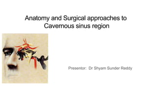
- 1. Anatomy and Surgical approaches to Cavernous sinus region Presentor: Dr Shyam Sunder Reddy
- 2. Schema of discussion Anatomy Surgical Approaches Historical perspective Research and Advances
- 3. Surgical Anatomy of CavernousSinus
- 4. Surgical anatomy of cavernous sinus is best explained under following headings – 1)Bony relationships 2) Dural relationships 3) Venous relationships 4) Neural relationships 5) Arterial relationships
- 5. BONY RELATIONSHIPS MEDIAL – -Middle clinoid process -Pituitary fossa -Body of sphenoid -Carotid sulcus (groove for intracavernous ICA at the lower margin of the sphenoid body) ANTERIOR – Optic strut/ Anterior clinoid process/ Lesser wing of sphenoid LATERAL – -Greater wing of sphenoid -Foramen -rotundum -ovale -spinosum POSTERIOR – Posterior clinoid process/ Dorsum sella/ Petrous apex/ Trigeminal impression
- 7. Floor & Medial wall – formed by single periosteal layer of dura, supero- medially it continues with dura of sella turcica. Roof, Lateral & Posterior wall- are double layered, formed by periosteal layer of dura + dura proper of middle & posterior fossa respectively. -roof medially continues with Diaphragma sella.
- 8. Venous Anatomy ORBIT DUR A TRANSVERSE SINUS JUGULAR BULB CIRCUL AR SINUS AFFERENT DRAINAGE – (IN) 1) Sphenoparietal sinus 2)Sup.Ophthalmic vein 3)Inf. Ophthalmic vein 4)Superficial Sylvian vein (middle cerebral vein) 5)Middle meningeal vein 6)Central retinal vein EFFERENT DRAINAGE – (OUT) 1) Sup. & Inf. Petrosal sinus 2)plexus of vein on ICA drains into Pterygoid plexus 3)Emissary veins of Sphenoid foramen, foramen ovale,
- 9. VENOUS SPACES WITHIN THE CAVERNOUS SINUS: wal l LATERAL COMP. -between the carotid and lateral sinus Thin space filled/ displaced by 5th N. tumor, ICA aneurysm. -Surgical appro. – posterolater./ subtemporal MEDIAL COMP. Between the pituitary and the carotid -Invaded by pituitary tumor. -Surgical appro. 1)superiorly- roof, medial to 3rd N. 2)inferiorly- sphenoid sinus / sella turcica ANTEROINFERIOR COMP. -Smallest, behind sup. Orbital fissure. invaded by orbital tumor. surgical appro. – Anterolaterally, POSTEROSUPERIOR COMP. – (largest space)between the ICA and post. half of roof of sinus Filled by sphenopetroclival meningioma/ clival chordoma. Surgical appro.– extradural, subtemp./Kawa se
- 10. Arterial Relationship 1)POST. VERTICAL SEGMENT – fixed by lateral fibrous ring. – Doesn't give-off branch. 2)POST. BEND – Meningohypopheseal trunk - give rise to 3 branches, i) Tentorial A. of Bernasconi & Cassinari– courses posterolaterally, supply tent./ tentorial meningioma; IIIrd IV th nerves ii) Inf. Hypopheseal A.– courses anteromedially, supply post. Pituitary, anastomose to opp. side. iii)Dorsal meningeal A.– courses posteroinferomedially,supply dura along upper clivus, VI nerve 3)HORIZONTAL SEG. – 2 arteries,-i)Inf. Cavernous sinus A.- ii)McConnell capsular A.- arises medial aspect,supply capsule of pituitary 4)ANT. BEND 5) ANT. VERTICAL SEG.- divides into MCA,ACA
- 12. Neural Relationships IIIrd N.- Runs ant.- lat. & inferiorly. -enters CS through ROOF, medial to ant. Petroclinoid lig. Runs in lateral wall of CS, inferolateral to ACP During drilling of ACP 3rd N. is vulnerable to injury. IVth N.- enters ROOF postero- lateral to IIIrd N. &inferomedial to free edge of tent Runs in lateral wall of CS ateroinferiorly enters in SOF SUPERIO R ORBIT AL FISSURE 3rd WITHIN OCCULOMOT OR CISTERN 4TH 6TH (MEDIA L TO V1) V1 IN THE MECKEL’ S CAVE TENTORI AL EDGE
- 13. Vth N.- enters through Meckel’s cave. V1 passes through lateral wall of CS Runs anteriorly & upwards, enters SOF V2 passes for a short distance in lateral wall of CS enters in f. rotundum VIth N.- enters to CS through Dorello’s canal runs anteriorly, inferolateral to ICA in the substance of CS lies medial to V1 enters in SOF Sympathetic fiber bundles, with carotid a. emerges from the foramen lacerum. Some of the fibers join the VIth nerve before ultimately being distributed to the V1 division to pupillodilator & ciliary ganglion sends symp. fibers long ciliary nerves HORNER’S SYN
- 14. 4th SPHENOPETRO SAL / GRUBER’S LIGAMENT 6th TRIGEMIN AL GANGLION REMOVED PETROLING UAL LIGAMENT (ICA passes underneath) GSP N V1 VIDIA N NERVE
- 15. EXPOSURE AFTER ANTERIOR PETROSECTOMY INTRAOP VIEW ANTERIOR POSTERIO R SUPERIOR CLIVUS INFERIO R PETROS AL SINUS V2 6TH IN THE DORELLO’S CANAL 4TH 3RD AICA PCOM SCA 7th, 8th
- 16. Surgical approaches to cavernous sinus Fronto temporal Extradural & Intradural Approaches Anterolateral Temporopolar transcavernous approaches Lateral Approach to posterior cavernous sinus(Rhomboid Approach)
- 17. Positionin g
- 19. FRONTOTEMPORAL EXTRADURAL & INTRADURAL APPROACH Initially developed by DOLENC ––as anteromedial transcavernous approach, for intracavernous aneurysm UNDERWENT SEVERAL MODIFICATIONS INDICATION ––Lesions confined to cavernous sinus/ with supratentorial extension ADVANTAGE --Can be combined with Middle fossa transpetrosal approach for excision of for posterior extension of tumor.
- 20. sylvian fissure dissection ICA exposure Intra petrous and intra cavernous segments
- 23. Lateral Approach to posterior cavernous sinus (Anterior Petrosectomy)
- 24. Lateral Approach to posterior cavernous sinus(Rhomboid Approach) --Bone to be drilled out in middle fossa is geometrically RHOMBOID SHAPE Intersection of GSPN to V3 Intersection of line projecting along the axis of GSPN to AE AE intersection with petrous ridge Porous trigeminus GSPN can be sectioned, to avoid VIIth N. retraction.
- 25. TECHNICAL CONSIDERATION OF INTRACAVERNOUS TUMOR RESECTIONINTRACAVERNOUS RESECTION- Well encapsulated & nonadherent tumors can be removed by 1)exposure of tumor capsule from surrounding tissue 2) debulking of tumor 3) sharp dissection of tumor capsule from surrounding tissue Invasive & adherent tumors can be removed by 1) interruption of tumor blood supply from periphery of
- 26. THANK YOU