SlideShare une entreprise Scribd logo
1  sur  5
1


              Automated Ejection Fraction Calculations
               from B-Mode (2-D) Echocardiography
                       Suhas Deshpande, Member, IEEE, IEEE-EMBS, CSULB, suhassd@ieee.org


                                                                      Heart is basically the pump for the human circulatory
   Abstract—Left Ventricular Ejection Fraction can be estimate       system. It provides the blood and thus oxygen and vital
to acceptable accuracy using 2-D echocardiography images. 2-D        nutrients from lungs to various parts of the body. The heart
Echocardiography due to its portability and versatility is used in   physiologically consists of four parts- left and right ventricles
emergency operations and ICUs. The estimation of LVEF can            and left and right Auricles. Right Auricle collects the
help the physians to get apriori knowledge of the start of
                                                                     deoxygenated blood from all the parts of the body. Right
conditions such as Coronary heart disease or Cardiomyopathy.
The ejection fraction can be evaluated by using the simson’s
                                                                     ventricle pumps the deoxygenated blood to lungs. The
approximation for calculating the volume of the 2-D image. The       Oxygenated blood reaches the left auricle and is pumped out
two methods tried here are Ellipsoidal and Stacked disk method.      to other parts of the body through left ventricle via the aorta.
                                                                     Thus the left ventricle performance describes the amount of
  Index Terms—Ejection Fraction, Echocardiography, Systole,          oxygen reaching the body.
Diastole
                                                                     A. Systole
                                                                        Systole is a phase of the cardiac cycle in which the heart
                       I. INTRODUCTION                               muscle contracts in a coordinated manner in response to a
                                                                     complex           endogenous autonomic physiologic electrical
H    eart diseases are by far the most common cause of deaths
     in US. In majority of the cases the death can be
prevented if there is a priori knowledge of some of the cardiac
                                                                     stimulus. Systole results in driving blood flow out of the heart
                                                                     and into the body and lungs. All four chambers of a human
                                                                     heart undergo systole and diastole in a timed fashion so that
functions. Left ventricular Ejection Fraction is one of the
                                                                     blood is propelled forward and backward through the
most important cardiac function which gives a good idea
                                                                     cardiovascular system.
about the cardiac problem in advance. The LVEF can be
estimated by using a 2D echocardiography, which is portable
and versatile, is widely used in emergency, operating, and           B. Diastole
intensive care department.                                              Diastole is the period of time when the heart fills with
                                                                     blood after systole. Ventricular diastole is the period during
                                                                     which the ventricles are relaxing, while atrial diastole is the
                  II. FUNCTIONING OF HEART                           period during which the atria are relaxing. During ventricular
                                                                     diastole, the pressure in the ventricles drops from the peak in
                                                                     systole to below the pressure in the left atrium, causing the
                                                                     mitral valve to open. This forces the blood from the atrium to
                                                                     flow into the ventricle.

                                                                                          III.   ECHOCARDIOGRAM
                                                                          Echocardiography is an ultrasound test done for cardiac
                                                                     assessment. The ultrasound waves are transmitted by a
                                                                     transmitter. The ultrasound waves are reflected off the cardiac
                                                                     walls. The computer maps the ECHOs on a line & then takes
                                                                     another sample on a different line thus building up a 2D
                                                                     picture approx 50 times a second. A pseudo image can be
                                                                     prepared on the basis of the received reflected ultrasound
                                                                     wave. This image, echocardiogram or ECHO can be used in
                                                                     diagnostics of the cardiac system. Since the system is simple
                                                                     in design and easy to carry it is very popular with the cardiac
                                                                     diagnosis.
Fig 1. Heart Anatomy and Blood Flow through Heart
                                                                          The frequencies commonly used 2-10MHz are much
                                                                     higher than the audible range of 2-18KHz. Higher
2

frequencies allow better resolution. Lateral resolution of a 5      B. Parasternal Long Axis
MHz is 2 mm compared to 3mm of 3MHz whilst axial
resolution varies from 0.5 mm to 1 mm) but tissue penetration
is poorer.
   The Echocardiogram can be optained by imaging the heart
by positioning the sensor at different position. Transthoracic
view and Parasternal view are commonly used. Trans
oesophagal view can be obtained by placing the sensor on a
catheter inside the oesophagus.


                                                                   Fig 4. Parasternal Long Axis (Image:National University of
                                                                                           Singapore)

                                                                     The transducer is placed in just to the left of the mid to
                                                                   upper sternal border. The right ventricular outflow region, the
                                                                   ventricular septum and the left atrium and ventricular are
                                                                   well visualized. This is one of the best views to obtain an M
                                                                   Mode and hence information on cardiac function.

 Fig 2. Acoustic Windows (Image University of Minnesota)            C. Parasternal Short Axis

  The 2D pictures are taken from the various echo windows
and give "standard views" to build up a complete picture of
the cardiac anatomy. Not all chambers are visible in every
view.

 A. Apical 4 Chamber


                                                                    Fig 5. Parasternal short Axis (Image:National University of
                                                                                             Singapore)

                                                                     From the parasternal long axis view the transduce is rotated
                                                                   90˚ to point towards the left shoulder. The aorta and coronary
                                                                   artery origins are seen well in cross section.

Fig 3. Apical 4 Chamber view (Image:National University of          D. Subcostal View
                       Singapore)

   The transducer is held at the apex of the heart and angled         This is a good view to see lovely images - especially in
towards the right shoulder. The 4 chambers are readily seen        babies as no ribs or lung tissue obscures the view. The atrial
and both the mitral and tricuspid valves. If the transducer is     septum is particularly well seen. Unfortunately             the
angled anteriorly then the aorta is also visualized. This view     transducer is the furthest from the heart and in older children
is shows the ventricular septum well to look for septal defects.   and adults the distance may be too great to allow detailed
                                                                   imaging.
3

                                                                   mitral valve closure; (c) as the instant of maximum cardiac
                                                                   dimension. End-systole is given by the instant preceding
                                                                   mitral valve opening or by the instant of minimum cardiac
                                                                   dimension.

                                                                                      IV. EJECTION FRACTION
                                                                     One of the key indicators of cardiac health is measurement
                                                                   of left ventricular (LV) volume and ejection fraction (EF).
                                                                   The ejection fraction determines the amount of blood pumped
                                                                   out of the ventricles with each cycle of the ECG.

                                                                   The most commonly applied models for the computation of
                                                                   the LV internal volume are derived from that proposed by
                                                                   Simpson. According to the criterion usually called
    Fig 6. Subcoastal view (Image:National University of           “Simpson‟s rule,” the LV is approximated by a stack of
                         Singapore)                                circular (or elliptical) disks whose centers are all in the major
                                                                   axis. The most important measurement of ventricular
                                                                   function is the LVEF (Left Ventricle Ejection Function),
 E. Arch View                                                      which is given by the normalized (nondimensional) difference
                                                                   between End-Diastolic Volume (EDV) and the End-Systolic
                                                                   Volume (ESV), both generally computed according to
                                                                   Simpson‟s rule. The simple formula defining this parameter
                                                                   is

                                                                                      LVEF =


                                                                       A. Stacked disk method

                                                                   In stacked disk method the left ventricle is divided into „n‟
 Fig 7. Arch view (Image:National University of Singapore)         disks. The number of disks „n‟ can be varied to get the
                                                                   required accuracy level. Each disk diameter is matched with
   This is obtained by sliding the transducer towards the upper    the respective contour of the left ventricle. Each disk is
sternal edge and suprasternal notch. It allows the ascending       considered to be an ideal circular shape. The LV volume is
aorta, arch and neck vessels to be imaged.                         estimated using the Simpson‟s equation


F. Importance
Echocardiography is the preferred method for the
documentation of cardiac function at rest. In particular, 2D
Transthoracic Echocardiography (TTE), because of its
portability and versatility, is widely used in emergency,
operating, and intensive care departments. The TTE
technique provides numerous highly significant quantitative
parameters. In fact, it has been demonstrated that cardiac risk
increases significantly when the values of certain parameters
are abnormal. A number of these parameters are connected to
properties of the LV. Thus LV dimensions, volumes, and wall        Figure 8. Stacked disc method for Ejection Fraction
thickness are widely used in clinical practice and research.       estimation
With regard to LV volume quantification, the most important
windows are the apical (4-chamber view and the 2-chamber
view) and the left parasternal (short-axis view at the papillary   Stacked-disk estimation of LV volume
muscle level). The various quantitative parameters are                                   = ((first disk area + last disk area)/2
generally measured at end-diastole and at end-systole. End-                                  + ∑in-1 disk area) x d
diastole can be defined in three ways: (a) as the onset of the     d = disk spacing; n = number of disks; disk are = *r2
QRS Complex in the ECG signal; (b) as the instant after
4


This is one of the basic volume estimation methods for most                         VI. SIMULATION & RESULTS
of applications.
                                                                      The image for the evaluation of Ejection Fraction was
    B. Ellipsoid method.                                            obtained from Phillips Ultrasound after email conversation
                                                                    with Dr. David Hull and Tibor Duliskovich. The images are
                                                                    extracted at end systole and end diastole.


Fig 9. The dimensional evaluation Ellipsoid method                              (a)                         ( b)
                                                                     Fig 10. Echocardiography images (a) End Diastole (b) End
  The left ventricle is assumed to be an ellipsoid. The volume                               Systole
of an ellipsoid can be given by the equation :
                                                                       The images were processed using the Mathworks
    V = 4π/3 abc ( a,b,c are the ellipsoid radii of the LV)         MATLAB©. The grayscale image was cropped to the region
                                                                    of interest, which is the Left Ventricle for this case. The
  In case of the Left ventricle a and c are measured from the       cropped image was filtered using a median filter, unmasking
Apical 4 chamber view. c can be measured from the                   filter for contrast enhastment. Edge detection was done to get
corresponding perpendicular plane (short axis view). It can be      the edge of the binary image.
assumed to be equal to D and the formula becomes

                         V = π/6 LD2

             V.   IMAGE ANALYSIS AND FILTERING

  The echocardiogram is an image created from the
ultrasound waves. Thus the recreated image contains a lot of
noise. Image is very blurred and often it is impossible to read
the image except for a trained people. Many filtering
techniques have been developed to filter the image to be                                (a)                 (b)
suitable for estimating the left ventricular volume.                       Fig 11. Filtered image (a) Systolic (b) Diastolic

   The Echocardiogram images generally have all frequencies
within a short region. It is useful to spread the frequency
spectrum throughout the range of the image. This helps in
thresholding to detect the region of interest (left ventricle in
this case). Histogram equalization is a technique to spread out
the frequencies in an image. Unsharp masking is a technique
used to enhance the image perception by actually using a
blurring effect. Technically, an unsharp mask is generally a
linear or nonlinear filter that amplifies high-frequency
components. Order statistical filters use the statistical data in
the specific order of the mask to filter the image. The median
filter, each pixel will be replaced by the median of the
neighboring pixels. Max(min) filter can be used to chek th
maximum(minimum) value in the neighboring                 pixels.         Fig 12. Edge detection (a) Systolic (b) Diastolic
Averaging filter finds the mean of the neighboring pixels.
The image is preferably converted to a binary image to                The edge detection came out to be a somewhat ellipsoid left
determine the dimensions of the image. This is done by first        ventricle in both systole and diastole.
contrast enhancing and then thresholding of the image.
5

                                                                                Imaging: From Nano to Macro - Proceedings, vol. 2006, pp. 97-100, 2006.
                                                                                   [6]            http://www.med.nus.edu.sg/paed/resources/cardiac_thumbnail/
Table 1. Ejection Fraction by Slotted disk method, Ellipsoid                    investigations/echo.htm
method and provided by Phillips                                                    [7] http://www.vhlab.umn.edu/atlas/echotutorial/echotutorial1.shtml
Method           Slotted Disk   Ellipsoid      Original                            [8] http://rwjms1.umdnj.edu/shindler/imageproc.html#edgefin3.m
                                               EF(Phillips)
Ejection         55.1%          61.45%              54%
fraction


  The End Systolic Volume and End Diastolic value was
calculated by calculating the pixels in the image. The pixels
along one axis give the dimension of the left ventricle along
that axis. For e.g to calculate the disk diameter the pixels
along the x axis were calculated. The number of disks was
chosen to be 20.


                          VII. CONCLUSION
  The ejection fraction calculated using the two methods is
around 54% which is the EF measured by the Phillips
Ultrasound (Data imprinted on the image). There are still
some discrepancies in the code and the image. The apical 2
chamber view would have been a better choice for the
estimation of the volume of LV volume. This is because the
ROI is the left ventricle. The repeatability is poor as the final
result depends much on the cropping of the image to get the
ROI as the left ventricle. There are some problems regarding
the cropping the image to the ROI. If the cropping border
touches the left ventricular edge, the estimation goes wrong.
This is because the ROI is not evaluated precisely due to
opening in the region. There are several discontinuities (black
subregions) present in the filtered image. These subregions
need to be removed to get an accurate EF measurement. A
better filtering approach may remove the discontinuities.


                    ACKNOWLEDGMENT
   I am thankful to Dr. Lobodzinsky for conducting the course
of Digital Image Processing which helped me to base my
study in the project. Further I would like to thank Dr. David
Hull and Tibor Duliskovich from Phillips Radiology
department to provide assistance in providing the images for
the project.

                              REFERENCES
   [1] Arthur J. Vander, “Cardiovascular Physiology,” Human Physiology: Th e
Mechanisms of Body Function, 9th edition: MGH, pp 375-399, 2004
   [2] Bonita Anderson, “Two dimensional echocardiographic measurements
and calculations,” Echocardiography: The Normal Examination and
Echocardiographic Measurements: Wiley, pp 96- 103, 2002
   [3] T. L. Szabo, "Improving ejection fraction estimation for 2d ultrasound
using a computer-generated cardiac model," Proceedings - IEEE Ultrasonics
Symposium, pp. 1757-1760, 2008.
   [4] U. Barcaro, "Automatic computation of left ventricle ejection fraction
from dynamic ultrasound images," Pattern Recognition and Image Analysis, vol.
18, pp. 351-358, 2008.
   [5] M. Jolly, "Assisted ejection fraction in B-mode and contrast
echocardiography," 2006 3rd IEEE International Symposium on Biomedical

Contenu connexe

Tendances

Bundle branch blocks
Bundle branch blocksBundle branch blocks
Bundle branch blocks
Adarsh
 
Hypertrophic cardiomyopathy
Hypertrophic cardiomyopathyHypertrophic cardiomyopathy
Hypertrophic cardiomyopathy
Fuad Farooq
 

Tendances (20)

Ventricular tachycardia
Ventricular tachycardiaVentricular tachycardia
Ventricular tachycardia
 
Bundle branch blocks
Bundle branch blocksBundle branch blocks
Bundle branch blocks
 
Long QT Syndrome
Long QT SyndromeLong QT Syndrome
Long QT Syndrome
 
Cardiac tumors
Cardiac tumorsCardiac tumors
Cardiac tumors
 
Basics of echo & principles of doppler echocardiography
Basics of echo & principles of doppler echocardiographyBasics of echo & principles of doppler echocardiography
Basics of echo & principles of doppler echocardiography
 
Mitral valve anatomy - ppt by kunwar sidharth
Mitral valve    anatomy - ppt by kunwar sidharthMitral valve    anatomy - ppt by kunwar sidharth
Mitral valve anatomy - ppt by kunwar sidharth
 
Tmt protocol
Tmt protocolTmt protocol
Tmt protocol
 
Stress echocardiography
Stress echocardiographyStress echocardiography
Stress echocardiography
 
LA Myxoma - For Medical Students
LA Myxoma - For Medical StudentsLA Myxoma - For Medical Students
LA Myxoma - For Medical Students
 
Constrictive pericarditis
Constrictive pericarditis Constrictive pericarditis
Constrictive pericarditis
 
Sudden cardiac death
Sudden cardiac deathSudden cardiac death
Sudden cardiac death
 
Right and left ventricular hypertrophy
Right and left ventricular hypertrophyRight and left ventricular hypertrophy
Right and left ventricular hypertrophy
 
LBBB
LBBBLBBB
LBBB
 
Restrictive cardiomyopathy
Restrictive cardiomyopathyRestrictive cardiomyopathy
Restrictive cardiomyopathy
 
Atrial Fibrillation
Atrial FibrillationAtrial Fibrillation
Atrial Fibrillation
 
Aortic stenosis
Aortic stenosis Aortic stenosis
Aortic stenosis
 
Aortic stenosis for post graduates
Aortic stenosis for post graduatesAortic stenosis for post graduates
Aortic stenosis for post graduates
 
Coronary anatomy and angiographic views
Coronary anatomy and angiographic viewsCoronary anatomy and angiographic views
Coronary anatomy and angiographic views
 
Hypertrophic cardiomyopathy
Hypertrophic cardiomyopathyHypertrophic cardiomyopathy
Hypertrophic cardiomyopathy
 
Aortic regurgitation
Aortic regurgitationAortic regurgitation
Aortic regurgitation
 

En vedette

Best Practices to Achieve Quality Pressure-Volume Loop Data in Large Animal M...
Best Practices to Achieve Quality Pressure-Volume Loop Data in Large Animal M...Best Practices to Achieve Quality Pressure-Volume Loop Data in Large Animal M...
Best Practices to Achieve Quality Pressure-Volume Loop Data in Large Animal M...
InsideScientific
 
Cardiomyopathies & Valvular Disorders - BMH/Tele
Cardiomyopathies & Valvular Disorders - BMH/TeleCardiomyopathies & Valvular Disorders - BMH/Tele
Cardiomyopathies & Valvular Disorders - BMH/Tele
TeleClinEd
 
Simple Interpretation of Pulmonary Function tests
Simple Interpretation of Pulmonary Function testsSimple Interpretation of Pulmonary Function tests
Simple Interpretation of Pulmonary Function tests
Gamal Agmy
 
Spirometry Interpretation
Spirometry Interpretation Spirometry Interpretation
Spirometry Interpretation
Ashraf ElAdawy
 
Angina pectoris presentation
Angina pectoris presentationAngina pectoris presentation
Angina pectoris presentation
Taher Haddad
 
Pulmonary Function Test
Pulmonary Function TestPulmonary Function Test
Pulmonary Function Test
gowri shanker
 

En vedette (19)

Understanding Ventricular Pressure-Volume Catheter Calibrations and Experimen...
Understanding Ventricular Pressure-Volume Catheter Calibrations and Experimen...Understanding Ventricular Pressure-Volume Catheter Calibrations and Experimen...
Understanding Ventricular Pressure-Volume Catheter Calibrations and Experimen...
 
Best-Practices to Achieve Quality PV Loop Data
Best-Practices to Achieve Quality PV Loop DataBest-Practices to Achieve Quality PV Loop Data
Best-Practices to Achieve Quality PV Loop Data
 
Best Practices to Achieve Quality Pressure-Volume Loop Data in Large Animal M...
Best Practices to Achieve Quality Pressure-Volume Loop Data in Large Animal M...Best Practices to Achieve Quality Pressure-Volume Loop Data in Large Animal M...
Best Practices to Achieve Quality Pressure-Volume Loop Data in Large Animal M...
 
Heart failure with preserved ejection fraction
Heart failure with preserved ejection fractionHeart failure with preserved ejection fraction
Heart failure with preserved ejection fraction
 
HFPEF
HFPEFHFPEF
HFPEF
 
ADH
ADHADH
ADH
 
Vasopressin
VasopressinVasopressin
Vasopressin
 
Anti Diuretic Hormone
Anti Diuretic HormoneAnti Diuretic Hormone
Anti Diuretic Hormone
 
Cardiomyopathies & Valvular Disorders - BMH/Tele
Cardiomyopathies & Valvular Disorders - BMH/TeleCardiomyopathies & Valvular Disorders - BMH/Tele
Cardiomyopathies & Valvular Disorders - BMH/Tele
 
Simple Interpretation of Pulmonary Function tests
Simple Interpretation of Pulmonary Function testsSimple Interpretation of Pulmonary Function tests
Simple Interpretation of Pulmonary Function tests
 
Spirometry Interpretation
Spirometry Interpretation Spirometry Interpretation
Spirometry Interpretation
 
Conduction System of the Heart
Conduction System of the HeartConduction System of the Heart
Conduction System of the Heart
 
Cardiomyopathies
CardiomyopathiesCardiomyopathies
Cardiomyopathies
 
Spirometry
SpirometrySpirometry
Spirometry
 
Angina pectoris presentation
Angina pectoris presentationAngina pectoris presentation
Angina pectoris presentation
 
Pulmonary & systemic circulation
Pulmonary & systemic circulationPulmonary & systemic circulation
Pulmonary & systemic circulation
 
Spirometry and peak flow metry in bronchial asthma
Spirometry and peak flow metry in bronchial asthmaSpirometry and peak flow metry in bronchial asthma
Spirometry and peak flow metry in bronchial asthma
 
Pulmonary Function Test
Pulmonary Function TestPulmonary Function Test
Pulmonary Function Test
 
Myocardial infarction
Myocardial infarctionMyocardial infarction
Myocardial infarction
 

Similaire à Ejection Fraction 2 D Echocardiography

basicechocardiography-170913182344 (2).pdf
basicechocardiography-170913182344 (2).pdfbasicechocardiography-170913182344 (2).pdf
basicechocardiography-170913182344 (2).pdf
littlealphonsa
 
Cardiovascular System.ppt
Cardiovascular System.pptCardiovascular System.ppt
Cardiovascular System.ppt
Shama
 
05 Cardiovascular System.ppt
05 Cardiovascular System.ppt05 Cardiovascular System.ppt
05 Cardiovascular System.ppt
Shama
 
Cardiology consultants in Aurangabad
Cardiology consultants in AurangabadCardiology consultants in Aurangabad
Cardiology consultants in Aurangabad
DrSumitShejol
 
Anatomy of the heart, Schleich
Anatomy of the heart, SchleichAnatomy of the heart, Schleich
Anatomy of the heart, Schleich
Tariq Abdulla
 

Similaire à Ejection Fraction 2 D Echocardiography (20)

EI 65 UNIT 5.ppt
EI 65 UNIT 5.pptEI 65 UNIT 5.ppt
EI 65 UNIT 5.ppt
 
Human Circulatory System/ Blood Vascular System
Human Circulatory System/ Blood Vascular System Human Circulatory System/ Blood Vascular System
Human Circulatory System/ Blood Vascular System
 
basicechocardiography-170913182344 (2).pdf
basicechocardiography-170913182344 (2).pdfbasicechocardiography-170913182344 (2).pdf
basicechocardiography-170913182344 (2).pdf
 
Basic echocardiography
Basic echocardiographyBasic echocardiography
Basic echocardiography
 
Diagnostic Imaging
Diagnostic ImagingDiagnostic Imaging
Diagnostic Imaging
 
Cardiovascular System.ppt
Cardiovascular System.pptCardiovascular System.ppt
Cardiovascular System.ppt
 
Cardiac Scan
Cardiac ScanCardiac Scan
Cardiac Scan
 
Heart and its disease
Heart and its diseaseHeart and its disease
Heart and its disease
 
Echo tee and tte
Echo tee and tteEcho tee and tte
Echo tee and tte
 
Denoising of ECG -- A discrete time approach using DWT
Denoising of ECG -- A discrete time approach using DWTDenoising of ECG -- A discrete time approach using DWT
Denoising of ECG -- A discrete time approach using DWT
 
Denoising of ECG -- A discrete time approach using DWT.
Denoising of ECG -- A discrete time approach using DWT.Denoising of ECG -- A discrete time approach using DWT.
Denoising of ECG -- A discrete time approach using DWT.
 
Physical assessment
Physical assessmentPhysical assessment
Physical assessment
 
Cardio vascular system.pptx
Cardio vascular system.pptxCardio vascular system.pptx
Cardio vascular system.pptx
 
05 Cardiovascular System.ppt
05 Cardiovascular System.ppt05 Cardiovascular System.ppt
05 Cardiovascular System.ppt
 
Cardiology consultants in Aurangabad
Cardiology consultants in AurangabadCardiology consultants in Aurangabad
Cardiology consultants in Aurangabad
 
Anatomy of the heart, Schleich
Anatomy of the heart, SchleichAnatomy of the heart, Schleich
Anatomy of the heart, Schleich
 
heart-180521025843.pdf
heart-180521025843.pdfheart-180521025843.pdf
heart-180521025843.pdf
 
Heart anatomy & physiology slide share, circulatory system pdf
Heart anatomy & physiology slide share, circulatory system pdfHeart anatomy & physiology slide share, circulatory system pdf
Heart anatomy & physiology slide share, circulatory system pdf
 
ANATOMY AND PHYSIOLOGY OF HEART
ANATOMY AND PHYSIOLOGY OF HEARTANATOMY AND PHYSIOLOGY OF HEART
ANATOMY AND PHYSIOLOGY OF HEART
 
Cardiovascular system
Cardiovascular systemCardiovascular system
Cardiovascular system
 

Plus de Suhas Deshpande (10)

Estimation of body mass distribution using projective image
Estimation of body mass distribution using projective imageEstimation of body mass distribution using projective image
Estimation of body mass distribution using projective image
 
Image Recontruction
Image RecontructionImage Recontruction
Image Recontruction
 
Frequency Image Processing
Frequency Image ProcessingFrequency Image Processing
Frequency Image Processing
 
Blood pH Measurement
Blood pH MeasurementBlood pH Measurement
Blood pH Measurement
 
Power and Electrical Safety in Hospitals
Power and Electrical Safety in HospitalsPower and Electrical Safety in Hospitals
Power and Electrical Safety in Hospitals
 
Spatial Image Processing
Spatial Image ProcessingSpatial Image Processing
Spatial Image Processing
 
Perspectives in blood flow measurement
Perspectives in blood flow measurementPerspectives in blood flow measurement
Perspectives in blood flow measurement
 
Equilibrioception
Equilibrioception Equilibrioception
Equilibrioception
 
Biometrics For Security Systems
Biometrics For Security SystemsBiometrics For Security Systems
Biometrics For Security Systems
 
Telesurgery
TelesurgeryTelesurgery
Telesurgery
 

Ejection Fraction 2 D Echocardiography

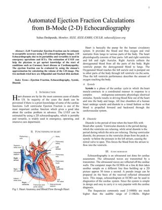
  • 1. 1 Automated Ejection Fraction Calculations from B-Mode (2-D) Echocardiography Suhas Deshpande, Member, IEEE, IEEE-EMBS, CSULB, suhassd@ieee.org  Heart is basically the pump for the human circulatory Abstract—Left Ventricular Ejection Fraction can be estimate system. It provides the blood and thus oxygen and vital to acceptable accuracy using 2-D echocardiography images. 2-D nutrients from lungs to various parts of the body. The heart Echocardiography due to its portability and versatility is used in physiologically consists of four parts- left and right ventricles emergency operations and ICUs. The estimation of LVEF can and left and right Auricles. Right Auricle collects the help the physians to get apriori knowledge of the start of deoxygenated blood from all the parts of the body. Right conditions such as Coronary heart disease or Cardiomyopathy. The ejection fraction can be evaluated by using the simson’s ventricle pumps the deoxygenated blood to lungs. The approximation for calculating the volume of the 2-D image. The Oxygenated blood reaches the left auricle and is pumped out two methods tried here are Ellipsoidal and Stacked disk method. to other parts of the body through left ventricle via the aorta. Thus the left ventricle performance describes the amount of Index Terms—Ejection Fraction, Echocardiography, Systole, oxygen reaching the body. Diastole A. Systole Systole is a phase of the cardiac cycle in which the heart I. INTRODUCTION muscle contracts in a coordinated manner in response to a complex endogenous autonomic physiologic electrical H eart diseases are by far the most common cause of deaths in US. In majority of the cases the death can be prevented if there is a priori knowledge of some of the cardiac stimulus. Systole results in driving blood flow out of the heart and into the body and lungs. All four chambers of a human heart undergo systole and diastole in a timed fashion so that functions. Left ventricular Ejection Fraction is one of the blood is propelled forward and backward through the most important cardiac function which gives a good idea cardiovascular system. about the cardiac problem in advance. The LVEF can be estimated by using a 2D echocardiography, which is portable and versatile, is widely used in emergency, operating, and B. Diastole intensive care department. Diastole is the period of time when the heart fills with blood after systole. Ventricular diastole is the period during which the ventricles are relaxing, while atrial diastole is the II. FUNCTIONING OF HEART period during which the atria are relaxing. During ventricular diastole, the pressure in the ventricles drops from the peak in systole to below the pressure in the left atrium, causing the mitral valve to open. This forces the blood from the atrium to flow into the ventricle. III. ECHOCARDIOGRAM Echocardiography is an ultrasound test done for cardiac assessment. The ultrasound waves are transmitted by a transmitter. The ultrasound waves are reflected off the cardiac walls. The computer maps the ECHOs on a line & then takes another sample on a different line thus building up a 2D picture approx 50 times a second. A pseudo image can be prepared on the basis of the received reflected ultrasound wave. This image, echocardiogram or ECHO can be used in diagnostics of the cardiac system. Since the system is simple in design and easy to carry it is very popular with the cardiac diagnosis. Fig 1. Heart Anatomy and Blood Flow through Heart The frequencies commonly used 2-10MHz are much higher than the audible range of 2-18KHz. Higher
  • 2. 2 frequencies allow better resolution. Lateral resolution of a 5 B. Parasternal Long Axis MHz is 2 mm compared to 3mm of 3MHz whilst axial resolution varies from 0.5 mm to 1 mm) but tissue penetration is poorer. The Echocardiogram can be optained by imaging the heart by positioning the sensor at different position. Transthoracic view and Parasternal view are commonly used. Trans oesophagal view can be obtained by placing the sensor on a catheter inside the oesophagus. Fig 4. Parasternal Long Axis (Image:National University of Singapore) The transducer is placed in just to the left of the mid to upper sternal border. The right ventricular outflow region, the ventricular septum and the left atrium and ventricular are well visualized. This is one of the best views to obtain an M Mode and hence information on cardiac function. Fig 2. Acoustic Windows (Image University of Minnesota) C. Parasternal Short Axis The 2D pictures are taken from the various echo windows and give "standard views" to build up a complete picture of the cardiac anatomy. Not all chambers are visible in every view. A. Apical 4 Chamber Fig 5. Parasternal short Axis (Image:National University of Singapore) From the parasternal long axis view the transduce is rotated 90˚ to point towards the left shoulder. The aorta and coronary artery origins are seen well in cross section. Fig 3. Apical 4 Chamber view (Image:National University of D. Subcostal View Singapore) The transducer is held at the apex of the heart and angled This is a good view to see lovely images - especially in towards the right shoulder. The 4 chambers are readily seen babies as no ribs or lung tissue obscures the view. The atrial and both the mitral and tricuspid valves. If the transducer is septum is particularly well seen. Unfortunately the angled anteriorly then the aorta is also visualized. This view transducer is the furthest from the heart and in older children is shows the ventricular septum well to look for septal defects. and adults the distance may be too great to allow detailed imaging.
  • 3. 3 mitral valve closure; (c) as the instant of maximum cardiac dimension. End-systole is given by the instant preceding mitral valve opening or by the instant of minimum cardiac dimension. IV. EJECTION FRACTION One of the key indicators of cardiac health is measurement of left ventricular (LV) volume and ejection fraction (EF). The ejection fraction determines the amount of blood pumped out of the ventricles with each cycle of the ECG. The most commonly applied models for the computation of the LV internal volume are derived from that proposed by Simpson. According to the criterion usually called Fig 6. Subcoastal view (Image:National University of “Simpson‟s rule,” the LV is approximated by a stack of Singapore) circular (or elliptical) disks whose centers are all in the major axis. The most important measurement of ventricular function is the LVEF (Left Ventricle Ejection Function), E. Arch View which is given by the normalized (nondimensional) difference between End-Diastolic Volume (EDV) and the End-Systolic Volume (ESV), both generally computed according to Simpson‟s rule. The simple formula defining this parameter is LVEF = A. Stacked disk method In stacked disk method the left ventricle is divided into „n‟ Fig 7. Arch view (Image:National University of Singapore) disks. The number of disks „n‟ can be varied to get the required accuracy level. Each disk diameter is matched with This is obtained by sliding the transducer towards the upper the respective contour of the left ventricle. Each disk is sternal edge and suprasternal notch. It allows the ascending considered to be an ideal circular shape. The LV volume is aorta, arch and neck vessels to be imaged. estimated using the Simpson‟s equation F. Importance Echocardiography is the preferred method for the documentation of cardiac function at rest. In particular, 2D Transthoracic Echocardiography (TTE), because of its portability and versatility, is widely used in emergency, operating, and intensive care departments. The TTE technique provides numerous highly significant quantitative parameters. In fact, it has been demonstrated that cardiac risk increases significantly when the values of certain parameters are abnormal. A number of these parameters are connected to properties of the LV. Thus LV dimensions, volumes, and wall Figure 8. Stacked disc method for Ejection Fraction thickness are widely used in clinical practice and research. estimation With regard to LV volume quantification, the most important windows are the apical (4-chamber view and the 2-chamber view) and the left parasternal (short-axis view at the papillary Stacked-disk estimation of LV volume muscle level). The various quantitative parameters are = ((first disk area + last disk area)/2 generally measured at end-diastole and at end-systole. End- + ∑in-1 disk area) x d diastole can be defined in three ways: (a) as the onset of the d = disk spacing; n = number of disks; disk are = *r2 QRS Complex in the ECG signal; (b) as the instant after
  • 4. 4 This is one of the basic volume estimation methods for most VI. SIMULATION & RESULTS of applications. The image for the evaluation of Ejection Fraction was B. Ellipsoid method. obtained from Phillips Ultrasound after email conversation with Dr. David Hull and Tibor Duliskovich. The images are extracted at end systole and end diastole. Fig 9. The dimensional evaluation Ellipsoid method (a) ( b) Fig 10. Echocardiography images (a) End Diastole (b) End The left ventricle is assumed to be an ellipsoid. The volume Systole of an ellipsoid can be given by the equation : The images were processed using the Mathworks V = 4π/3 abc ( a,b,c are the ellipsoid radii of the LV) MATLAB©. The grayscale image was cropped to the region of interest, which is the Left Ventricle for this case. The In case of the Left ventricle a and c are measured from the cropped image was filtered using a median filter, unmasking Apical 4 chamber view. c can be measured from the filter for contrast enhastment. Edge detection was done to get corresponding perpendicular plane (short axis view). It can be the edge of the binary image. assumed to be equal to D and the formula becomes V = π/6 LD2 V. IMAGE ANALYSIS AND FILTERING The echocardiogram is an image created from the ultrasound waves. Thus the recreated image contains a lot of noise. Image is very blurred and often it is impossible to read the image except for a trained people. Many filtering techniques have been developed to filter the image to be (a) (b) suitable for estimating the left ventricular volume. Fig 11. Filtered image (a) Systolic (b) Diastolic The Echocardiogram images generally have all frequencies within a short region. It is useful to spread the frequency spectrum throughout the range of the image. This helps in thresholding to detect the region of interest (left ventricle in this case). Histogram equalization is a technique to spread out the frequencies in an image. Unsharp masking is a technique used to enhance the image perception by actually using a blurring effect. Technically, an unsharp mask is generally a linear or nonlinear filter that amplifies high-frequency components. Order statistical filters use the statistical data in the specific order of the mask to filter the image. The median filter, each pixel will be replaced by the median of the neighboring pixels. Max(min) filter can be used to chek th maximum(minimum) value in the neighboring pixels. Fig 12. Edge detection (a) Systolic (b) Diastolic Averaging filter finds the mean of the neighboring pixels. The image is preferably converted to a binary image to The edge detection came out to be a somewhat ellipsoid left determine the dimensions of the image. This is done by first ventricle in both systole and diastole. contrast enhancing and then thresholding of the image.
  • 5. 5 Imaging: From Nano to Macro - Proceedings, vol. 2006, pp. 97-100, 2006. [6] http://www.med.nus.edu.sg/paed/resources/cardiac_thumbnail/ Table 1. Ejection Fraction by Slotted disk method, Ellipsoid investigations/echo.htm method and provided by Phillips [7] http://www.vhlab.umn.edu/atlas/echotutorial/echotutorial1.shtml Method Slotted Disk Ellipsoid Original [8] http://rwjms1.umdnj.edu/shindler/imageproc.html#edgefin3.m EF(Phillips) Ejection 55.1% 61.45% 54% fraction The End Systolic Volume and End Diastolic value was calculated by calculating the pixels in the image. The pixels along one axis give the dimension of the left ventricle along that axis. For e.g to calculate the disk diameter the pixels along the x axis were calculated. The number of disks was chosen to be 20. VII. CONCLUSION The ejection fraction calculated using the two methods is around 54% which is the EF measured by the Phillips Ultrasound (Data imprinted on the image). There are still some discrepancies in the code and the image. The apical 2 chamber view would have been a better choice for the estimation of the volume of LV volume. This is because the ROI is the left ventricle. The repeatability is poor as the final result depends much on the cropping of the image to get the ROI as the left ventricle. There are some problems regarding the cropping the image to the ROI. If the cropping border touches the left ventricular edge, the estimation goes wrong. This is because the ROI is not evaluated precisely due to opening in the region. There are several discontinuities (black subregions) present in the filtered image. These subregions need to be removed to get an accurate EF measurement. A better filtering approach may remove the discontinuities. ACKNOWLEDGMENT I am thankful to Dr. Lobodzinsky for conducting the course of Digital Image Processing which helped me to base my study in the project. Further I would like to thank Dr. David Hull and Tibor Duliskovich from Phillips Radiology department to provide assistance in providing the images for the project. REFERENCES [1] Arthur J. Vander, “Cardiovascular Physiology,” Human Physiology: Th e Mechanisms of Body Function, 9th edition: MGH, pp 375-399, 2004 [2] Bonita Anderson, “Two dimensional echocardiographic measurements and calculations,” Echocardiography: The Normal Examination and Echocardiographic Measurements: Wiley, pp 96- 103, 2002 [3] T. L. Szabo, "Improving ejection fraction estimation for 2d ultrasound using a computer-generated cardiac model," Proceedings - IEEE Ultrasonics Symposium, pp. 1757-1760, 2008. [4] U. Barcaro, "Automatic computation of left ventricle ejection fraction from dynamic ultrasound images," Pattern Recognition and Image Analysis, vol. 18, pp. 351-358, 2008. [5] M. Jolly, "Assisted ejection fraction in B-mode and contrast echocardiography," 2006 3rd IEEE International Symposium on Biomedical