Plateforme de protéomique et d'analyse des protéines par spectrométrie de masse - UCL - Lab'inSight Proteomics
•
1 like•485 views

description of the laboratory skills (09/03/2010)

Recommended
Recommended
More Related Content
Similar to Plateforme de protéomique et d'analyse des protéines par spectrométrie de masse - UCL - Lab'inSight Proteomics
Similar to Plateforme de protéomique et d'analyse des protéines par spectrométrie de masse - UCL - Lab'inSight Proteomics (20)
More from Réseau LIEU (Liaison Entreprises-Universités)
More from Réseau LIEU (Liaison Entreprises-Universités) (20)
Recently uploaded
Recently uploaded (20)
Plateforme de protéomique et d'analyse des protéines par spectrométrie de masse - UCL - Lab'inSight Proteomics
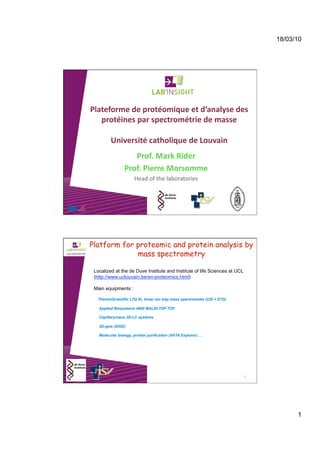
- 1. 18/03/10 1 Plateforme de protéomique et d’analyse des protéines par spectrométrie de masse Université catholique de Louvain Prof. Mark Rider Prof. Pierre Morsomme Head of the laboratories Platform for proteomic and protein analysis by mass spectrometry 2 Localized at the de Duve Institute and Institute of life Sciences at UCL (http://www.uclouvain.be/en-proteomics.html) Main equipments : ThermoScientific LTQ XL linear ion trap mass spectrometer (CID + ETD) Applied Biosystems 4800 MALDI-TOF-TOF Capillary/nano 2D-LC systems 2D-gels (DIGE) Molecular biology, protein purification (AKTA Explorer) …
- 2. 18/03/10 2 MASSPROT plaBorm in Woluwe Historical overview 3 1997 MS set up in the HORM Unit by Mark Rider Early work on protein and phosphorylation site identification LCQ Classic 2002 Upgrade to a more sensitive MS First LC-MS experiments LCQ Deca XP Plus LTQ XL + ETD 2005 Capillary/nano 2D-LC system Proteomic gel-free work flow 2008 Linear ion trap with Electron Transfer Dissociation Gas-phase radical ion fragmentation to preserve labile PTMs top-down capabilities MASSPROT plaBorm in Louvain‐la‐Neuve Historical overview 4 1993 Protein sequencer 1996 2D-gels 2007 2D-LC-MALDI-TOF-TOF
- 3. 18/03/10 3 Platform for proteomic and protein analysis by mass spectrometry 5 – Protein expression, purificaKon and characterizaKon – Proteome analysis – DifferenKal profiling and quanKficaKon of protein expression – Post‐TranslaKonal ModificaKons discovery Localized at the de Duve Institute and Institute of life Sciences at UCL (http://www.uclouvain.be/en-proteomics.html) Main equipments : ThermoScientific LTQ XL linear ion trap mass spectrometer (CID + ETD) Applied Biosystems 4800 MALDI-TOF-TOF Applications: Know-how of the platform 6 – Protein expression and purificaKon – Expression in bacteria, yeast (S.cerevisiae, P.pastoris) and plants – Soluble and membrane proteins purificaAon Morsomme et al., J. Biol. Chem. 2002, 277, 29608
- 4. 18/03/10 4 Expression of a functional antibody in Nicotiana tabacum plants and culture cells 7 – Expression in plants – Transient / stable – ConsAtuAve/inducible Navarre et al., Transgenic Res. 2006, 15, 325 Expression of a functional antibody in Nicotiana tabacum plants and culture cells 8 De Muynck et al., Transgenic Res. 2009, 18, 467
- 5. 18/03/10 5 Expression of a functional antibody in Nicotiana tabacum plants and culture cells 9 NS0 SR1 BY2 HC LC Know-how of the platform 10 – Protein expression and purificaKon – Expression in bacteria, yeast and plants – Soluble and membrane proteins purificaAon – Proteome analysis – 2D‐gels + gel‐free (1D, 2D or 3D LC‐MS) – Soluble and membrane proteins – Non‐model species
- 6. 18/03/10 6 Proteome of non-model species: example of Nicotiana tabacum BY2 cell culture 11 56 % of identification Duby et al., Proteomics 2010, in press 795 1422 Know-how of the platform 12 – Protein expression and purificaKon – Proteome analysis – DifferenKal profiling and quanKficaKon of protein expression – DIGE (2D‐gels) – Isotopic labeling (iTraq, O18…) – Label free quanAficaAon with spectral counts soluble proteins: E. Coli periplasm, glycosomes from Trypanosomes (Vertommen et al., Mol Biochem Parasitol. 2008,158:189) (Vertommen et al., Proteomics. 2009, 9:2432) membrane proteins : E. Coli outer membrane (Leverrier et al., Proteomics. 2010, 10:771) biomarkers of neurodegenerative diseases in human
- 7. 18/03/10 7 Know-how of the platform 13 – Protein expression and purificaKon – Proteome analysis – DifferenKal profiling and quanKficaKon of protein expression – Post‐TranslaKonal ModificaKons discovery phosphorylation signal transduction glycation diabetes oxidation oxidative stress acetylation epigenetics ubiquitylation glutathionylation disulfide bridge glycosylation … 14 IdenKficaKon of Fructosamine‐3‐phosphate residues in human hemoglobin The aim of this work was to identify the fructosamine residues on hemoglobin that are removed as a result of the action of FN3K in intact erythrocytes
- 8. 18/03/10 8 15 Delpierrre G et al. J. Biol. Chem. 2004;279:27613 Three‐dimensional structure of human oxy‐hemoglobin showing the posiKon of glycated residues First mass-spectrometric identification of glycated residues in human hemoglobin that are phosphorylated by FN3K Nine different fructosamine residues phosphorylated by FN3K were identified The physiological importance of deglycation is unknown at present. Deglycation might prevent the further conversion of fructosamines into advanced glycation products Platform for proteomic and protein analysis by mass spectrometry 16 – Protein expression and purificaKon – Expression in bacteria, yeast and plants – Soluble and membrane proteins purificaAon – Proteome analysis – 2D‐gels + gel‐free (1D, 2D or 3D LC‐MS) – Soluble and membrane proteins – Non‐model species – DifferenKal profiling and quanKficaKon of protein expression – DIGE (2D‐gels) – Isotopic labeling (iTraq, O18…) – Label free quanAficaAon with spectral counts – Post‐TranslaKonal ModificaKons discovery – phosphorylaAon , glycaAon, acetylaAon, ubiquitylaAon, glutathionylaAon, disulfide bridge, glycosylaAon … http://www.uclouvain.be/en-proteomics.html
- 9. 18/03/10 9 17 Aknowledgements Mark rider Didier Vertommen Pierre Morsomme Marc Boutry Hervé Degand Anne-Marie Faber http://www.uclouvain.be/en-proteomics.html