

Recommandé
Contenu connexe
Tendances
Tendances (20)
En vedette
Similaire à Minarcik robbins 2013_ch22-female
Similaire à Minarcik robbins 2013_ch22-female (20)
Plus de Elsa von Licy
Plus de Elsa von Licy (20)
Dernier
Dernier (20)
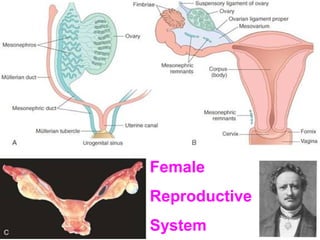
Minarcik robbins 2013_ch22-female
- 2. TOPICS • TODAY (Part I) • NEXT CLASS (Part II) – Vulva – Vagina – Cervix, uterus – Body, uterus – Tubes – Ovaries – Placenta
- 3. VULVA • Synonymous with EXTERNAL genitalia • Everything ANTERIOR to the INTROITUS • Usual classification of Degen., Inflam., Neopl. • Common Diseases: – BARTHOLIN Cyst – Vulvar Vestibulitis – Deg./Inflam. Epithelial: LICHEN diseases – BENIGN tumors: Condyloma(ta) – MALIGNANT tumors: VIN, SCC
- 4. Result from Inflammation/Obstruction of the Bartholin glands (i.e., greater vestibular glands) Often result in abscesses Surgical removal is curative when local procedures are inadequate or often recurrent NEVER become malignant
- 5. VULVAR VESTIBULITIS, assoc. w. vulvodynia
- 6. “LICHEN” DISORDERS LICHEN Sclerosu(i)s (atrophic skin) LICHEN Simplex Chronicus (hypertrophic skin) Common features of FIBROSIS and INFLAMMATION
- 10. The types of lichen lesions which show HYPER-plastic mucosal changes are often regarded as being “potentially” pre-malignant
- 11. CONDYLOMA(TA)
- 12. VIN, SCC • Like condylomas, HIGHLY linked to HPV • VIN=changes leading to SCCin-situ, look like “plaques” • BEYOND VIN = INFILTRATION
- 13. VIN
- 17. VAGINA • CONGENITAL: Parallel uterus anomalies • INFLAMMATORY – PRE-menopausal: STD – POST-menopausal: ATROPHY • BENIGN: Hidradenoma, Condyloma • MALIGNANT: VIN, INFILTRATING SCC
- 18. CONGENITAL • Imperforate hymen (hematocolpos) • Atresia • Absence (agenesis) • Septate • Double vag/uterus (didelphys)
- 20. • 90% VAGINITIS • Bacterial Vaginitis is the most common cause of vaginitis, • • accounting for 50% of vaginitis cases. As previously mentioned, BV is caused by an overgrowth of organisms such as Gardnerella vaginalis (gram-variable coccobacillus), Mobiluncus species, Mycoplasma hominis, and Peptostreptococcus species. Risk factors include pregnancy, intrauterine device (IUD) use, and frequent douching. Candida species (C albicans, C tropicalis, and C glabrata) are airborne fungi that are natural inhabitants of the vagina in as many as 50% of women, and vaginal candidiasis is the second most common cause of vaginitis. Risk factors include oral contraceptive use, IUD use, young age at first intercourse, increased frequency of intercourse, receptive cunnilingus, diabetes, HIV or other immunocompromised states, chronic antibiotic use, and pregnancy. T. vaginalis infection, the third most common cause of vaginitis, is caused by trichomonads. These organisms are flagellated protozoans. Trichomonads primarily infect vaginal epithelium, and they less commonly infect the endocervix, urethra, and Bartholin and Skene glands. Trichomonads are transmitted sexually and can be identified in as many as 80% of male partners of infected women. Risk factors include tobacco use, unprotected intercourse with multiple sexual partners, and the use of an IUD.
- 22. VAGINAL NEOPLASIA • VIN • INFILTRATING SCC • ADENOSIS (D.E.S.) • ADENOCARCINOMA (Di-Ethyl-Stilbestrol)
- 23. VIN
- 24. NORMAL VIN
- 25. SCC
- 37. DYSPLASIA / CIN / SIL
- 39. INFILTRATION
- 40. How have we “CURED” Cervical Carcinoma?
- 41. ENDOMETRIUM • • • • • • • FUNCTIONAL HISTOLOGY D.U.B. (Dysfunctional Uterine Bleeding) INFLAMMATION ADENOMYOSIS/ENDOMETRIOSIS POLYPS/HYPERPLASIA ADENOCARCINOMA and/or STROMAL LEIOMYOMYOMAS, -SARCOMAS • MITOSES differentiate benign from malignant
- 43. MITOSES (Glandular and Stromal) = VACUOLES/SECRETION = PRE-ovulatory POST-ovulatory
- 44. DYSFUNCTIONAL UTERINE BLEEDING (DUB) • Anovulatory Cycle • Inadequate Luteal Phase • Oral Contraceptives • Menopause • Post-Menopause
- 45. ENDOMETRITIS • PID • Post-partum Sepsis • BCP’s • TB • IUD’s
- 46. ADENOMYOSIS • Defined as normal endometrial glands deep within the myometrium
- 48. ENDOMETRIOSIS Defined as normal endometrial glands OUTSIDE the confines of the myometrium Reverse menstruation vs. Embryologic “rest” theories EXTREMELY common cause of cyclical abdominal/pelvic pain Broad Ligament, Ovary (“chocolate cysts”), Peritoneum, Bowel, Umbilicus
- 50. “CHOCOLATE” CYST
- 56. Adenocarcinoma of the Endometrium = Carcinoma of the Uterus
- 59. • • • • • • • • • ADENOCARCINOMA of the ENDOMETRIUM Papillary, Polypoid Clear Cell Adeno-Squamous Mucinous Serous Preceded by hyperplasia, dysplasia (EIN) Estrogenic, DES effects Ass. w.: obesity, diabetes, hypertension, infertility Stromal “sarcomatous” conditions can co-exist, i.e., “adenosarcoma”
- 60. GRADING and STAGING • GRADING – 1, 2, 3 – Well, Moderate, Poor • STAGING – (I) Corpus – (II) Corpus + Cervix – (III) Beyond uterus, but inside true pelvis – (IV) Outside true pelvis or involving bladder or rectal mucosa
- 62. SALPINGITIS/PID GC and CHLAMYDIA PYOSALPINX PERITONITIS TUBO-OVARIAN ADHESIONS STERILITY INFERTILITY
- 63. Peritubal CYSTS • Endometriosis • Hydatid Cysts of Morgagni (Mullerian rests) Para-, Peritubal)
- 67. DISEASES of OVARIES • DEGENERATIVE? • INFLAMMATORY? • CYSTS • TUMORS – – – – Müllerian (“Germinal”) Germ Cell Sex Cord/Stromal Metastatic
- 68. DISEASES of PREGNANCY •EARLY Pregnancy •LATE Pregnancy
- 69. DISEASES of PLACENTA • ANOMALIES • “BENIGN” tumors (MOLES) • MALIGNANT tumors (CHORIOCARCINOMA)
- 73. Everything you can see or feel is lined by serosa (i.e., mesothelial cells, visceral and parietal
- 76. • • • • • • • • • • • • TERMS “Germinal” Epithelium (Mesothelium) Ovum (Oocyte) Tunica Albuginea Primordial Follicle Primary Follicle Mature “Graffian” follicle (antral or secondary) Granulosa cells ( Estrogen) Thecal cells ( Estrogen) Corpus luteum ( Progesterone) “Atretic” follicle Corpus Albicans “Stroma”
- 81. B=GRANULOSA D=THECA INTERNA E=THECA EXTERNA
- 84. ESTROGEN • • • • • Controlled by FSH and LH Develop, Lactate Breast Lobules Proliferate Endometrial Glands “Cardioprotective” “Bone Mass” protective
- 85. PROGESTERONE • • • • Controlled by FSH and LH SECRETE Endometrial Glands IMPLANTATION of the blastocyst Lactation
- 89. POLY-Cystic Ovarian Disease (Stein-Leventhal syndrome) 5% Prevalence Anovulation Oligomenorrhea Obesity Hirsutism
- 91. OVARIAN TUMORS • MÜLLERIAN (MAJORITY) – – – – – – – – Serous (Benign, Borderline, Malignant) Mucinous (Benign, Borderline, Malignant) Endometroid (Benign, Borderline, Malignant) Adenosarcoma (Carcinoma AND Sarcoma) Mesodermal Mixed (MULTIPHASIC Sarcoma) Clear Cell Brenner (almost always benign) Transitional (almost always look like Brenner) Germ Cell (Not surprisingly, like males) • • SEX-CORD/STROMAL • METASTATIC
- 92. OVARIAN TUMORS • • • • Solid vs. Cystic Functional vs. NON-functional Benign vs. Malignant First clinical presentation may be ascites, in carcinomas. • Malignant ascites in a woman is ovarian cancer until proven otherwise • CA-125 is THE important tumor marker in ovarian cancer, especially as a follow up.
- 93. SEROUS, BENIGN
- 94. MUCINOUS, BENIGN
- 98. PSAMMOMA bodies are dried up papillae of papillary adenocarcinomas, usually in the thyroid, but in ANY papillary adenocarcinoma
- 100. OTHER MÜLLERIAN • ENDOMETRIOD, malignant – (looks like endometrium) • CLEAR CELL, malignant – (clear cells, reminiscent of renal clear cell ca.) • CYSTADENOFIBROMA, benign – (BENIGN “FIBROUS” COMPONENT) • BRENNER TUMOR, benign – (transitional cell nests) • CARCINOMA with SARCOMA – (adenosarcoma, mixed Müllerian)
- 101. “GERM CELL” Tumors • Teratomas (usually benign in ovary), i.e., “mature” cystic teratoma or dermoid cyst • “Immature” teratomas are regarded as malignant • Dysgerminoma (look exactly like the testicular seminoma), malignant • Endodermal Sinus (Yolk Sac), malignant, Just like testicular • Choriocarcinoma, malignant, just like testicular
- 107. ENDODERMAL SINUS TUMOR, aka YOLK SAC TUMOR
- 108. CHORIOCARCINOMA, Just like testis or placenta
- 109. SEX-CORD/STROMAL TUMORS • Chiefly benign and NON-cystic, i.e., “solid”, often functional (hyper-estrogen-ism) • Granulosa-Theca • Fibroma-Theca • Sertoli-Leydig (Androblastoma)
- 110. CALL-EXNER BODIES
- 111. B=GRANULOSA D=THECA INTERNA E=THECA EXTERNA
- 113. DISEASES of PREGNANCY •EARLY Pregnancy •LATE Pregnancy
- 114. EARLY PREGNANCY • SPONTANEOUS ABORTION • ECTOPIC PREGNANCY
- 115. Spontaneous Abortion • 15% - 35% • Fetal Causes – Usually Genetic • Maternal Causes (placental, uterus infections or trauma) – Toxo, Mycoplasma, Listeria – Trauma
- 116. Ectopic Pregnancy • Chiefly TUBAL, but ovarian or abdominal rare • 1% OF NORMAL WOMEN • 35%-50% OF WOMEN with previous SALPINGITIS/PID • + HCG, Abdominal pain, 1st trimester, ultrasound
- 118. LATE PREGNANCY • PLACENTAL ANOMALIES • TWIN PLACENTAS • PLACENTAL INFLAMMATIONS • TOXEMIA (ECLAMPSIA/PREECLAMPSIA) • INTRAUTERINE GROWTH RETARDATION
- 119. PLACENTAL ANOMALIES • • • • Accessory Lobes Bipartite Placenta Circumvallate Placenta Placenta Accreta, chorion going DIRECTLY to the myometrium
- 123. CIRCUMVALLATE
- 124. PLACENTA ACCRETA NO DECIDUA BETWEEN VILLI AND MYOMETRIUM
- 125. MRI of Placenta PREVIA, or LOW-LYING placenta, usually anatomically normal, but just lies LOWER than it should.
- 127. TOXEMIA of PREGNANCY (PRE-eclampsia) • Hypertension • Proteinuria • Edema • Related to Placental Ischemia, but MANY theories • Risk for DIC, convulsions (eclampsia)
- 128. Intrauterine Growth Retardation • Fetal causes: Genetic, malformations • Maternal Causes, vascular diseases, toxemia, infections, placental diseases • Placenta size (350-700g) ~ Fetal size (7.5 lb)
- 129. Placental Infections • Villitis vs. chorionamnionitis vs. funisitis • ASCENDING vs. hematogenous • ASCENDING are usually bacterial, and chorionamnionitis • HEMATOGENOUS are often TORCH, and villitis
- 130. Placental Neoplasms, i.e. gestational trophoblastic disease • Benign: MOLES (Hydatidiform moles) • Malignant: CHORIOCARCINOMA • BOTH are associated with increased or persistent levels of the placental hormone HCG
- 132. (Hydatid)-iform Mole • 1/1000 in USA • 1% in Indonesia • Also called NON-invasive mole in its most common benign variant, but can also be “invasive” • Complete (2% chorioCA incidence) or partial (0% incidence) • Grapelike clusters, i.e., swollen villi
- 134. The MAIN thing differentiating benign from malignant from worrisome trophoblastic neoplasms is INVASIVENESS of the trophoblast
Notes de l'éditeur
- Johannes Müller
- Müllerian first, everything else second!
- Bartholin glands in women are analogous to Cowper’s (bulbourethral) glands in men. They are also called GREATER vestibular glands. Inflamation or obstruction to the ducts of these glands can cause cyst formation.
- The GREATER vestibular glands are located on the LOWER (posterior) wall of the vagina. The vestibular glands (NOT greater vestibular) are located on the UPPER (anterior) wall of the vagina.
- “LSA” is the buzz word that all the cool people use who would like you to think they know a lot about this disease Lichen Sclerosus et Atrophicus
- “Sclerosus” refers to fibrosis of the underlying dermis. “Atrophicus” refers to thin epidermis. “Lichen” is Greek for “crusty”
- This type of “lichenoid” disorder has HYPER-plastic epidermis, NOT atrophic., like most of the SKIN lichenoid disorders do. The “hyperplasia” is felt to be related to “itching” or mechanical epidermal irritation.
- This is probably because of the “promoter” concept again. It also sounds like more of a legal disclaimer, because itching shouldn’t cause cancer, right?
- Yep, HPV again! (usually types 6 and 11)
- VIN, CIN, PIN, VIN. (why a double “V”? (Vulvar,Vaginal too), all represent PRE-cancerous changes, vulva, cervix, prostate, vagina, respectively. Soon you’ll see EIN, too for the endometrium.
- Very LOW grade VIN looks almost like normal skin, very HIGH grade VIN is regarded as carcinoma-in-situ, i.e., cancer, but hasn’t infiltrated yet. Identify the areas of VIN on this picture. Factures such as loss of maturation pattern, nuclear aberrations such as enlargement, hyperchromasia, pleomorphism, mitoses differentiate LOW fro HIGH.
- INFILTRATING squamous cell carcinoma. Any doubt?
- Any doubt? INFILTRATING squamous cell carcinoma!
- Radical vulvectomy specimen? How do you know this is malignant melanoma? Where is the pigment? Would you order a S-100 and HMB-45 immunostain? Answer: YES Why are some of the WORST melanomas not grossly (or even microscopically) pigmented?
- Atresia, double vagina, double uterus.
- Bacteria, Candida, Trichomonas are the common causes of vaginitis
- Note the trich’s flagella, wet prep, SEM in lower right.
- A specific subtype of adenocarcinoma ("Clear Cell") occurs in a small percent of women (termed "DES-Daughters") born between 1938 and 1973 (later outside the United States) that were exposed to the drug diethylstilbestrol (DES) in utero
- Yep, you guessed it. Caused by HPV again, usually.
- Squamous metaplasia of cervical glands. Can you understand how this might be difficult to differentiate from infiltrating squamous cell carcinoma?
- Cancer or metaplasia? Ans: Cancer Why?
- The old name “botryoid” rhabdomyosarcoma, is still often used. Sarcoma botryoides, or botryoid sarcoma. IS “botryoid” the Greek root word for “grape bunches”? Ans: YES Can rhabdomyosarcomas arise from mature skeletal muscle? ANS: NO Skeletal muscle precursor cells? YES
- Colposcopist’s view. Where, precisely, is the squamocolumnar junction?
- What is this? This is the visual LEAP between gross and miscrsocopic, We can also see the PRECISE squamocolumnar junction microscopically too.
- What is this? How is an N/C ratio reflective of serum estrogen levels? Is a PAP smear a great test of estrogen level? Ans: YES
- This is precisely the squamous metaplastic process? Where else might squamous metaplasia occur and why?
- MANY neutrophils often accompany mature squamous cells on pre-premenopausal pap smears, Do NOT be fooled into thinking some inflammatory process is going on which needs to be treated.
- What is your diagnosis? Cancer? Breech presentation? HPV? Polyp? Answer: Polyp
- Glands and fibrous stroma of a typical cervical polyp. Because it has GLANDS, would you call it an ENDO-cervical polyp, rather than ECTO-? Ans: YES Are most cervical polyps ENDO_cervical? Ans: YES
- Note that the journey from normal epithelium to carcinoma is a GRADUAL one, often many years too. Sometimes, only MONTHS!!!!!
- Which one is “worse:? Which one is more convincingly HPV? Why? (Ans: Koilocytosis)
- Would you expect microinfiltration to originate from a carcinoma-in-situ appearing epithelium? Ans: YES The “infiltration” on the right may NOT look grossly infiltrated.
- Normal, CIN-I, CIN-II, CIN-III. Which is which?
- A classic by Noyes!
- PRE-ovulatory = proliferative endometrium. POST-ovulatory = secretory endometrium. If you diagnose “SECRETORY ENDOMETRIUM”, what have you proven? Ans: the woman has ovulated
- What is the greatest concern in POST menopausal bleeding? Ans: CARCINOMA
- What is the difference between adenomyosis and endometriosis? Is adenomyosis also called endometriosis “IN-terna”? Ans: YES Is endometriosis called endometriosis EX-terna? Ans: YES
- What are these hemorrhagic areas in the thickened myometrium? Ans: ENDOMETRIOSIS
- Endometriosis, rectal.
- “Chocolate” refers to the consistency of the hemorrhage, nothing more. So therefore, not all “chocolate” cysts are due to endometriosis.
- Endometrial “polyp”, colposcopist’s view and gross specimen.
- Endometrial “polyp”, microscopic view. Because endometrial polyps are really excesses of estrogen targeted tissue, i.e., endometrium, are they related to estrogen excesses? Ans : YES Are they related to hyperplastic endometrium? Ans: YES Can they be confused with hyperplastic endometrium on a D&C specimen? Ans: YES Although they can be functional glands, i.e., show cyclic changes, most are in postmenopausal women.
- Polyps vs hyperplasia? Does it matter? In each case it is a functional hyperplastic proliferation of endometrium.
- Intrauterine leiomyoma. Classically, leiomyomas are: 1) Subserosal, 2) Mural, 3) Submucosal
- Just as NUCLEOLI differentiate benign from malignant prostate glands, what differentiates benign from malignant smooth muscle tumors? Ans: MITOSES per high power fields. NOT pleomorphism, NOT hyperchromasia, NOT nuclear size, …………………………………..But MITOSES!!!
- Associated with: HYPERESTROGENISM OBESITY DIABETES HYPERTENSION INFERTILITY Obviously, Endometrial hyperplasia (EIN), endometrial intraepithelial neoplasia
- INVASIVENESS is KEY feature to differentiate endometrial hyperplasia from endometrial carcinoma.
- What are the usual glandular features of adenocarcinoma vs. “benign” glands?
- These are merely adjectives, HOWEVER, some types are better than others, prognostically. HOWEVER, grading and staging are of utmost importance, independent of whatever of these “adjectives” you use.
- Primary germ cells, male or female, first arise in the yolk sac and migrate to the genital ridge, which is in close proximity to the mesonephros. Eventually, retroperitoneal testes migrate through the inguinal canal to the scrotum, covered by peritoneum. Ovaries stay in the pelvis, and are covered by serosa, and are therefore intraperitoneal, but POSTERIOR to the fallopian tubes.
- The CORTEX is the site of developing follicles. The MEDULLA is relatively free of developing follicles, and rich in connective tissue (stroma) and blood vessels.
- Major internal female genitalia structures, landmarks, and interrelationships. In which ligament does the ovarian artery lie? Through which structure does the round ligament travel. Normally the uterus is a bit ANTE-VERTED and ANTE-FLEXED
- Major internal female genitalia structures, landmarks, and interrelationships.
- GREAT whole mount to demonstrate overall cortex vs. medullary differentiation.
- Name the main structures.
- OOCYTE PRIMORDIAL FOLLICLE (simple squamous covering) PRIMARY FOLLICLE (cuboidal epithelial covering)
- Zona pellucida, arrow, becomes “atretic” follicle. Is this a primary follicle? Ans: YES Why?
- Secondary = Graffian = Antral follicle Where is the antrum?
- Find the cumulus oophorus, liquor folliculi, and corona radiata
- Granulosa and theca INTERNA cells make estrogen.
- LUTEAL cells, under LUTEINIZING hormone and FSH too, make progesterone. LUTEUM means YELLOW. Why? Why is ANYTHING bright yellow? A corpus luteum of pregnancy is considerably larger than a regular, NON-pregnancy, corpus luteum, often, perhaps about a half or third the size of the ovary itself.
- Corpus albicans. ALBA means WHITE. Why is it white?
- Most common PRE-menopausal cyst.
- Any EXTREMELY yellow cyst of a premenopausal ovary, is regarded as luteal in origin. Very common PRE-menopausal cyst
- Although the cortical area of the normal ovary contains cysts, i.e., various stages of follicular development, true PCOD (PolyCystic Ovarian Disease, or Stein-Leventhall) ovaries are BIGGER (2x) than normal premenopaosal ovaries and have “true” cysts, NON-ovulatory, NOT just stages of follicular development. Is a “cyst” a “tumor” (i.e. swelling) in the classical sense of the word, like a bump on the head. Is a cyst usually a true neoplasm? Ans: Of course not!
- Always think of true ovarian tumors as following the normal anatomy/histology in these FOUR groups---mullerian, germ, sex-cord, metastatic. In contrast to the testicle, the ovary DOES occasionally get metastases.
- Gross, microscopic, physiologic, behavioral classification factors for ovarian tumors.
- The HUGEST tumors ever reported in human beings (50-100 lbs.?) are frequently benign mucinous ovarian tumors.
- Q: What other adjective can we give to this tumor besides serous? Ans: Papillary Benign or malignant? Ans: Donno
- Close up of papillae
- Why is this serous and NOT mucinous?
- PSAMMOMA bodies Ovary, thyroid, breast, meningiomas, any place where papillary adenocarcinomas are found.
- Serous and mucinous
- Less common Müllerian tumors, all malignant except for one (Brenner)
- I TOLD you this looks the same as TESTICULAR germ cell tumors.
- Dermoid “cyst” = BENIGN CYSTIC TERATOMA, BY FAR the most common ovarian NEOPLASM of younger women, usually BENIGN
- Whether the teratomatous elements are “mature” or “immature” determine, greatly, the behavior of the teratoma, i.e., benign or malignant.
- IMMATURE looking neural tissue. This is much more likely to behave badly (i.e., malignant) than a mature one. Often, you might see retinal tissue, like you see here.
- Female dysgerminomas are IDENTICAL in appearance to male seminomas, i.e., germ cells + lymphocytes. You’d have to tell the pathologist whether this was a male or female in order for him to diagnose seminoma vs. dysgerminoma.
- Schiller-Duvall Body, just like in the testis yolk sac tumor!
- EXACTLY the same as a malignant HCG producing testicular choriocarcinoma or a malignant HCG producing placental choriocarcinoma
- “Sex cord” = “stroma” MANY are functional, i.e., associated with hyper estrogenism (or androgenism)
- Call-Exner bodies are virtually diagnostic of granulosa cell tumors. Q: Do they remind you of “rosettes”? Ans: YES
- Q: Would a “thecoma” derived from theca INTERNA be more likely to be functional than a thecoma derived from theca EXTERNA? Ans: YES Why? Note the “theca” has both a vesicular and spindle cell appearance. The juicy vesicular cells, theca interna, and tumors derived from them, can secrete estrogen. The spindly theca externa cells, usually do not, and may look simply like fibromas.
- Many thecomas look white and fibrous, That is why the term fibrothecoma is often used? Is a fibrothecoma or fibroma less likely to be functional than a thecoma? Ans: YES Why? Ans: It is derived from characteristically NON-estrogen producing cells.
- Not separated 3 ways, as in trimesters, but TWO ways, early and late.
- Most tubal pregnancies occur in week 5-8 of pregnancy, does this one look a but later? Would you think the hemorrhage might be retrograde before it ruptures the muscular wall?
- Normal ~500 gram placenta.
- Might this umbilical cord insertion be a little eccentric? YES, Velamentous? NO
- Accessory placental lobe. An extreme lobe might be called a BI-partite placenta.
- In a circumvallate placenta the amnionic, i.e., amniotic, membranes “thicken” or “double back”. Incidence: 1% Associations: Abruptions, Oligohydramnios
- You might guess this kind of placenta would be VERY difficult to remove, and remnants (retained POC) might result in endometritis. If the decidua is a cushion between placenta and uterus, then accreta means loss of cushion. ACC-reta, IN-creta, PER-creta, in order of severity.
- And don’t forget placenta “abruptio” or premature separation of placenta with hemorrhage (i.e., hematoma)
- Twin zygosity (mon- or di-) is related to the number of CHORIONS, NOT amnions or umbilical cords!
- Toxemia of pregnancy occurs in an amazing 6% of all pregnancies. Toxemia is also called PRE-eclampsia. When PRE-eclampsia is particularly severe and associated with more serious systemic effects such as DIC or convulsions, it is called ECLAMPSIA.
- Wee now return full circle back to chapter 10, diseases of childhood!
- What does TORCH stand for? T-oxo O-ther R-ubella C-MV (Do you see the BASOPHILIC intranuclear inclusion in the above villitis pic?) H-erpes
- Syncytial cells are FUSED, CYTO-trophoblastic cells are deeper stem cells. Is this chorionic villus mature or IM-mature? Ans: mature Why? Ans: It has blood vessels in its core. If it was IMMATURE, it would NOT need secondary blood vessels and can diffuse oxygen and nutrients WITHOUT secondary blood vessel formation.
- In COMPLETE moles, ALL the villi are swollen. They turn into choriocarcinomas 2% of the time. In PARTIAL moles, only some are. They NEVER turn into choriocarcinomas.
- NOTE trophoblast looks NORMAL, i.e., NON-invasive and NON-proliferative, and NON atypical. How does the TROPHOBLASTIC layer look on the right, uniform and thin, or invasive and thick? Ans: Uniform.
- Choriocarcinoma. Note invasive trophoblast.
- Choriocarcinoma. Note extreme pleomorphism of trophoblastic cells.