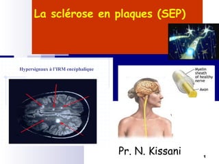
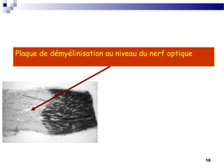
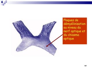
This document provides an overview of the approach to diagnosing and classifying ataxia. It discusses the various causes of ataxia including hereditary, non-hereditary, and those affecting the cerebellum, sensory pathways, frontal lobe, and thalamus. The diagnostic approach involves a thorough history, neurological exam, rating scales, neuroimaging, genetic testing, and other laboratory tests. Hereditary ataxias are classified including autosomal dominant cerebellar ataxias (SCA types 1-31), autosomal recessive ataxias (Friedreich's ataxia, ataxia telangiectasia), X-linked ataxias, and mitochondrial ataxias.
This document discusses the neurologic manifestations of HIV/AIDS in India. Some key points include:
- Opportunistic infections like cryptococcal meningitis and tuberculosis account for the majority (around 70%) of neurologic events seen in HIV patients in India.
- Conditions like progressive multifocal leukoencephalopathy and myelopathy are relatively rare compared to Western countries.
- Common neurologic manifestations include meningitis, mass lesions in the brain, and various neuropathies.
- The pattern of neurologic involvement tends to change as the CD4 count declines, with more severe complications occurring at very low CD4 levels.
This document discusses different types of ataxia, including their causes and clinical features. It begins by defining ataxia as a lack of muscle coordination during voluntary movements. It then covers various forms of ataxia such as cerebellar ataxia (the most common type), sensory ataxia affecting proprioception, cortical ataxia involving the frontal lobe, and thalamic ataxia. The document also discusses the classification of cerebellar ataxias into hereditary forms like autosomal dominant and recessive types, as well as the clinical assessment and diagnostic approach for ataxia.
This document provides an overview of ataxia, including its definition, causes, clinical features, classifications, hereditary forms, and spinocerebellar ataxia (SCA). Key points include: Ataxia is caused by dysfunction of the cerebellum and its pathways, resulting in loss of coordination. Common clinical features are gait and limb ataxia, dysarthria, and gaze abnormalities. Causes include genetic, paraneoplastic, infectious, autoimmune, and others. Hereditary forms include autosomal dominant and recessive SCAs, Friedreich's ataxia, and more. SCA is the most common hereditary ataxia and subtypes are distinguished by their clinical
Walking aids
Occupational Therapy
To learn how to use assistive devices like
canes, walkers, or wheelchairs
To learn techniques to help with daily
activities like dressing, bathing, etc.
Braces or splints to prevent contractures
Adaptive equipment to help with tasks
Surgery
Counseling
Selective dorsal rhizotomy to reduce leg
spasticity
To help cope with the emotional impact and
adjust to physical changes
Orthopedic surgery for contractures or
deformities
Support groups
Hereditary neuropathies are a diverse group of inherited conditions affecting the peripheral nervous system. They are frequently underdiagnosed due to their indolent onset over decades and lack of clear family history in some cases. Charcot-Marie-Tooth disease is the most common inherited neuropathy, with two main types - CMT1 characterized by demyelination and CMT2 characterized by axonal loss. CMT1 results from mutations affecting myelin protein zero or peripheral myelin protein 22 genes, causing demyelination and onion bulb formation. Accurate diagnosis relies on detailed family history, neurological examination, and electrodiagnostic testing to distinguish inherited from acquired neuropathies.
This document provides an overview of the approach to evaluating and diagnosing ataxia. It begins with definitions of ataxia and discusses tests to differentiate various systems that can cause ataxia-mimicking symptoms. It then covers approaches to evaluating cerebellar ataxia, including assessing mode of onset, progression, focal vs symmetric involvement, and localizing the lesion. Common etiologies of acquired, inherited, autosomal dominant and recessive ataxias are summarized. The document provides a step-wise algorithm for evaluating and categorizing ataxia.
The document discusses how HIV can infect the nervous system and cause various neurological syndromes. It notes that HIV enters the nervous system early during initial infection and may cause immediate or delayed symptoms. Both acute and chronic infections can result in issues like meningitis, dementia, transverse myelitis, peripheral neuropathies, and others. The document provides details on clinical features, evaluations, differential diagnoses, treatments, and more for the various neurological conditions associated with HIV.
Intravenous Immunoglobulin in Neurology Ade Wijaya
Intravenous immunoglobulin (IVIg) works as an immunomodulatory agent and can treat several autoimmune neurological diseases. It has been approved by the FDA to treat chronic inflammatory demyelinating polyneuropathy, multifocal motor neuropathy, and Guillain-Barré syndrome. IVIg has also shown clinical benefits for other conditions like myasthenia gravis, neuromyelitis optica, and multiple sclerosis based on controlled clinical trials. Potential side effects include headaches, fever, and fatigue.
GEMC- Sickle Cell Disease: Special Considerations in Pediatrics- Resident Tra...Open.Michigan
This is a lecture by Hannah Smith, MD from the Ghana Emergency Medicine Collaborative. To download the editable version (in PPT), to access additional learning modules, or to learn more about the project, see http://openmi.ch/em-gemc. Unless otherwise noted, this material is made available under the terms of the Creative Commons Attribution Share Alike-3.0 License: http://creativecommons.org/licenses/by-sa/3.0/.
This document provides a historical overview of epilepsy from ancient times to the modern era. Some key points:
- Epilepsy has been documented as far back as ancient Mesopotamia in the 10th century BCE, with accurate descriptions of seizures. Hippocrates in the 5th century BCE was the first to argue epilepsy had a physical rather than supernatural cause located in the brain.
- Advances continued through Islamic, Renaissance, and 19th century physicians with improved classification of seizure types and localization of epileptic foci. The late 19th century saw dedicated epilepsy hospitals established.
- Major technological advances included the EEG in the 1920s allowing precise localization, and effective anti-seizure drugs
1. Acute bacterial meningitis is commonly caused by hematogenous spread of bacteria from other sites of infection to the meninges. Common causative organisms vary by age.
2. Clinical manifestations range from sudden onset of shock and coma to more gradual onset of fever and nonspecific CNS symptoms. Signs include meningeal irritation and increased intracranial pressure. Lumbar puncture reveals pleocytosis and abnormal CSF parameters.
3. Encephalitis is caused by a variety of viruses transmitted by arthropods or direct person-to-person spread. It presents with abrupt fever and neurological deficits that can include seizures or coma. Diagnosis involves CSF analysis and serology. Outcomes range from
Oligodendrocytes produce myelin sheaths around axons that insulate and speed up nerve conduction. Demyelinating disorders involve damage to existing myelin sheaths, while dysmyelinating disorders impair myelin formation. Common demyelinating conditions include multiple sclerosis, acute disseminated encephalomyelitis, and neuromyelitis optica spectrum disorder. Diagnostic tools include neurological exam, MRI, and lumbar puncture. Differential diagnosis requires considering alternative causes such as infection, autoimmune disease, or metabolic derangement.
This document provides an overview of herpes simplex encephalitis (HSE). It discusses the etiology, epidemiology, pathophysiology, clinical manifestations, diagnosis and treatment of HSE. Key points include: HSE is most commonly caused by HSV-1 and is a leading cause of viral encephalitis in children; presentation varies from non-specific symptoms to seizures, altered mental status and focal neurological deficits; diagnosis involves CSF analysis, neuroimaging and PCR testing; and treatment involves high-dose intravenous acyclovir administered for at least 21 days. Recurrence of HSE is possible due to viral reactivation or immune-mediated processes.
neurodegeneration due to braiin iron accumulationSachin Adukia
This document provides an overview of neurodegeneration due to brain iron accumulation (NBIA). It defines NBIA as a heterogeneous group of inherited neurodegenerative disorders characterized by extrapyramidal movement disorders and abnormal iron accumulation in the basal ganglia. It then describes several specific disorders that fall under the NBIA classification, including their typical clinical presentations, imaging features, pathology findings, and treatment approaches. Key disorders discussed include PKAN, PLAN, MPAN, and BPAN. The document provides detailed information on the genetic causes and characteristic signs of each condition.
Sjogren Syndrome- a complex CNS disease Diana Girnita
This document discusses central nervous system involvement in Sjögren's syndrome. It begins with a brief history of Sjögren's syndrome and then discusses prevalence of central nervous system manifestations, which range from 10-60% in primary Sjögren's syndrome. Common central nervous system manifestations include headaches, cognitive deficits, mood disorders, spinal cord involvement, optic neuritis, and focal or multifocal brain lesions. Testing such as CSF analysis, MRI, EEG, and cerebral angiography can provide additional information, though findings may be nonspecific. Autoantibodies such as anti-Ro/SSA antibodies may have prognostic value in assessing central nervous system disease severity in Sjögren's syndrome patients.
Acute infections of the nervous system like bacterial meningitis can be life-threatening if not recognized and treated early. The document discusses various acute infections including bacterial meningitis, viral meningitis, encephalitis, and fungal infections. It provides details on the clinical presentation, diagnosis, and management of bacterial meningitis, which is often characterized by the classic triad of fever, headache, and neck stiffness, and requires prompt lumbar puncture and antibiotic treatment to identify the pathogen and prevent complications.
examination of the peripheral ocular systemrzgar hamed
1. The document discusses examination of the peripheral ocular motor system, including ocular alignment and diplopia. It describes how the visual axes are normally aligned and how misalignments can cause diplopia.
2. Methods for testing ocular motor function are provided, including tests for eye movement range, corneal light reflection testing, and cover-uncover testing to detect heterotropias or heterophorias.
3. The types of eye movements and deviations are defined, including terms for different types of strabismus like esotropia, exotropia, and vertical deviations. Analysis of cover-uncover testing helps differentiate between monocular and alternating deviations.
Cerebral microbleeds are small brain hemorrhages detected by MRI that are caused by leakage of blood from damaged small vessel walls. They are increasingly recognized in patients with cerebrovascular disease, Alzheimer's disease, vascular cognitive impairment, and normal elderly populations. Microbleeds in lobar regions may indicate cerebral amyloid angiopathy and link vascular and amyloid neuropathologies, while deep or infratentorial microbleeds often reflect hypertensive vasculopathy. Detection of microbleeds provides insight into cerebral small vessel disease and its relationship to cognitive impairment and dementia.
Sydenham's chorea is a movement disorder that is the most common form of autoimmune chorea. It typically develops in children between ages 5-15 after a Group A beta-hemolytic streptococcal infection and is part of rheumatic fever. Symptoms include generalized chorea and other neurological symptoms like motor impersistence. Diagnosis involves looking for elevated antistreptococcal titers and imaging may show basal ganglia enlargement. Treatment considers severity, requirements, availability, cost, evidence and side effects. It is thought to be caused by molecular mimicry between streptococcal and human proteins that leads to an autoimmune response.
This document discusses the approach to evaluating and diagnosing myelopathy. It begins by defining myelopathy as spinal cord, meningeal, or perimeningeal damage or dysfunction. It then lists signs that strongly indicate or are consistent with but not diagnostic of myelopathy. Alternative diagnoses are also discussed. Common causes of acute myelopathy are then summarized, including multiple sclerosis, spinal cord infarction, and transverse myelitis. Features suggesting infectious etiology and patterns of spinal cord involvement are outlined. The document concludes by discussing compressive myelopathies and pearls for localizing spinal cord lesions.
The document discusses the extrapyramidal system, which is a neural network located in the brain that is part of the motor system. It centers around the modulation and regulation of anterior horn cells. Key structures include the cortex, basal ganglia, and structures that connect them like the substantia nigra. Imbalances can result in syndromes like Parkinson's disease, characterized by tremors, rigidity, and postural instability. The document outlines other extrapyramidal system disorders and their associated involuntary movements.
This document discusses autoimmune epilepsy, noting that some epilepsies may have an autoimmune basis which has changed their evaluation and management. It outlines that autoimmune epilepsy can present as recent-onset cryptogenic epilepsy, status epilepticus, or subacute onset seizures. Supporting features include a viral prodrome, antecedent psychiatric symptoms, drug resistance, or a history of autoimmunity or cancer. Treatment involves immunotherapy to reduce seizures, though antiepileptic drugs may still be used acutely or for maintenance in some cases. Testing for neural-specific antibodies can provide evidence of an autoimmune basis.
Recent guidelines for management of neurocystcercosisNeurologyKota
This document summarizes key information about neurocysticercosis (NCC), a parasitic infection caused by the larval stage of the pork tapeworm Taenia solium. It discusses the clinical manifestations of NCC including seizures, headaches, hydrocephalus and neurological deficits depending on the location and number of cysts. Diagnosis involves imaging like CT and MRI along with serological tests. Treatment involves antiparasitic drugs along with corticosteroids to prevent inflammation from dying cysts. Prognosis depends on the degree and location of infection, with most patients experiencing resolution of symptoms if the lesions resolve. Prevention focuses on improved animal husbandry, meat inspection and sanitation.
Quatrième rencontre sur les syndromes secs et la maladie de gougerot.Khadija Moussayer
Le syndrome de Gougerot-Sjögren (SGS) est une maladie auto-immune qui se manifeste essentiellement par une sécheresse des muqueuses, principalement buccales et oculaires, appelée autrement syndrome sec. Cette diminution des sécrétions lacrymales et salivaires est un motif fréquent de plaintes, elle concerne le quart des personnes âgées de plus de 60 ans au Maroc. Parmi les causes multiples du syndrome sec figurent l’atrophie des glandes exocrines liée à l’âge et l’utilisation de certains médicaments. La maladie de Gougerot-Sjögren est une de ses causes les plus emblématiques.
Véritable "syndrome carrefour" au sein des maladies auto-immunes, le syndrome de Gougerot-Sjögren touche entre 0,2 et 0,5 % de la population, à près de 90 % des femmes et surtout autour de la cinquantaine. Son diagnostic est parfois délicat alors même que les tests utilisés pour la confirmation d’abord d’un syndrome sec et ensuite du SGS souffrent encore d’un manque de standardisation. Les manifestations fonctionnelles du syndrome sec dépendent de la muqueuse atteinte :
- La sécheresse salivaire ou xérostomie : se manifeste par une sensation de bouche sèche, une soif accrue avec ingestion répétée des liquides au cours des repas, des difficultés à la mastication, des douleurs de la bouche ou des problèmes dentaires. Dans les cas extrêmes, l’élocution est entravée, les aliments adhèrent à la langue nécessitant alors la prise d’eau pour les décoller, ou encore la langue se colle au palais au réveil. L’examen de la bouche peut révéler une fissuration de la bouche ou des lèvres ainsi qu’un aspect dépapillé de la langue et vernissé de la bouche. Une parotidomégalie, un signe important dans le syndrome sec en rapport avec la maladie de Sjogren, s’observe dans 50% des cas de la maladie.
- La sécheresse oculaire ou xérophtalmie : sa fréquence est d’une grande ampleur puisqu’elle concerne 15 à 20% des personnes âgées de plus de 70 ans et représente jusqu’à 30% de motifs de consultation en ophtalmologie. La xérophtalmie est en rapport avec un déficit quantitatif des larmes ou avec une altération qualitative du film lacrymal. Ses manifestations sont variables, allant d’une sensation de corps étranger, de brûlure et de rougeur oculaire, à une gêne à la lumière et une vision trouble ou paradoxalement à des larmoiements.
Etiologies et Classifications des Accidents Ischémiques Cérébrauxsaddek khellaf
un cours qui élucide d'une façon globale les différentes étiologies des accidents vasculaires ischémiques, puis présente les principales classifications qui on permis d'élaborer (au plus précisément possible) un lien de causalité directe de telle ou telle pathologie a l'AVC ischémique installé.
3. 3
I- INTRODUCTION
La sclérose en plaques (SEP) est une affection neurologique
fréquente débutant le plus souvent chez l'adulte jeune.
Elle se caractérise par la survenue de poussées cliniques
correspondant à l’apparition successive de foyers de
démyélinisation disséminés (plaques)au sein de la substance
blanche du système nerveux central .
La SEP n'est pas une maladie primitivement axonale, mais les
atteintes de l'axone sont plus importantes et plus précoces que
les conceptions classiques ne le laissaient supposer
Le caractère imprévisible des poussées et la dispersion des
lésions rendent compte de l'extrême polymorphisme clinique de
l'affection.
L'étiologie de la SEP, encore mal connue, relèverait de la
conjonction d’un facteur lié à l'environnement et d’un terrain
génétique prédisposé, conduisant à l’émergence de réactions
auto-immunitaires dirigées contre la myéline du SNC.
4. 4
II- EPIDEMIOLOGIE et ETIOLOGIE
Environ 40000 personnes atteintes en France :
prévalence 60/10 5
80 à 100/105
dans les pays scandinaves et 20/105
en
nord Afrique.
Constitue la maladie neurologique handicapante la plus
hospitalisée.
Les 2/3 des SEP débutent entre 20 et 40 ans.
La maladie est 2 fois plus fréquente chez la femme.
A Marrakech, sur 10 ans, environ 140 cas.
5. 5
Distribution géographique inégale de la maladie, fréquente dans les
pays tempérés, où son incidence augmente du sud vers le nord.
L'étude des déplacements de population montre que la prévalence
de la SEP n’est pas modifiée pour les sujets ayant immigré après
l’âge de 15 ans.
National Multiple Sclerosis Society, 1993
7. 7
III-ETIOPATHOGENIE
SEP, à ce jour ne connaît pas de cause précise,
Elle repose comme même sur des anomalie immunologique.
Les hypothèses avancées :
infection par un virus latent dont l'activation provoque une
réponse immunitaire secondaire,
disposition génétique liée aux groupes tissulaires mais sans
respect des lois mendeliennes,
incidence du climat avec une fréquence plus forte dans les
climats tempérés.
L’implication du système immunitaire dans la SEP est démontré
par l’augmentation de la synthèse intrethécale des
immunoglobulines IgG, IgA et IgM selon un profil oligoclonal
Ainsi, l’étiologie de la SEP apparaît multifactorielle, en
rapport probable avec un évènement lié à l’environnement,
de nature possiblement virale, entraînant dans une tranche
d’âge déterminée, et chez des sujets génétiquement
prédisposés, une réaction auto-immune dirigée contre la
myéline du SNC.
8. 8
IV-ANATOMIE PATHOLOGIQUE
Caractéristiques des plaques de
démyélinisation:
taille variable, contour bien limité,
réparties de manière aléatoire au sein
de la substance blanche du SNC avec
une prédominance pour les régions
périventriculaires, le centre ovale, les
hémisphères cérébelleux, les cordons
postérieurs et antérolatéraux de la
moelle, et les nerfs optiques. Les
plaques sont en général plus nombreuses
que ce que la clinique aurait pu laisser
prévoir.
Plaque de démyélinisation dans la SB
(vue nécropsique)
9. 9
Plaque de démyélinisation dans la SB
(vue nécropsique)
existence de
plaques
asymptomatiques.
elles peuvent
être repérées du
vivant du malade
par l’IRM ou les
potentiels
évoqués (PEV,
PES et PEA).
10. 10
A la phase aiguë de la démyélinisation:
infiltrat de cellules mononuclées, dont en
particulier des macrophages phagocytant les
produits de dégradation de la myéline.
A la phase chronique:
raréfaction des oligodendrocytes, et une gliose
astrocytaire.
La démyélinisation est responsable:
ralentissement ou d’un blocage de la conduction
nerveuse, souvent réversibles en début de
maladie.
12. 12
V - ETUDE CLINIQUE
Le diagnostic de SEP repose sur:
dissémination spatiale et temporelle des lésions.
Le terrain (adulte de 20 à 40 ans),
et le caractère inflammatoire du LCR +++
Insister sur l’association de symptômes
neurologiques ne pouvant s’expliquer par
une lésion unique.
dans le cas contraire, l’IRM et les potentiels
évoqués peuvent apporter la preuve d’une
atteinte multifocale.
La dissémination temporelle signifie l’existence
de poussées antérieures régressives
Place de l’interrogatoire;
baisse de l’acuité visuelle,
paresthésies?, etc....).
13. 13
Les manifestations cliniques de la SEP
sont extrêmement polymorphes,
affectant le plus souvent:
les nerfs optiques (NORB),
les voies pyramidales
la sensibilité,
les fonctions cérébelleuses
et vestibulaires,
les nerfs oculo-moteurs,
et les fonctions génito-sphinctériennes.
14. 14
1) La névrite optique rétro-bulbaire
(NORB) +++
*Inaugurale de la maladie dans 1/3 des cas,
et alors souvent isolée.
Permet d’affirmer la multifocalité des
lésions lorsqu’il existe d’autres symptômes
neurologiques.
*Correspond à une démyélinisation des
fibres maculaires du nerf optique, elle
peut être asymptomatique alors
détectée que par les potentiels évoqués
visuels (PEV).
15. 15
Sur le plan séméiologique +++
a) baisse rapide de l'acuité visuelle d'un oeil
(durant quelques heures ou jours);
b) flou visuel, scotome central qui sera objectivé
par la campimétrie (CV), ou d'une perte de la vision
des couleurs témoignant d'une dyschromatopsie;
c) douleurs orbitaires et péri-orbitaires
inconstantes, accentuées par la mobilisation de
l’œil;
d) fond d'œil normal à la phase aiguë dans la
majorité des cas "le malade ne voit rien,
l'ophtalmologiste non plus"
Les PEV sont abolis à ce stade (du fait de la
démyélinisation).
18. 18
2) L’atteinte pyramidale
- Pratiquement constante dans les formes évoluées,
et révélatrice de la maladie dans 10 à 20% des cas.
- Responsable d’un déficit moteur affectant le plus
souvent les 2 membres inférieurs (paraparésie),
L’intensité de ce déficit est aussi très variable
(simple fatigabilité à la marche, au tableau de
myélite transverse).
- un véritable sd pyramidal:
- ROT très pathologiques
- signe de Babinski
- ou de Hoffmann,
- et une abolition des réflexes cutanés abdominaux.
- Ou simple irritation pyramidale:
- exagération des ROT,
19. 19
3) L’atteinte sensitive
a- Elle relève avant tout d’une atteinte
lemniscale prédominant au niveau des
cordons postérieurs de la moelle, et
responsable de :
Signes fonctionnels :
Paresthésies, évocatrices par leurs caractères
particuliers à type de sensation d'étau, de
cuirasse, de peau épaissie, de vêtements trop
serrés, etc..
20. 20
il ne s’y associe pas nécessairement de déficit de la
sensibilité+++ (SP<<<<SF).
- Signes physiques :
- Signe de Lhermitte.
- Une ataxie proprioceptive avec signe de Romberg.
- Une perte du sens de la position et du mouvement des
articulations, une hypopallesthésie, et une astéréognosie.
21. 21
4) L’atteinte cérébelleuse
- De nature statique et cinétique. Souvent
précoce, bilatérale et asymétrique
Responsable d’une dysarthrie + tremblement
intentionnel souvent invalidant, et d’une ataxie
aggravant les troubles de la marche, souvent
liée à l’atteinte sensori-motrice
marche cérébello-ataxo spasmodique.
22. 22
Les autres atteintes
5) L’atteinte vestibulaire
De nature centrale, responsable d’un syndrome
vestibulaire dysharmonieux, néanmoins intense à la
phase aiguë.
6) Paralysies oculo-motrices
- Responsable d’une diplopie parfois ressentie comme une
vision floue ou une simple "fatigue visuelle".
- L’atteinte la plus fréquente est celle de la bandelette
longitudinale postérieure (BLP) ophtalmoplégie
internucléaire.
7) Atteinte des autres nerfs crâniens
- Une parésie faciale périphérique (VII) est possible
Les voies auditives sont souvent démyélinisées,
23. 23
8) Troubles génito-sphinctériens
- Par leur impact psychologique et leur très grande
fréquence, ils constituent une des principales menaces de
la maladie, ils sont à type de:
*pollakiurie,
*mictions impérieuses,
*incontinence urinaire,
*rétention aiguë d’urine, de constipation et d’impuissance.
9) Troubles psychiques et intellectuels
Etat anxio-dépressif réactionnel fréquent,
Euphorie puérile
10) Manifestations paroxystiques
- Crises spinales sensitivo-motrices
- Phénomène d’Uhthoff.
24. 24
VI- FORMES CLINIQUES
Evolutives:
Rémittente: avec uniquement des poussées et rémissions
Progressive: correspond typiquement à une paraparésie évoluant d'un
seul tenant, d'âge de début souvent tardif (entre 50 et 60 ans),
Rémittente secondairement progressive
Progressive secondairement Rémittente
Selon lq richesse des symptômes:
Le syndrome clinique isolé syndrome clinique isolé est un premier
épisode de démyélinisation d’origine inflammatoire touchant le
système nerveux, autrement dit une poussée que l’on observe pour la
première fois et qui représente pour la personne atteinte un risque
accru de sclérose en plaques (SP)..
26. 26
1-L’IRM
L encéphalique est beaucoup plus sensible que le scanner
pour la détection des plaques.
L’IRM est par ailleurs la seule modalité d’imagerie adaptée à
l’exploration du tronc cérébral et de la moelle épinière.
Les plaques apparaissent comme des hypersignaux sur les
séquences pondérées en T2, situés dans la substance
blanche, et en particulier dans les régions périventriculaires
et de la fosse cérébrale postérieure. Elles apparaissent iso-
ou hypointenses sur les séquences pondérées en T1. Elles
peuvent s’accompagner d’une prise de gadolinium (traduisant
caractère " actif " des plaques, mais n’entraînent pas
d’effet de masse.
A l’inverse, certaines poussées ne s’accompagnent
d’aucune anomalie explicative.
Les hypersignaux en T2 de la susbtance blanche ne sont
cependant absolument pas spécifiques des plaques de SEP,
et peuvent se voir dans de nombreuses autres situations
pathologiques.
27. 27
L'IRM permet avant tout d’affirmer la
dissémination spatiale des lésions devant une
séméiologie compatible avec un processus lésionnel
unique.
Les séquences Flair distinguent mieux les
hypersignaux périventriculaires.
Hypersignaux à l’IRM encéphalique
30. 30
2) La ponction lombaire (après FO ou
imagerie cérébrale excluant un
processus expansif):
- Normale dans 10 à 40% des cas,
- Hypercytose modérée dans 30 à 40%
lors des poussées, (5 à 30 cellules/mm3
de type lymphocytaire).
- Hyperprotéinorachie modérée
(0.40 à 0.60 g/l) dans un tiers des cas.
31. 31
Hypergammaglobulinémie à l'électrophorèse
du LCR, et profil oligoclonal des gammaglobulines à
l'immunoelectrophorèse, non retrouvé dans le sang,
signant l’origine intrethécale de la synthèse des
immunoglobulines (profil exsudatif). (signe cardinal de
l'affection).
La synthèse intrathècale et oligoclonale
d’immunglobulines n’est pas pathognomique de SEP +++
peut s’observer dans d’autres affections,
infectieuses en particulier : neuroborreliose, neuroSIDA.
Inflammatoire: neurobehçet,
33. 33
3) Les potentiels évoqués (PE)
La démyélinisation produit un
ralentissement de la conduction avec
désynchronisation des volées de
potentiels d'action. Ce trouble
fonctionnel ne se manifeste pas toujours
cliniquement mais peut être décelé au
niveau des voies sensori-motrices par
l'enregistrement des potentiels évoqués:
visuels (PEV),
auditifs (PEA),
somesthésiques (PES),
et moteurs (PEM).
35. 35
L'intérêt des PE est double :
Prouver la dissémination spatiale des lésions
lorsque la séméiologie ne le permet pas.
Attester de l'organicité de manifestations
purement subjectives, présentes ou passées, en
révélant une anomalie des PE de la voie
concernée.
Les PEV obtenus par inversion d'un damier sont
à ce titre les plus utiles.
Le retard de latence des PE n'est pas
spécifique de la SEP.
36. 36
4)Les autres examens paracliniques
Exploration urodynamique,
Bilan sanguin à visée diagnostique
différentielle (Se référer au Dc.
différentiel).
37. 37
VIII- DIAGNOSTIC POSITIF
Les derniers critères diagnostiques de la SEP de 2010
Dissémination spatiale
1 lésion T2 dans au moins deux des
quatre territoires du système nerveux
central considérés comme
caractéristiques de SEP : - juxtacortical,
- périventriculaire, - sous-tentoriel -
médullaire * * : En cas de syndrome
médullaire ou du tronc cérébral, les
lésions symptomatiques sont exclues des
critères diagnostiques et ne participent
pas au compte des lésions
-Une nouvelle lésion en T2 et/ou une
lésion prenant le gadolinium sur une IRM
de suivi quel que soit le moment de l’IRM
initiale.
- La présence simultanée de lésions
asymptomatiques rehaussées et
nonrehaussées par le gadolinium à
n’importe quel moment.
Dissémination temporelle
39. 39
IX - DIAGNOSTIC DIFFERENTIEL
La sclérose en plaques peut tout simuler,
cependant le diagnostic différentiel se pose
en pratique dans les trois circonstances
cliniques suivantes :
1) Lors d'une 1ère manifestation mono ou
pauci-symptomatique :
- Processus vasculaire en cas d'installation
brutale sans signe inflammatoire à la PL.
- Cause infectieuse :virus neurotrope,
Lyme, HIV, HTLV1, syphilis, tuberculose.
40. 40
2) Devant une séméiologie mono- ou pauci-symptomatique
progressive, sans signes inflammatoires à la PL, sans arguments
électrophysiologiques de dissémination spatiale, et sans réponse
favorable au traitement corticoïde :
-Etiologie expansive ou compressive en IRM (tumeur de la
fosse postérieure, anomalies de la charnière occipito-
vertébrale, myélopathie cervicarthrosique).
-Affection carentielle sclérose combinée de la moelle par
carence en vitamine B12 ou en acide folique.
-Affection hérédo-dégénérative (en dernier lieu) en cas de
contexte familial, de syndrome polymalformatif (cyphoscoliose,
pied creux).
41. 41
3) Devant une évolution par poussées ou des
épisodes récidivants :
Maladie de Behçet,
lupus érythémateux disséminé,
syndrome de Gougerot-Sjögren (Sd sec),
d’angiomes
et de cavernomes.
42. 42
X- EVOLUTION et PRONOSTIC
La forme rémittente :
Les poussées sont résolutives (spontanément ou sous
traitement corticoïde).
Evolution spontanée le plus souvent favorable vers la
récupération complète de l’acuité visuelle en quelques
semaines.
La grossesse (surtout le post-partum), et les
infections virales peuvent favoriser les poussées.
Au fur et à mesure de l'évolution les poussées
franches diminuent d'intensité mais chacune d'entre
elles aggrave le déficit antérieur. L'affection est
alors entrée dans sa phase progressive avec handicap
permanent,
43. 43
La forme progressive : est de plus mauvais
pronostic (vital et surtout fonctionnel).
Le pronostic est difficile à établir dès le
début de l'affection, l'existence de
séquelles neurologique dès la première
poussée, des poussées rapprochées, et une
évolution rapidement progressive du déficit
neurologique permanent sont plutôt
défavorables.
44. 44
XI – TRAITEMENT
1) Prise en charge d’une poussée invalidante :
Hospitalisation et repos au lit
Corticothérapie en " flash " par voie intra-
veineuse (ex : cure de 3 jours de Solumédrol®
en perfusion : 1000 mg pendant 3 jours, et
relais per os par la méthylprédnisolone. Ce
traitement diminue la durée et la gravité de la
poussée sans modifier l'évolution générale de
la maladie.
Il nécessite le respect des contre-indications
et précautions d'emploi habituelles.
45. 45
2) Traitement de fond
Vise à prévenir les poussées et à bloquer l'évolution du processus
inflammatoire.
a) Les interférons (récemment disponibles au Maroc) :
Les interférons ß-1b (Bêtaféron®) et ß-1a
(Avonex®, Rebif®) ont fait la preuve de leur efficacité. Leur impact
sur le pronostic à long terme reste incertain. Ces traitements coûteux
nécessitent :
des injection sous-cutanée pour (Bêtaféron®), et intra-musculaire
pour (Avonex®), avec respect des contre-indications.
Les molécules de la famille des Interférons bêta sont efficaces sur
la fréquence des poussées et sur la progression des lésions IRM,
mais pas sur durée des poussées, ni la progression de la maladie
b) Les traitement de 2è intention:
GyTysabri injectbale
c) traitements par voie orale: teriflunomide commercialisé sous le
nom d’Aubagio; le diméthyl fumarate, commercialisé sous le nom
de Tecfidera.
46. d) Les immunosupresseurs:
l'azathioprine (Imurel®) il semble qu'à
la phase des poussées évolutives ce
traitement puisse stabiliser la maladie,
il présente l'avantage d'être en général
bien supporté.
le cyclophosphamide (Endoxan®) et le
méthotrexate ont pu être proposés dans
les formes progressives sévères.
46
47. 47
3) Traitement symptomatique
Capital +++++
La rééducation neurologique et la
kinésithérapie favorisent la récupération
d'une fonction optimale après une
poussée;
la rééducation vésicale aide à prévenir
infections et lithiases.
Lutte contre la spasticité : traitement
très progressif devant parfois respecter
la conservation d’une hypertonie
nécessaire à la compensation du déficit
moteur.
Baclofen (Lioresal ® per os); -Dantrolène
(Dantrium®) ;
Diazepam (Valium®) -Parfois
Baclofen intrathècal
48. 48
Lutte contre la constipation : Régime adapté, laxatifs.
Lutte contre les troubles vésico-sphinctériens :
Bilan uro-dynamique systématique
Ditropan® si mictions impérieuses ou Vasobral® si RAU, voir
auto-sondages
Prévention des infections urinaires par des apports d’eau
importants, l’acidification des urines (vitamine C), et la
pratique régulière d’un ECBU.
Lutte contre les phénomènes paroxystiques et névralgiques en
particulier :
Carbamazépine (Tégrétol®)
Lutte contre les douleurs chroniques et contre les phénomènes
anxio-dépressifs et d’exclusion sociale :
Antidépresseurs tricycliques (Anafranil® ou Laroxyl®) et
soutien psychologique.
Lutte contre le tremblement cérébelleux :- Rivotril®,
Avlocardyl®, Dépakine®
Prévention des complications du décubitus chez les patients
grabataires.
49. 49
En conclusion
La SEP doit être connue de tout médecin généraliste,
Elle peut se présenter sous différents aspects
sémiologiques (tous renvoyant vers une atteinte de la
substance blanche) et est parfois déroutante,
L’IRM reste l’examen paraclinique de choix,
Son traitement comporte 3 volets fondamentaux,
dont celui de fond est en perpétuelle rénovation du fait
des recherches dans ce domaines, ce qui ne pourrait
qu’améliorer le pronostic des patients.